Regeneration and repair Flashcards
What processes are involved in wound healing?
- Injury
- Haemostasis – limitation of blood loss
- Formation of blood clot to limit loss of blood
- Inflammation
- Regeneration or repair
Where do new cells come from
Stem cells
Stem cells
can become specialised cells e.g. neuron, cardiac myocyte, macrophage
Can self-renew
Replace dead/damaged cells
types of stem cell
totipotent
multipotent
unipotent
Totipotent
– produce all cell type e.g. embryonic stem cells
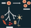
Multipotent-
become several cell types e.g. haematopoietic stem cells
Unipotent
- produce one cell type
E.g. all the epithelial stem cells will only ever become epithelial cells
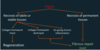
Regernation is: (2)
- The regrowth of cells
- Can be physiological
Regeneration: regrwoth of cells
Minimal evidence of injury (no scar)
- Only possible with minor injuries e.g. superficial skin incision/abrasion
- The regrowth of cells
regernation: can be physiologal
e.g. production of white cells in bone marrow (leucocytosis)

where are stem cells found
- In the skin
- Epidermis- basal level
- Unipotent stem cells which divide and differentiae into squamous cell
- Intestinal mucosa
- Bottom of crypts
- Unipotent stem cells producing simple columnar cells
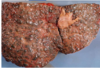
- Liver
- Between hepatocytes

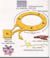
Cells can be organised into 3 different groups depending on their proliferative activity. These are:
Labile
Stable
Permanent
labile tissue
- These cells are short lived, and can easily be replaced by replication and maturation of stem cells.
- This means these tissues have a high reproductive capacity.
- For example, epithelial cells (such as those in the gastrointestinal tract) and haematopoietic tissue
stable tissue
- These cells normally have a slow rate of cell replication.
- However, they can divide rapidly when required.
- Hepatocytes, renal tubular cells and pancreas are examples of stable cells.
Permanent
- These cells are unable to undergo effective replication.
- left cell cycle and cannot re-enter
- Only a few stem cells are present.
- Neurones, skeletal muscle and cardiac muscle are an example of permanent cells.
which tissue types can regenerate
within labile and stable tissues when tissue damage isn’t extensive.
The presence of stem cells makes this possible, as they can divide and differentiate to replace the lost cells.
control of regeneration
Cell to cell communication occurs via local mediators such as growth factors, hormones or by direct cell-cell or cell-stroma contact. This communication allows control of regeneration.
Contact inhibition
Isolated cells replicate until they encounter other cells or ECM
- Cadherins bind between cells
- Inhibiting further proliferation

growth factors
Growth factors are polypeptides which are coded for by proto-oncogenes. The act in an autocrine (acting on the cell itself that secretes the growth hormone) or paracrine (acting on cells a short distance away) manner. They stimulate or inhibit cell proliferation through binding to specific receptors to stimulate gene transcription.

examples of growth factors
- Epidermal growth factor
- Vascular endothelial growth fact
- Platelet derived growth factor
- Tumour necrosis factor
Epidermal growth factor
induces mitosis in epitheilial cells, hepatocytes and fibroblasts
Vascular endothelial growth factor
induces developemnt of blood vessels in tumours, chronic inflammation and wound healing
platelet derived growth factor
causes migration and proliferation of fibroblasts, smooth muscle cells and monocytes
tumour necrosis factor
causes migration and proliferation of fibroblasts and secretion of collagenase
fibrous repair is the opposite of
regeneration
when does fibrous repair take place instead of regeneration
- necrosis of permanent tissue
- Labile or stable tissue
- collagen framwork destroyed
- on-going chronic inflammation
define fibrosis
replacemnt of functioning tissue with a scar
outline fibrous repair (formation of a scar)
- Bleeding- blood clot formation
-
Inflammation
- Acute then chronic digestion of the blood clot, damaged tissue and foreign body
- Minutes- days
-
Proliferation
- Of capillaries (angiogenesis)
- Fibroblast
- Myofibroblasts
- Extracellular matrix
- Collectively turns to granulation tissue
-
Remodelling
- Maturation of scar
- Reduced cell population
- Increased collagen
- Myofibroblasts contract
- Fibrous scar (weeks- years)
- Maturation of scar
function of granulation tissue
fills the gap; capillaries supply oxygen and nutrient; contracts and closes the defect
cells invovled in fibrous repair
Neutrophils
macrophages
lymphocytes
endothelial cells
fibroblasts
myofibroblassts
neutrophils and macrophages
phagocytosis and release of mediators
lymphocytes
eliminates pathogens and coordinates other cells
endothelial cells
proliferation and angiogenesis
fibroblast structure
- Spindle shaped nucleus
- Cytoplasmic extensions- interdigitate with each other

fibroblasts function
- Secrete collagen and elastin
- Form ECM
myofibroblasts
- Secrete collagen and elastin
- Form ECM

how are myofibroblasts like smooth muscle
can contract like smooth muscle due to expression of intracellular actin
collagen is the most common
protein in the body
29 different types
type 1 collagen
bones, ligaments, tendons, skin, sclera, cornea, vessels
type II
cartiliginous tissue
type III
skin, ligaments, blood vessels and internal organs
type IV
basement membrane, lens, glomerular filtration
type 5
skeletal muscle, blood vessels, lung, bone, tendon
collagen function
Provides extracellular framework
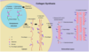
synthesis of collagen
- Pre-procollagen – polypeptide alpha chain produced in the ER of fibroblasts
- Undergoes Vitamin C dependent hydroxylation
- Pro-collagen alpha chains are then cross-lined to form triple helix in the cytoplasm of the (myo)fibroblast
- Procollagen secreted outside the cell and C and N terminals of procollagen cleaved à tropocollagen in ECM
- Tropocollagen become crosslinked to form microfibrils, fibrils and collagen fibres
acquired defective collagen disease
Scurvy
- Vitamin C deficiency
- Inadequate hydroxylation of pre-procollagen
- Defective triple helix= defective collagen
- Unable to heal wounds
- Tendency to bleed
- Tooth loss

inherited defective collagen disease
ehlers-danlos syndrome and osteogenesis imperfecta
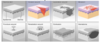
the skin can heal in two ways
by ….
- primary intention
- secondary intention
primary intention
- Incised wound
- Apposed edges (sutured)
- Minimal blood clot and granulation tissue
- Epidermis regenerates
- Dermis undergoes fibrous repair
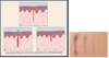
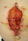
Secondary intention
- Significant tissue loss
- Unopposed edges
- Infection
- Ulcers
- Abscess
- Abundant clot, inflammation and granulation tissue
- Considerable wound contraction required (myofibroblasts)
- Dermis requires significant repair
- Epidermis regenerates from edges

Outline fracture healing
1) Haematoma surrounds the injury- granulation tissue forms, angiogenesis
2) Soft callus forms (1 week)
- Fibrous tissue and cartilage
- Woven bone begins to form
3) Hard callus (several weeks)
* Woven bone gradually organised into lamellar bone- osteoclasts
4) Remodelling (months- years)
* Lamellar bone remodelled to original outline of bone

Factors influencing wound healing can be
local or systemic
Factors influencing wound healing: local
Size
Location
Mechanical stress
Blood supply
Local infection
Foreign bodies
Factors influencing wound healing: systemic
- Age- older people take longer to heal
- Anaemia, hypoxia, hypovolaemia
- Obesity
- Diabetes- bacteria love sugary blood
- Drugs
- Vitamin deficiency
- Malnutrition
complications of fibrous repair
- Wound dehiscence
- excessive fibrosis
- adhesions
- loss of functions
- disruption of architecture
- excessive scar contraction
wound dehiscence
- Not enough collagen being laid down
- Occurs in:
- Obesity
- Increased pressure from inside by subcutaneous fat
- Elderly
- Malnutrition
- Steroid use
- Obesity
excessive fibrosis
Keloid scar
Too much collagen and elastin exceeding the edges of the wound

excessive scar contraction
Constriction of tubes
Fixed flexion deformities (contractures)

Adhesions
- Fibrous bands of collagen and elastin (typically seen in abdominal cavity after operation)
- Post-operative adhesions
- Can cause obstruction of tubes
loss of function
- Replacement of specialised tissue by fibrous tissue
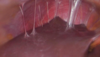
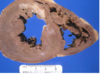
- E.g. in the heart after MI
- Develop arrythmia due to fibrosis tissue not being able to carry electrical conduction
Disruption of architecture
E.g. long term alcohol abuse and the liver