Neoplasia 1 Flashcards
a tumour
a swelling (any clinical detectable lump or swelling)
neoplasm
‘ a neoplasm is an abnormal growth of cells that persists after the initial stimulus is removed’ - new growth - just one type of tumour

oncology
study of tumours and neoplasm
hyperplasia
increase in cell number

hypertrophy
increase in cell size
regeneration
return to normal mechanisms e.g. the liver
- stimulated by GF

benign neoplasia
gross and microscopic appearances are considered to be innocent, implying that it will remain localised and not spread to other sites
- less well differentiated
cancer
a malignant neoplasm
neoplasm
an abnormal growth of cells that persists after the initial stimulus and invades surrounding tissues with the potential to spread to distant sites
metastasis
where a malignant neoplasm has spread from its original site to a new non-contiguous site
dysplasia
A pre-neoplastic alteration in which the cells how disordered tissue organisation.
characteristics of dysplasia
Reversible
Can exhibit considerable pleomorphism, with large hyperchromatic nuclei and high nuclear to cytoplasmic ratio
If detected early it can be prevented from progressing to cancer
summary of how a tumour can be defined

primary site
original location of malignant neoplasm
secondary site
place to which it has spread
how do we tell the difference between benign and malignant tumours
- Behaviour of the tumour
- E.g. if its metastases = probably malignant
- Appears different to the naked eye
- Differentiation
behaviour of benign neoplasms
- remain confined to their site of origin and do not produce metastases
- Grow in a confined area- capsule
- Pushing outer margin
- Rarely dangerous (depends on location)

where could a benign tumour be dangerous
in the brain- confined space
behaviour of malignant neoplasms
- invade and have the potential to metastasise
- Irregular outer margin and shape
- May have ulcerations and necrosis
- Infiltrative
- Grow very rapidly

definititon of differentitation
process of becoming different by growth or differentiation
differentitation and benign tumours
closely resemble the parent tissue- well differentiated
differentiation and malignant neoplasms
range from well to poorly differentiated dependent on how closely they resemble the cell origin

cells with no resemblance to any tissue are called
anaplastic
characteristics of poorly differentiated cells (6)
- Increased nuclear size
- Increases nuclear to cytoplasmic size
- Increased nuclear staining (hyperchromasia)
- Increased mitotic figures
- Abnormal mitotic figures
- Variation in size and shape of cells and nuclei (pleomorphism)
what term do clinicians use to indicate differentition
grade
- a high grade tumour is usually poorly differentiated
melanoma
- poorly differneited
- pigment= melanin

summary of behaviour of benign tumour
- grows locally
- retained functions of their cells of origin
histology of benign tumour
- resembles cells/tissue of origin
- few mitoses
- normal or mild increase in nuclear to cytoplasmic ration
- cells are unifrom throughout the tumour
summary of behaviour of malignant tumour
- expansive and invasive
- potential to metastasise
- less likely to retain functions of cells of origin and may soemtimes acquired unexpected functions
histology of malignant tumour
- failure ot fully differentiate
- many mitoses
- high nuclear to cytoplasmic ration
- cells/ nuceli vary in size and shape (pleomorphism)
pleomorphism
cells/ nuceli vary in size and shape
example of grade
grade 1- well differentiated
grade 3- poorly differnetiated

gradding pattern for the breast
Gleasons pattern
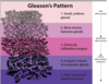
the higher the grade
the less liekly to survive

dysplasia and differentiation

Altered differentiation
Not yet invasive
Mild, moderate and severe- worsening differentiation
Dysplasia in the cervice
CIN1- mild dysplasia
Malignant changes just affecting lower third of epithelium
CIN2- moderate dysplasia
Lower 2/3rd
CIN3- severe dysplasia
Full thickness of the epithelium
why do we get neoplasia (2)
- Cacrinogenesis
- Non-lethal genetic damage (can be genetic, can be spontaneous, can be environmental)
mutations and neoplasia
- Accumulated mutations in somatic cells
- Mutations are caused by initiators- mutagenic agents
- Promoters then cause cell proliferation
- A tumour is formed by the clonal expansion of a single precursor cell that has incurred genetic damage
give 4 intiators
- Chemicals
- smoking
- alcohol
- diet and obesity
- Infectious agents
- HOV
- Radiation
- Inherited mutation
progression of a normal cell to neoplastic cells due to mutation
1) normal cell population
2) Intiator causes mutation in one cell
3) Mitosis of cell and vertical transmsision of mutation
4) most cells start to have mutation

germline mutations
e.g. BRCA1 skips out this progressing stage*
a selection of cell is monoclonal if
they all originated from a single founding cell
neoplasms emerges from
this group of cells by a process called progression
- characterised by an accumulation of more mutations
- mutations may give the cell advanatges such as faster growth

4 classes of normal regulatory genes
- Growth promoting proto-oncogenes
- Growth inhibiting tumour suppressor genes
- Genes that regulated programmed cell death – apoptosis
- Genes involved in DNA repair
proto-oncogenes
- participate in signalling pathwyas which drive proliferation
- ‘increase in function’ mutation
- when mutated become Oncogenes
oncoge
oncogenes
- are created by mutations in proto-oncogenes and encode proteins called oncoproteins that have the ability to promote cell growth in the absence of normal growth promoting signals
- They can transform cells despite a normal copy of the same gene
- Oncogenes are dominant over their normal counterparts (allele)
oncogene mutations can be targeted by
inhibitors eg.g. BRAF inhibitor
- therefoe eimportnant to identify oncogene causing cancer

tumour suppressor genes
- Normal function is to stop cell proliferation
- Generally cause a loss of function
- In most instances both alleles must be damaged for transformation to occur
- Abnormalities in these genes leads to failure of growth inhibition (e.g. TP53)
apoptosis regulating genes
- May acquire abnormalities that result in less cell death and enhanced survival of mutated cells
- E.g. follicular B cell lymphoma
DNA repair genes
- Loss of function mutations
- Contribute indirectly to carcinogenesis
- Impair the ability of cell to recognise and repair non-lethal genetic damage in other genes
- As a result affected cells acquire mutations at an accelerated rate, a state referred to as a mutator phenotype and is marked by genomic instability
