Protozoa 1: Intestinal Protozoan Disease Flashcards
What contributes most to diarrhoeal illnesses in tropics?
WASH - lack of water, sanitation, hygiene
Which organisms are most likely to cause tropical diarrhoeal illness?
• Parasites
o Entamoeba Histolytica [subacute – weeks]
o Schistosoma
o Balantidium Coli [associated with pigs in Philippines]
• Bacteria
o Shigella [rapid – more fulminant then amoebiasis]
o [Some E Coli, Campylobacter, Salmonella]
o C Diff
Which organism causes amoebiasis and how does it most commonly present?
Entamoeba Histolytica is an intestinal parasite.
The other entamoeba do not cause disease.
What is the incidence of amoebiasis and who is most at risk?
- Majority of infection asymptomatic
- 40 - 50M cases a year of symptomatic amoebiasis
- 40k - 110k deaths a year
- Faeco-oral transmission
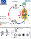
What is the life-cycle of amoeba?
- Four-nucleated cyst ingested
- Eight amoebic trophozoites released
- The trophozoites then reside in the colon feeding on bacteria. They multiply by binary fission.
- The trophozoite is hugely motile and when invasive contains ingested red blood cells (if these found in stool = diagnostic).
- As they move towards the end of the colon, stool becomes more solid, less to feed on for the trophozoites so they shed their wall and cysts into the stool
- Cysts can survive days to weeks
- Only E hystolytica can cause invasive disease
- Trophozoites therefore found in diarrhoeal stool, while cysts found in formed stool
- Cysts are killed only by harsh conditions. Trophozoites are non infective

How do amoebae cause disease?
- Bind to host intestinal cells via galactose-binding lectin
- Amoebapores disrupt target cell causing apoptosis
- Amoeba phagocytose the dead cell -> develop mucosal ulcers
- “bind-lyse-eat”
- Causes inflammatory response
What is the clinical presentation of amoebiasis?
Most cases asymptomatic
Clinical Features of Intestinal Amoebiasis
- Incubation can range from days to years! It can present >decade post-infection.
- Most people are asymptomatic.
- Spectrum of disease ranges from asymptomatic to fulminant necrotising colitis.
- Symptoms include diarrhoea that may contain mucus and blood, abdominal pain, rectal ulceration. In the immunosuppressed the picture can be more florid/toxic.
- Complications include:
- Amoebic abscess – most commonly liver
- Amoeboma: inflammatory mass in ileocaecam that can cause obstruction
- GI haemorrhage, Colitis – from mild to fulminant, Strictures, Peritonitis, Skin ulceration.
What are potential consequences of amoebiasis?
- Amoebic abscess (mostly liver)
- Amoeboma (chronic inflammatory mass, ileocaecal region)
- Haemorrhage
- Peritonitis
- Post-dysenteric ulcerative colitis
- Skin ulceration (usually perianal)
- Stricture
What are the clinical features of amoebic liver abscess?
- E Histolytica can invade the liver to form a single abscess > multiple abscesses
- Again days to decades may follow infection
- Right lobe 4x more common then left and 10x more common in M > F
- A minority have a clear history of proceeding dysentery.
- Complications include: rupture into lung/pleura/pericardium, peritoneum and haematogenous seeding into any other organ
Clinical Features
- Fever, sweats, rigors may occur. RUQ pain. No Jaundice.
- Can have reactive right-sided pleural effusion with associated symptoms
- Clinically it cannot be confused with hydatid cyst (these x2 pathologies can cause some anxiety in differentiating them on CT), which is slow growing with pt typically presenting with distended abdomen, resultant abdominal pain or incidental finding. Amoebic liver abscess pts are toxic.
Extra-intestinal amoebiasis can be caused by infection with Entamoeba histolytica

How do you diagnose amoebiasis?
- Cysts in the stool don’t help as indistinguishable from non-pathogenic E. dispar
- Fresh stool sample with trophozoites that have ingested RBCs are diagnostic.
- Stool antigen testing is sensitive/specific. However, it remains+ lifelong.
-
Serology is sensitive but often unhelpful in endemic areas.
- Amoebic serology in amoebic liver abscess is the preferred diagnostic method
- Colonoscopy shows ulceration and biopsy results stain parasites magenta.
- Imaging

How do you manage amoebiasis?
Treatment
- Fluid/electrolyte balance, maintaining haemodynamics, etc.
- Metronidazole for 1 week.
- Followed by Amoebicide: Diloxanide or Paromomycin for 1 week.
- In reality – a patient with a liver abscess will be treated with empirical antibiotics as well to cover for possible bacterial aetiology.
- Surgery may be indicated if perforation etc.
- Indications for ALA aspiration: any abscess in left lobe of liver must be aspirated immediately to avoid rupture into pericardium, failure to respond to medical treatment in 2-3 days, impending rupture, marked diaphragm elevation, severe pain, large abscesses >10cm. In practice most are aspirated for diagnostic testing.
Which organism causes giardiasis?
Giardia duodenalis, Giardia lamblia, Giardia intestinalis are all the same thing
- Giardia is a flagella protozoa that inhabits the small bowel
- The trophozoite stage is a flattened 15μm pear-shaped thing with 4 pairs of flagella
- They then encyst into an oval 10μm containing four small nuclei

How is giardiasis transmitted?
- Faeco-oral
- Mostly infected water
- Cysts may also be ingested on food
- Lack of WASH - water, sanitation, hygiene
- The cysts can survive outside the body for several week udner favourable conditions
What is the life cycle of giardia?
Cysts are resistant forms and are responsible for transmission of giardiasis. Both cysts and trophozoites can be found in the feces (diagnostic stages) . The cysts are hardy and can survive several months in cold water. Infection occurs by the ingestion of cysts in contaminated water, food, or by the fecal-oral route (hands or fomites) . In the small intestine, excystation releases trophozoites (each cyst produces two trophozoites) . Trophozoites multiply by longitudinal binary fission, remaining in the lumen of the proximal small bowel where they can be free or attached to the mucosa by a ventral sucking disk . Encystation occurs as the parasites transit toward the colon. The cyst is the stage found most commonly in nondiarrheal feces . Because the cysts are infectious when passed in the stool or shortly afterward, person-to-person transmission is possible. While animals are infected with Giardia, their importance as a reservoir is unclear

How do giardiae cause disease?
- Trophozoites adhere to intestinal wall
- Interfere with enzyme activity and disrupt brush border
- Stimulate inflammatory cytokine response
- Trophozoites encyst as the pass along bowel
- Cyst is oval and contains four small nucle and a central refractile axoneme.
- Cysts are infective as soon as they are passed
- Excyst in the upper GI tract and liberate trophozoites in the newly infected host

What is the clinical presentation of giardiasis?
Incubation period is usually around a week but can be longer
- Most infections are asymptomatic
- Classic diarrhoea, abdominal camps, flatulence, bloating, eggy taste (taste of rotten eggs), significant weight loss
- There is never blood in the diarrhoea, which can be water-greasy
- Most become asymptomatic after a period of time
- Lactose intolerance can follow in spite of giardia eradication
How do you investigate for giardia?
- Stool microscopy for cysts -> higher sensitive if concentrate sample
- Stool antigen testing -> available for giardia, entamoeba, cryptosporidium
- Stool PCR assays
- “String test”
Essentially you want to find evidence of cysts or trophozoites
How do you treat giardiasis?
- Rehydrate
- Metronidazole TDS for 5-days or Albendazole will reduce severity and duration of persistent symptoms.
- Tinidazole effective as a single dose of 2g for adults
- Nitazonaxide another option
What are the three intestinal coccidia infection?
- Cryptosporidium Hominis
- Cyclospora
- Cystoisospora belli
Features of cryptosporidium
- Cryptosporidium have global distribution and is faecal-oral spread
- They are implicated in waterborne epidemics of diarrhoeal illness (C. hominis in humans and C. parvum in broader range of animals)
- Resistant to chlorination and small enough to pass through filtration systems
- They are responsible for childhood diarrhoea, travellers’ diarrhoea, protracted symptoms in immunocompromised, waterborne outbreaks in both developed and developing countries. Does usually affect immunosuppressed/infants.
- Diarrhoea usually lasts 1-2 weeks but as with all can go on for several weeks.
- Most common chronic diarrhoeal illness in HIV+ pts
Pathophysiology
- oocyst -> releases four sporozoites -> invade epithelial cells -> may invade colon and biliary tree
- ability to produce thin-walled oocysts that maintain its life-cycle within the host (‘internal autoinfection’), or to produce thick-walled oocysts that are excreted in faeces
Investigation
- Faecal microscopy: acid-fast oocysts that are 5μm
- Specific antigen-assays are more sensitive
Management
- Supportive and initiation of ART

Features of cyclospora
- Spread by faecal-oral route but the cyst that passes in stool is not infective for 1-2 weeks until it has developed – so contamination is through environment.
- Symptoms vary from asymptomatic to flu-like symptoms with diarrhoea of a rather giardia-like nature. The diarrhoea can last a month and come and go.
- Diagnosis:
o The oocysts are slightly larger then cryptosporidium being 10μm.
o They also stain pink on modified ZN stain (ugh!).
• Treatment: Co-trimoxazole.
Features of cystoisospora
- The least common of the intestinal coccidian infections
- Occurs worldwide but usually affects the immunosuppressed
- Acute diarrhoea that can go on for weeks causing weight loss.
- Uniquely, eosinophilia may be present.
- Diagnosis:
o The largest oocysts of them being about 25μm and elliptical
o They stain with acid-fast
• Treatment: Co-trimoxazole


