pelvis viscera 1 of 2 Flashcards
transverse the wall of the bladder in an oblique direction. How does this assist function?
ureters
- transverses the wall of the bladder in the oblique direction
- two thing inhibit refluc of urine into the ureter
- pressure from a filling bladder
- bladder contraction during micturition
- two thing inhibit refluc of urine into the ureter

posterior to vas deferens(male) or pass below uterine artery(female)
retroperitoneal in both sexes
ureters

what arteries supply the plvic region of the ureter?
- arteries
- male
- inferior vesicular artery
- female
- uterine and vaginal
- male
what are are the veins and lymphatic structure/desitination for the palvic portion of the ureter
- veins
- vesical venous plexus around inferior end
- sex dpendent
- male
- inferior vesicular
- female
- uterine and vaginal
- male
- lymphatic
- internal iliac nodes
describe the GVE, parasympathetic, and GVA
- Sympathetic
- T11-T12/ L1-L2
- superior hypogastric plexus
- inferior hypogastric plexus
- T11-T12/ L1-L2
- parasympathetic
- S2-S4
- pelvic slanchnic nerve
- S2-S4
- visceral afferents
- travel with
- pain with sympathetic and parasympathetic
- travel with
Why would the ureter be at risk durin a hysterectomy?
ureter is at risk during hysterectomy because uterine artery pass over it

loin to groin pain is a reflection on which nerve fibers?
How could a ureter be involved?
T11-L2
ureters are innervated by GVA fibers that accompany GVE sympatheric fibers from the T11-L2 spinal cord levels.
Passage of a kidney stone produces loin-groin pain (abdomen->pelvis)
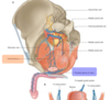
explain and diagram the urinary bladder
- components
- body
- fondus(base)
- spex
- neck(surroung the urethra)

where does the urethra meet the bladder?
urethra meets the bladder at the angles of the smooth walled trigone

this remnant runs upward from the apex of the bladder. What happens if it does not seal off?
median umbilical ligament (remnant of urachus) run upward from apex of the bladder
if the lumen of the urachus persists in newborn, urine leaks from umbilicus
what are the ligaments that support urinary bladder? sex specific
- male
- puboprostatic ligament
- female
- pubvesical ligaments
explain the urethral structture and distinguish between sexes
- female
- short, mostly in perineum
- empties into vestibule
- external urethral sphincter (perineum)
- male
- preprostatic
- internal uretthral sphincter
- prostatic part
- where urinary and reproductivetract merge
- membrenous part
- enternal urethral sphincter
- spongy part
- ends in navicular fossa
- preprostatic

differentiate the location of the urinary bladder between age groups
urinary bladder is in
- adult
- in pelvis
- can be damaged in pelvis trauma when empty
- child
- abdomen
- can rupture if full in abdominal trauma
shorter urethra and opeining in vesitbule leaves females…what about hospilization?
increase susceptible to bladder infection (cystitis)
make it easier for passage of catheters and cystoscopes/wall of urthral bulb can be damaged in males

why is bladder cancer a concern?
can invade ureter, rectum, uterus, seminal vesicles and prostat as well as lateral wall of pelvic cavity.
what arteries/veins are located anteriosuperior to the bladder?
superior vesical artery and vein

artery/vein found near fundus and neck of bladder and pelvic urethra
sex specific
- male
- inferior vesical artery and vein
- female
- vaginal artery and vein

what drains the fundus of the bladder and proximal urethra? how?
vesical veinous plexus
- drain fundus of the bladder and proximal urethra
-
Inferior vesical/vaginal veins
- to the internal ilac vein
-
sacral veins** into the **vertebral venous plexuses
- may provide a route for tumor metastasis to the vertebral column, pelvic bones and skull
-
Inferior vesical/vaginal veins
- male
- vesical veinous plexus is continuous with the
- prostatic venous plexus
- dorsal vein of penis
- vesical veinous plexus is continuous with the
- female
- communicates with
- uterovaginal venous plexus
- dorsal vein of clitoris
- communicates with
describe the nerve supply of urinarybladder and proximal urethra. differentiate only between sympathetic and parasympathetic
sex
- male
- vesical pplexus for bladder and prostatic plexus
- female
- vesical plexus for bladder and proximal urethra
visceral difference
- visceral efferents
- parasympathetic
- contraction of detrusor muscle
- male
- inhibition of internal urethral sphincter
- sympathetic
- male
- contraction of internal urethral sphincter
- prevent urination in shy male- when the sympathetic tone is high
- contraction of internal urethral sphincter
- male
- parasympathetic
- visceral afferents - pain trave laccordin to pain line
- parasympathetic
- fundus and neck of bladder and proximal urethra
- sympathetic
- anteriosuperior portions of bladder
- parasympathetic
how does urine pass to by the ureters to the bladder?
peristaltic contractions of the ureters via - increase in parasympathetic tone
explain the nerves involved in the process of micturation reflex
mituriction
- internal pressure ofthe filling bladder close ureters to prevent backflow
- bladder wall stretching stimulate GVA fibers (S2-S4) going to spinal cord
-
parasympatheticGVE fibers S2-S4 cause urine to flow in urethra
- stimulation of detrusor muscle in wall of bladder
- male only
- relaxation of the internal urethral sphincter
voiding
- relaxation of levator ani during urination (GSE)
- abdominal muscle contracted (GSE) leading to an increase in bladder pressure
stimulation of detrusor muscle in walls of bladder is caused by?
parasympathetic fibers S2-S4 cause uring to flow in urethra
- sitmulation of detrusor muscle in walls of bladder
- male only
- relaxation of the interna lurethral sphincter
what does voiding involve?
- relaxation of the levator ani during urination (GSE)
- abdominal muscle contracted (GSE) increasing the bladder pressure
-
pudendal nerve (GSE
- volunatary relaxation of external urethral sphincter
- branches of pudendal nerve lead to release of urine outside of the body
- male only
- final drops pushed out of urethra by conraction of bulbospongiosus muscle controlled by branches of pudendal nerve(GSE)


