pelvic walls and cavities 1 of 2 Flashcards
what forms the pelvis?
pelvis is part of the trunk with in the girdle formed by 2 hip bones, divided by a plevic brim.
- greater pelvis/ false pelvis
- inferior abdomen
- lesser pelvis/ true pelvis
- encloses pelvic cavity
- contains
- urinary elements
- gastrointestinal and reproductive system
- inlet(brim) and outlet
what divides the pelvis?
pelvic brim
differentiate between the pelivis and the perinuem
- pelvis
- False/greater pelvis
- inferior abdomen
- true/lesser pelvis
- encloses pelvic cavity containing elements of the
- urinary, gastrointestinal and reproductive system
- inlet adn outlet
- encloses pelvic cavity containing elements of the
- False/greater pelvis
- perineum
- area between the thighs, inferior to floor of the pelvic cavity
- bounded by pelvic outlet
-
contains
- perineal mucles and glands
- anal canal
- lower vagina
- portion of the urethra
- external genitalia
what lies inferior to the floow of the pelvic cavity?
perineum
what is the perineum bound by?
pelvic outlet
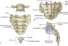
what does the green line represent?
the boundry between the pelvis and perineum
what are the roles of the bony pelvis? what are the borders?
roles in relation to pelvis and perineum
- protect and support viscera
- attachment for muscles, fascia and erecrile bodies of genitalia
borders
- 2 hip bones (os cozae, innominate bones)
- sacrum
- coccyx
what is the Os COxae composed of ?
- ilium
- ischium
- pubis
acetabulum- fusion of all three regions of the os coxae.
- cavity forms the hip joint iwth head of femur
- to become one bone between 16 & 18 years ols
which portions of the bony pelvis lie in the same axis?
Anterior supreior Iliac spine and the pubic symphysis lie in the same vertical plane
coccyx and the upper margin of pubic symphysis lie in the same horizontal plane
coccyx
- how many vertebrae?
- articulate
coccyx
- 3-4 fused vertevrae
- articulate with sacrum (sacrococcygeal joint) via cornua
- two horns that project upward to the sacrum
- be sure to differentiate between the sacral and coccyx coruna
sacrum
- vertebrae?
- articulate
- canal?
- foramina?
- important super structures
- 5 fused vetebrae
- S1-S5
- articulate with
- L5
- saccral coruna and the coccyx coruna
- sacral canal and haitus:
- cauda equina
- anterior and posterior sacral foramina
- where anterior and posterior rami of sacral spinal nerves exit
- super structures to ID
- promontory-anterior part of the vetebral body that projects forward
- ala- wing like transverse processes on S1
- anterior part of the vetebral body that projects forward
promontory-anterior part of the vetebral body that projects forward
wing like transverse processes on S1
ala- wing like transverse processes on S1
describe the lumosacral joints. Types of joints? parts? support?
Lumbsacral joint= joint between sacrum and lumbar 5
- zygapophyseal joints (synovial)
- occurs beterrn adjacent and superior articular processes
- intervertebral disc between L5 and S1 (symphisis)
supported by
- iliolumber ligaments
- transverse process to the ilium
- lumbosacral ligaments
- transverse process to the sacrum
list the ligaments and their attatchment involved in the lumbosacral joints
- iliolumber ligaments
- transverse process to the ilium
- lumbosacral ligaments
- transverse process to the sacrum
joint between the sacrum and ilium.
sacroliliac joint- two protions
- synovial potion
- iliac and sacral protion- articular surfaces
- anteriorsacroiliac ligament
- syndesmotic (fibrous) portion
- iliac and sacral tuberosities
- interosseous sacroiliac ligament (STRONGEST)
ligaments
- anterior sacro-iliac ligament
- interosseous sacro-iliac ligament
- STONGEST
- posterior sacro-iliac ligament
What the following structures belong to and are they synovial, syndesmostic, symphysis, fibrocartilagenous?
- zygopophyseal joint
- articular surface of iliac and sacral
- intervertebral disc between L5-S1
- interpubic disc
- iliac and sacral tuberosities
- lumbosacral joints- 2 joints
- zygopophyseal - SYNOVIAL
- inttervertebral disc between L5 and S1 - SYMPHYSIS
- sacroiliac joint- two portions to the joint
- articular surfaces-SYNOVIAL PORTION
- iliac and sacral tuberosities - SYNDESMOTIC (FIBROUS) PORTION
- pubic symphysis
- articular surfaceces on body of the pubic bone- FIBROCARTILAGENOUS JOINT
- interpubic disk-pad of fibrocartilage that joins the bones
- articular surfaceces on body of the pubic bone- FIBROCARTILAGENOUS JOINT
describe the ligaments and joints involved with the pubic symphysis
- pubic symphysis
- interpubic disc- fibrocartilaginous structure that joins the articular surfaces on body of pubic bones
- ligaments
- superior
- superior pubic ligament
- inferior
- inferior pubic ligament
- superior
which ligament forms the greater sciatic foramen?
sacrospinous ligament
- attatches
- anterior surface of the sacrum
- to ischial spine
what ligament generates the lesser sciatic foramen?
sacrotuberous ligament
- attaches
- lateral margin of the sacrum
- to ishcial tuberosity
weight of the body causes rotation at what location? what stops the formation of lordosis?
weight of the body causes the inferior sarum to rotate superiorly att the sacro-iliac joint.
this tendency (lordosis) is opposed by ligaments: sacrospinous and sacrotuberous
- ligaments prevent upward tiliting of sacrum
describe th border involved with the pelvic inlet
- sacral promontory
- S1
- sacral alae
- linea terminalis
- arcuate line
- pecten pubis aka:pectineal line
- pubic crest and pubic symphysis
describe the perimeter of the pelvic outlet
pelvic outlet
- pubic symphisis
- ischiopubic rami
- ischial tuberosities
- sacrotuberous ligament
- coccyx
describe the following for malw and female
- greater pelvis
- lesser pelvis
- pelvic inlet
- pelvic outlet
- sacrum
- ischial spines
- pubic arch & subpubic angle
What causes the relaxation of the pelvic ligaments and increased joint mobility? What is the purpose of this?
the homrone relaxin
purpose: increase in pelvis diameter to facilitate childbirth but may cause back pain and pelvic pain
Back pain and pelvic pain associated with pregnancy is caused by what? explain the process
Rlaxin->relaxes the pelvic ligament and increases the mobility of joints.
this is all to increase the pelvic diamter and facilitate childbirth
what is measured as to consider the capacity for childbearing?
-
Interspinous distance
- the narroest part of birth canal
- increases 10-15% in pregnancy
-
lesser poelvis AP diameter
- narrowest fixed part of the birth canal
- determine possibility of vaginal birth
A woman can expect what to increase in distance for childbearing? What is the doctor measuring when they state the baby will not be able to be birthed vaginally?
- the interspinous distance increases 10-15% in pregnancy
- the child has exceeded the lesser pelvis AP diameter
- the narrowest part of the birth canal
what is the sacral hiatus used for during childbirth?
epidural anesthesia
Where is the caudal epidural usually placed for childbirth?
sacral hiatus used in caudal epidural anesthesia
A patient presents from a car accident with a straddle injury. What has been damaged?
fracture of 4 pelvic rami.
A older genetlemen feels himself slipping from the ladder and falls on his feet from 10 feet up. what part of the pelvis is in jeapordy of damage?
acetabulum fracture: trasmitted from lower limbs
pelvic fractures can lead to 3 distinguished areas of concern. What are they
- straddle injury-fracture of 4 pelvic rami
- car accident
- fracture of acetabulum- transmitted from lower limbs
- fall on feet
- may cause injury to pelvic soft tissues, organs nerves and vessels
what are two common issues with teh sacroiliac joint?
- degenerative changes
- may become fibrous and ossify
- inflammatory changes
- associated with injury, infection (intravenous drug use) or part of some inflammatory diseases
describe the following structures in the formation of the pelvic cavity
- anterior wall
- posterior wall
- lateral walls
- pelvic floow
- roof
- anterior wall
- pubic bones and pubic symphysis
- bones = bodies and rami
- pubic bones and pubic symphysis
- posterior wall
- sacrum/coccyx, piriformis muscle and pelvic fasciae
- lateral walls
- hip bone, obturator membrane, obturator internus muscle** + fasciae, **sacrospinous ligament and sacrotuberous ligament
- pelvic floor
- formed by the muscular pelvic diaphragm (levator and coccygeus muscles) and its fasciae
- the pelvic diaphragm separates the pelvic cavity from the perineum
- formed by the muscular pelvic diaphragm (levator and coccygeus muscles) and its fasciae
- roof
- the pelvic inlet is open and continuous with the abdominal cavity
list the following for the piriformis
- boundary
- origin
- insertion
- innervation
- action
- boundary
- posterior wall
- exit via greater sciatic foramen
- posterior wall
- origin
- anterior sacrum (foramina S2-S4)
- insertion
- greater trochanter of femur
- innervation
- nerve to the piriformis L5-S2
- action
- thigh abduction and external rotation stabilize hip joint
list the following for the obturator internus
- boundary
- origin
- insertion
- innervation
- action
- boundary
- lateral wall
- exit via lesser sciatic foramen
- lateral wall
- origin
- ilium and ischium pelvic surface: obturator membrane
- insertion
- greater trochanter of femur
- innervation
- nerve to the obturator internus (L5 -S1)
- action
- external rotation of thigh stabilize hip joint
damage to the L5-S1 will leave which of the muscles of the pelvic wall paralyzed and partially paralyzed? what action will affected?
- piriformis
- nerves: L5-S2
- partially affected
- action
- thigh abduction and external rotation
- nerves: L5-S2
- obturator internus
- nerve: L5-S1
- fully paralyzed
- action
- external rotation of thigh stabilize hip joint
- nerve: L5-S1
define the route of the superior gluteal nerve
through the greater sciatic foramen, above the piriformis
define the route of the sciatic nerve, what other nerves travel with this nerve?
Greater sciatic foramen, below the piriformis
- all nerves:
- sciatic nerve
- inferior gluteal
- posterior femoral cutaneous
- qudaratus femoris nerves
define the route of the pudendal nerve? what other nerve travels with this?
lesser sciatic foramen
- this includes pudendal nerve and obturator internus
define the route of the obtorator nerve
obtorator canal, not through greater or lesser sciatic foramen
what are the muscles of the pelvic wall?
- levator ani
- iliococcygeus
- pubococcygeus
- puborectalis
- coccygeus
define the following for the levator ani
- the muscle contributions
- boundary
- origin
- insertion
- innervation
- action
- contributions
- iliococcygeus
- pubococcygeus
- puborectalis
- boundary
- floor
- origin
- body of pubis
- tendinous arch of obturator fascia
- ischial spine
- insertion
- perineal body
- coccyx
- annococcygeal ligament
- innervation
- S4- nerve to levator ani
- S2-S4-inferior rectal nerves
- Coccygeal plexus
- action
- support pelvic viscera and resist tintra-abdominal pressure
define the following for coccygeus
- boundary
- origin
- insertion
- innervation
- action
- boundary
- floow-over sacrospinous ligament
- origin
- ishial spine
- insertion
- sacrum
- innervation
- Br of S4 and S5 spinal nerves
- action
- helps support pelvic viscera
- flexes coccyx
forms a u shpaed sling ano rectojunction. name adescribe the functions behind this muscle
puborectalis
- forms a U shaped muscular sling
- passes posterior to the anorectal junction
- responsible for
- anorectal flexure
- playes a major role in maintaining fecal acontinence
- mechanism
- relaxation of the puborectalis allows the rectum to attaina amore vertical position, therby enabling a more direct path for feces to be eliminatted
what appertture is walled by the levator anus?
anal aperture
what are the borders of the anterior pelvic opening?
urogenital hiatus
levator anus-pubic symphisis
low estrogen affects what part the pelivs and how? What else affects this area?
plevic floor may
- atrophy because reduced estrogen, in postmenapause
- weaken in connective tissue disease
- childbearing
how does childbearing affect the pelvic floor?
- can alter the strength and elasticity of the pelvic diaphragm
- can damage nerves in the pelvic region
- can produuce tears and muscle detachment
- MOST OFTEN TORNpubococcygeus &puborectalis
a weakening in the pelvic floor can lead to what pathologies?
- prolapse of internal pelvic organs
- intolerance
- feca
- urinary
What can be done to combat the pelvic floor?
kegel exercises can be used to strengthen the pelvic floor and reduce incontinence and prevent prolapse
what is the primary blood supply to the pellvis?
internal iliac- from the common iliac
Describe the posterior division of the internal iliac artery
note primary blood supplyto pelvis: internal iliac
- iliolumbar
- feeds the iliac, lumbar and spinal branches: posterior abdominal wall and cauda equina
- lateral sacral
- supply posterior pelvic wall, vertebral sacral canal, muscle and skin posterior to sacrum
- superior gluteal
- passes between lumbosacral trunk and S1
- exits throug hgreater sciatic foramen to supply gluteal area and pelvic wall.
describe the following arteries
- arises from abdominal aorta atL1
- arises during bifurcation of the aorta into 2 common iliac arteries at LV4
- passes between lumbosacral trunk and S1
- exits between roots of s2andS3(or S1 and S2)to gluteal area and pelvic wall
- Ovarian /testicular artery
- arises from abdominal aorta
- median sacral artery
- at bifurcation of the aorta into 2 common iliac arteries of LV4
- Superior gluteal artery
- passes between lumbosacral trunk and S1
- exits through greater sciatic foramen to supply gluteal area and pelvic wall
- Inferior gluteal artery
- exits between roots S2 and S3 (or S1 and S2) to gluteal area and pelvic wall
which aretery is a direct continuation of the inferior mesenteric artery?
superior rectal artery
- direct continuation of the inferior mesenteric artery
describe the anterior division of internal iliac artery
- umbilical artery
- superior vesicular artery to bladder and ureter
- medial umbilical ligament (remnant artery to placenta)
- obturator artery
- exits throug hobturator canal to middle thigh
- vaginal artery (female)/inferior vesical artery (male)
- to bladder, prostate, seminal glands
- middle rectal artery
- internal pudendal artery
- courses with pudendal nerve into perineum
- inferior gluteal artery exits between roots of S2 and S3 (or S1 and S2) to gluteal area and pelvic wall
- uterine artery (female)- not shown in image
describe the vasculature of the male pelvis
descrie the vasculature of the female pelvis
Describe a femoral hernia and the dangers involved in the surgical repair.
Aberrant obturator artery** or **accessory obturaor artery** arises from the **inferior epigastric artery
- br of external iliac artery
- descends across the pelvic brim to obtorator canal
- These arteries run close to the femoral ring and is in danger during the surgical repair of a femoral hernia
Desceribe the venous formation around organs- 4 examples
Form communicating plexuses of veinsaround pelvic organs
- vesical venous plexus
- prostatic venous plexus
- uterovaginal venous plexus
- rectal venous plexus
describe the drainage of the pelvis
- main drainage of the pelvis
- internal iliac vein
- drains into the common iliac vein, which continues as the inferior vena cava at the L5
- internal iliac vein
- others
- inferior mesenteric vein (portal system)
- superior rectal vein
- inferior vena cava
- median sacral vein
- right ovarian/right testicular vein
- (left via renal vein)
- vertebral venous plexus
- pathway for pelvic caner to metastasize to spinal cord and brain
- inferior mesenteric vein (portal system)
Discuss lymph drainage from the pelvis
- primarily
- ovaries/testes
- primarily
- internal and external iliac nodes that draing into common iliac nodes
- lymp from ovaries and testes drain into
- lateral aortic (lumbar nodes)** that drain into the **preaortic nodes
- eventually drains via the thoracic duct into the left brachiocephalic vein
describe the primary drainage of the pelvis
internal and external iliac nodes drain into common iliac nodes
describe the drainage from the ovaries and testes
lateral aortic (lumbar nodes) that drain into preaortic nodes
