Pathology - GI Flashcards
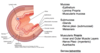
Name all the histologic layers of the gut wall.
Mucosa includes epithelium, lamina propria, and muscularis mucosa
Submucosa includes glands (Brunner’s in the duodenum), and submucosal nerve plexi
Muscularis externa aka propria (inner circular, outer longitudinal) w/ myenteric (Auerbach’s) plexus
Serosa/adventitia
How can you tell the difference between small intestine and colon histologically?
Small intestine will have more prominent villi, while the colon is more flat-topped.
Where in the GI tract is this? Name the cell types.

Small intestine

What is the function of paneth cells in the small intestine?
Make anti-microbial shit like lysozyme and defensins
Name one distinguishing hisotlogic feature in the duodenum, jejunum, and ileum, respectively.
Duodenum: submucosal (Brunner’s) glands that make alkaline mucus
Jejunum: plicae circulares
Ileum: Peyer’s patches (lymphoid nodules)
What abnormal antibodies are associated with Celiac disease?
Anti-endomysial, anti-gliadin, and anti-tissue gransglutaminase antibodies.
What skin finding is associated with Celiac disease?
Herpetitis dermatiformis

Which of these histologic sections was taken from a patient with Celiac disease? Explain.

Bottom was from Celiac disease - villi are blunted like a lawnmower came along
Aside from blunted villi, what other histopathologic finding is seen in Celiac disease?
Intraepithelial lymphocytes

Are small bowel neoplasms common?
No
What is one possible congenital cause of sudden onset of hematochezia in a child less than 2 years old?
Meckel’s diverticulum
What is the “rule of 2s” regarding Meckel’s diverticulum? How does it cause GI bleeding?
Rule of 2s: happens in 2% of the population, presents in kids 2 years or younger, 2 within 2 feel of the ileocecal valve, 2 inches long.
Causes bleeding cuz it often contains gastric mucosa that makes acid, which escapes the diverticulum to the surrounding small bowel, which isn’t used to acid, causing ulceration -> bleeding
Where along the GI tract is this?

Colon

What is the name of the nerve plexus found within the submucosa of the wall of the GI tract?
Meissner’s plexus
What is the name of the nerve plexus located between the two muscular layers of the muscularis externa/propria?
Auerbach’s/myenteric plexus
Name it, do it.

Meissner’s plexus
Knowing that ulcerative colitis primarily affects the mucosa and submucosa, explain why there is a risk of developing toxic megacolon.
Damage of the submucosa can damage Meissner’s plexi -> messed up peristalsis -> gut fills up with stuff and bursts
Compare UC with Crohn’s with regards to their GI tract involvement (location, pattern, etc.).
UC: involves rectum and ascends proximally, continuously; only involves mucosa and submucosa.
Crohn’s: happens anywhere along the GI tract (but most often seen in the terminal ileum), inflammation is transmural.
Which disease puts one at the greatest risk of GI carcinoma: UC, or Crohn’s?
Ulcerative colitis
Which inflammatory bowel disease is a “string sign” on imaging associated with?
What about these associations…?
Pseudopolyps
Creeping fat
Sclerosing cholangitis
Lead pipe on imaging
Granulomas
Cobblestone mucosa
Rectal involvement
Strictures
Skip lesions
Crohn’s disease
UC: pseudopolyps, primary sclerosing cholangitis, lead pipe on imaging, rectal involvement
Crohn’s: skip lesions, creeping fat, strictures, granulomas, cobblestone mucosa
Describe the GI wall layer involvement of diverticular disease.
The mucosa bulges out
What is Hirschsprung disease and how dose it present?
Failure of neural crest migration -> absence of nerve plexi in the distal colon -> poo gets backed up and infants present with megacolon.

Name three genes that are often mutated in colon cancer.
APC, K-ras, p53
What are the three types of colonic adenomas? Which type is more likely to progress to an invasive disease?
Tubular, villous, tubulovillous. Villous is more likely to progress to invasive disease.


