Oral Structures- Lab Flashcards
(22 cards)
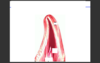
What is this structure?
What is the arrow pointing to?
Where is the muscle, transitional epithelium., lamina propria,

Lip
Hair Follicle
Center of side, bottom left of figure, bottom border of lip.
What is this structure?
List the dark pink and bottom layer?

Inner Lip
Non-Keratinized Strat. Sq. Ep
CT (Muscle Tissue)
What is this structure?
What covers this structure?
What tissue type makes the basket-weaving appearance?

Tongue
Stratified Squamos Ep.
Muscles in interlacing groups
What is this structure?
What are the arrows poing to?
What is the light pink tissue in the left corner?

Filiform Papillae
Filiform Papillae
Fat (Adipose)
What is the arrow point it?

Fungiform Papillae
Ep. covering this shows little keratinization
What is this structure?
What are the little arrows point to?
What are the large arrows pointing to?

Circumvallate Papillae
Taste Buds
Duct of Serous Gland (Von Ebner)
Notice the moat travels all the way around the gland.
What is this structure?
What are the arrows?
What is the large blue arrow?

Circumvallate Papillae
Taste Buds
von Ebner Gland
What is this structure?
What is the arrow at the top?
What are the arrows in the middle?

Taste Buds
Taste Pore
Darker Nuclei -> supporting cells;
ligher staining nuclei -> sensory cells
What is this structure?
What is the arrow point to?

Parotid Gland
Interlobular Duct
What is this structure?
What are the white material?

Parotid Gland
Fat
What is the structure?
What are the light pink kiwi structures?
What are the dark purple structures? Why do these stain darker?
What is the large blue arrow?

Parotid Gland
Striated Duct
Secretory Units; RER in their cytoplasm
Small intercalated duct
What is the structure?
What type of secretaory units do these have?

Submandibular Gland
Serous mixed in whith minor mucous secretory
What is this structure?
What type of secretory unit does this tissue have?

Sublingual Gland
Mucous mixed with minor serous
What type of tissue is this?
What are the arrows pointing to?

Submandibular Gland
Serous Demilunes
What are the three structures?
What contributes to enamel?
Enamel
Dentin
Pulp Cavity
Lines of Retzius
Where is the
Pulp
Dentin
Alveolar Bone
Layer of Cementum
Peridontal Ligament
Enamel

White portions inside
solid pink portion in center
left side
thin layer betweend entin and alveolar bone
two black arros
Does not exist due to decalcification
What is this structure?
What is the black arrow pointing to and what type of tissue lines it?
What is the blue arrow pointing to?

Tooth
Gingival Sulcus; non-keratinized stratified squamos ep.
outside of gingiva is lined by completely keratinized dtrat. sq. ep.
What is this tissue?
What are the three structures?
What are the arrows pointing to?

Tooth
Peridontal Ligament
Cementum
Dentin
Sharpley’s Fibers
What is this tissue?
What are the 4 structures?

Tooth-Root
Alveolar Bone
Peridontal Ligament
Cementum (Cementocytes inside)
Dentin
What is the structure?
What are the 6 structures?

Developing Tooth
Pulp
Odontoblasts
Predentin (Green Arrow)
Dentin
Enamel (Black arrow)
Ameloblasts (Produces Enamel)
What is this structure?
What are the 4 structures?

Developing Tooth
Dentin
small arrows (Odontoblastic layer of cells making dentin)
large arrows ( where enamel has leached out as a result of decalcifying tooth)
Pulp Cavity
