Obnoxious First Midterm Questions Flashcards
What makes up the pachymeninx?
Both dural mater layers: periosteal and meningeal
-meningeal layer is tougher, with more collagen fibers than the periosteal
What makes up the leptomeninx?
leptomeninx = both the pia mater and arachnoid layers
What is the difference between the epidural space in the brain and spinal cord?
In the brain, the epidural space is a potential space that contains the meningeal vessels and only fills during bleeding of, for example the middle meningeal artery.
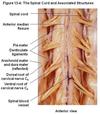
The spinal epidural space is a natural space between the spinal dural sac and the vertebral periosteum (endorachis). This area contains the internal vertebral venous plexus and adipose tissue. Use for epidural injections in anesthesiology
What is the subdural space?
Another potential space that, in the skull, transmits the superior cerebral veins. With trauma, it can be the site of slow-bleeding subdural hematomas that may take weeks to show symptoms
In the spinal cord, it is also a potential/virtual/artifical space that under normal physiological conditions should not really exist
If the brain herniates through the foramen magnum in the case of rising intracranial pressure, what parts of the brain are affected?
The medulla and tonsils of the cerebellum may be squeezed into the foramen magnum in this case, affecting the cardio and respiratory centers which may lead to death
What are the subarachnoid spaces?
Between pia mater and arachnoid layer, surrounding entire brain and spinal cord, these are normal physiological spaces through which CSF flows, along with cerebral arteries and veins
dilations in the subarachnoid space = cisterns
What are the borders of the Lemniscal Trigone?
Anteriorly: Brachium of Inferior Collicle
Posteriorly/Inferiorly: Superior Cerebellar Peduncle
Laterally: Lateral Sulcus of Midbrain + Cerebral Peduncle
What are the borders of the anterior horn of the lateral ventricle?
Lateral wall: head of caudate nucleus.
Medial wall: the septum pellucidum.
Floor/anterior wall/roof: the radiation of corpus callosum (the radiation of rostrum -
genu - and body)
What are the borders of the central part of the lateral ventricle?
Central part of lateral ventricle: located posterior to interventricular foramen, and found at level of Splenium of Corpus Callosum
Borders:
Roof: corpus callosum
Floor: dorsal aspect of thalamus, covered by the lamina affixa
Lateral wall: body of the caudate nucleus
Medial wall: choroid lamina epithelialis of the lateral ventricle
What are the borders of the posterior horn of the lateral ventricles?
aka occipital horn
Lateral wall: tapetum of the corpus callosum.
• The other walls are formed by the occipital white matter.
Medial wall: two longitudinal elevations are seen: the bulb of the posterior horn, formed by the
radiation of the corpus callosum (forceps major) and the calcar avis, produced by the calcarine fissure.
Floor: the collateral trigone (caused by the collateral sulcus) is found.
What are the borders of the inferior horn of the lateral ventricles?
aka temporal horn
Floor: hippocampus and collateral eminence (caused by the collateral sulcus).
Roof and lateral wall: formed by the white substance of the hemisphere (radiation of splenium
corporis callosi: tapetum) and along its medial border are the stria terminalis and tail of caudate nucleus.
Anteriorly: the amygdaloid nucleus bulges into the anterior end of the horn.
Medial wall: choroid lamina epithelialis is attached to the
fimbria of hippocampus and to the stria terminalis.
What are the borders of the 3rd ventricle?
Lateral: thalamus, hypothalamus
Posterior: habenular & posterior commissures, suprapineal and pineal recesses
Anterior: columns of fornix, anterior commissure, lamina terminalis, triangular recess
Floor: hypothalamus with optic and infundibular recesses
Roof: Choroid Tela and Plexus
What are the teniae in the brain?
What is the tenia thalami?
The teniae are the attachment sites of epithelial choroid lamina
The tenia thalami is where the choroid lamina epithelialis of the 3rd ventricle is attached to the thalamus

What veins does the cavernous sinus flow to?
What veins flow into the cavernous sinus?
Cavernous sinus flows to the superior and inferior petrosal sinuses
Veins that flow into it:
- Middle cerebral vein
- Sphenoparietal sinus
- Superior opthalmic vein
- inferior opthalmic vein
Branches of what nerves innervate the cranial dura mater?
Branches of the 3 parts of the Trigeminal nerve innervate most of the dura mater, except the posterior cranial fossa which is innervated by the Vagus nerve
What nerves innervate the anterior cranial fossa part of the dura mater?
The anterior meningeal branches, part of the ethmoidal nerve, which comes from V1 (Opthalmic)
What nerves innervate the middle cranial fossa part of the dura mater?
Branches of V2 and V3 (Maxillary and Mandibular branches of Trigeminal)
- the maxillary nerve accompanies the anterior branch of the middle meningeal artery
- the mandibular nerve exits the foramen ovale, but then its recurrent branch curves up to come through the foramen spinosum, accompanying the posterior branch of the middle meningeal artery
What nerve innervates the posterior cranial fossa part of the dura mater?
How does this differ from the tentorium cerebelli (and some of the posterior falx cerebri)?
Branches of the Vagus nerve innervate the posterior cranial fossa
However, the tentorium cerebelli is innervated by the tentorial nerve, which comes from the Opthalmic branch of trigeminal (V1)
What vein do the superior and inferior petrosal sinuses drain into?
-> sigmoid sinus
Where does the inferior sagittal sinus drain into?
First to the straight sinus, then the confluence of sinuses
Not confluence of sinuses directly!
What is the Brodmann number for the precentral gyrus?
Brodmann Number 4,6
Primary Motor Area
What is the Brodmann number of the postcentral gyrus?
3,1,2
Primary Somatosensory Area
What is significant about the superior temporal gyrus?
Contains Wernicke’s speech area
What is significant about the triangular and opercular parts of the inferior frontal gyrus?
They contain Broca’s speech area