Nose Flashcards

1
Q

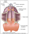
list the following with respect to the external nose

  1. nasal bones
  2. frontal processes of the ______
  3. join the nasal septal cartilage in the midline
  4. cartilage
  5. name of the air space housed by the external nose
A

external nose

  1. 2 nasal nasal bones
  2. frontal processes of the maxillae
  3. 2 lateral nasal septal cartilages
    1. join the nasal septal cartilage in the midline
  4. 2 c-shapped major alar carttilages and 2-4 minor alar cartilages

air space housed by the external nose is known as the vestibule

2
Q

muscles of the external nose are innervated

A
  1. muscles of the external nose are innervated by facial nerve
  2. skin of external nose is innervated by
    1. trigeminal nerve
      1. infratrochlearV1
      2. external nasal V1
      3. infraorbital V2
  3. external nose is supplied blood via
    1. facial - lateral nasal
    2. ophthalmis a - dorsal nasal and external nasal
3
Q

skin of external nose is innervated by

A
  1. muscles of the external nose are innervated by facial nerve
  2. skin of external nose is innervated by
    1. trigeminal nerve
      1. infratrochlearV1
      2. external nasal V1
      3. infraorbital V2
  3. external nose is supplied blood via
    1. facial - lateral nasal
    2. ophthalmis a - dorsal nasal and external nasal
4
Q

external nose is supplied blood via

A
  1. muscles of the external nose are innervated by facial nerve
  2. skin of external nose is innervated by
    1. trigeminal nerve
      1. infratrochlearV1
      2. external nasal V1
      3. infraorbital V2
  3. external nose is supplied blood via
    1. facial - lateral nasal
    2. ophthalmis a - dorsal nasal and external nasal
5
Q

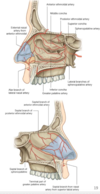
descfribe the bones involved in the formation of the nose and nasal cavity

A
6
Q

describe the lateral skeleton of the nasal wall

A
  1. nasal skeleton
    1. lateral nasal wall
      1. ethmoid
      2. inferior concha
      3. maxillary
      4. lacimal
      5. palatine
      6. sphenoid
      7. nasal
      8. frontal/alar
      9. lateral septal cartilages
    2. medial nasal wall
      1. ethmoid
      2. vomer
      3. nasal
      4. frontal
      5. sphenoid
      6. maxillary
      7. palatine
      8. septal cartilage
    3. floor
      1. macillary
      2. palatine
    4. roof
      1. ethmoid
      2. nasal
      3. frontal
      4. sphenoid
      5. comer/palatine
      6. septal and alar cartilages
7
Q

describethe medial nasal skeletal wall

A
  1. nasal skeleton
    1. lateral nasal wall
      1. ethmoid
      2. inferior concha
      3. maxillary
      4. lacimal
      5. palatine
      6. sphenoid
      7. nasal
      8. frontal/alar
      9. lateral septal cartilages
    2. medial nasal wall
      1. ethmoid
      2. vomer
      3. nasal
      4. frontal
      5. sphenoid
      6. maxillary
      7. palatine
      8. septal cartilage
    3. floor
      1. macillary
      2. palatine
    4. roof
      1. ethmoid
      2. nasal
      3. frontal
      4. sphenoid
      5. comer/palatine
      6. septal and alar cartilages
8
Q

describethe nasal fllor of the nasal skeleton

A
  1. nasal skeleton
    1. lateral nasal wall
      1. ethmoid
      2. inferior concha
      3. maxillary
      4. lacimal
      5. palatine
      6. sphenoid
      7. nasal
      8. frontal/alar
      9. lateral septal cartilages
    2. medial nasal wall
      1. ethmoid
      2. vomer
      3. nasal
      4. frontal
      5. sphenoid
      6. maxillary
      7. palatine
      8. septal cartilage
    3. floor
      1. maxillary
      2. palatine
    4. roof
      1. ethmoid
      2. nasal
      3. frontal
      4. sphenoid
      5. comer/palatine
      6. septal and alar cartilages
9
Q

describe the roof of the nasal skeleton

A
  1. nasal skeleton
    1. lateral nasal wall
      1. ethmoid
      2. inferior concha
      3. maxillary
      4. lacimal
      5. palatine
      6. sphenoid
      7. nasal
      8. frontal/alar
      9. lateral septal cartilages
    2. medial nasal wall
      1. ethmoid
      2. vomer
      3. nasal
      4. frontal
      5. sphenoid
      6. maxillary
      7. palatine
      8. septal cartilage
    3. floor
      1. macillary
      2. palatine
    4. roof
      1. ethmoid
      2. nasal
      3. frontal
      4. sphenoid
      5. comer/palatine
      6. septal and alar cartilages
10
Q

describe the nasal passages and divisions

A

nasal septum divides the chamber of the nose into

  1. two nasal passages (or nasal cavities),
    1. each with anterior (nares) and
    2. posterior openings
11
Q

opening to the nasal canities, on the cranium.

name and description

A

nasal aperture aka piriform aperture

  1. on the cranium
  2. opening to the nasal cavities
  3. the inferior margin of the nasal aperture has a bony prominence known as the anterior nasal spine (maxillae)
  4. posterior nasal apertures
    1. choanae -2x’s
      1. the posterior openings of the nasala passages
    2. together, the choanae form the posterior nasal apertures that open into the nasopharynx
12
Q

what generates the posterior opening of the nasal passages?

A

nasal aperture aka piriform aperture

  1. on the cranium
  2. opening to the nasal cavities
  3. the inferior margin of the nasal aperture has a bony prominence known as the anterior nasal spine (maxillae)
  4. posterior nasal apertures
    1. choanae -2x’s
      1. the posterior openings of the nasala passages
    2. together, the choanae form the posterior nasal apertures that open into the nasopharynx
13
Q

Ethmoid bone forms the majority of the _____ and superior portion of the ____ _____ with respect to the nasal cavity

A

ethmoid bone

  1. details
    1. forms the majority of the ROOF of the nasal cavities and the superior portion of the LATERAL WALL
    2. perpendicular plate forms the suprior portions of the nasal septum
    3. cribriform pplatte forms the boundary with cranial cavity
    4. superior and middle nasal conchae and ethmoid air cells (sinuses) are partt of the ethmoid
14
Q

what forms the superior portion of the nasal septum

A

ethmoid bone

  1. details
    1. forms the majority of the ROOF of the nasal cavities and the superior portion of the LATERAL WALL
    2. perpendicular plate forms the suprior portions of the nasal septum
    3. cribriform pplatte forms the boundary with cranial cavity
    4. superior and middle nasal conchae and ethmoid air cells (sinuses) are partt of the ethmoid
15
Q

forms the boundary of the cranial cavity, witth respect to the nasal cavity.

A

ethmoid bone

  1. details
    1. forms the majority of the ROOF of the nasal cavities and the superior portion of the LATERAL WALL
    2. perpendicular plate forms the suprior portions of the nasal septum
    3. cribriform pplatte forms the boundary with cranial cavity
    4. superior and middle nasal conchae and ethmoid air cells (sinuses) are partt of the ethmoid
16
Q

superior and middle nasal conchae and air cells (sinuses) are part of the _____

A

ethmoid bone

  1. details
    1. forms the majority of the ROOF of the nasal cavities and the superior portion of the LATERAL WALL
    2. perpendicular plate forms the suprior portions of the nasal septum
    3. cribriform pplatte forms the boundary with cranial cavity
    4. superior and middle nasal conchae and ethmoid air cells (sinuses) are partt of the ethmoid
17
Q

what are the 3 scroll shaped structures that project into the nasal caviy?

A

conchae and meatus

  1. each lateral wall has 3 scroll-shpaed structures that project into the nasal cavity - aka turbinattes
    1. inferior nasal conchae
    2. middle nasal conchae
    3. superior nasal conchae
  2. function
    1. primarily - incrase the surface area of the nasal mucosa
  3. meatus
    1. air space lateral and inferior to each concha
    2. house openings for communication channels with the paranasal sinuses and the orbit
18
Q

what is the function of the turbinates?

A

conchae and meatus

  1. each lateral wall has 3 scroll-shpaed structures that project into the nasal cavity - aka tturbinattes
    1. inferior nasal conchae
    2. middle nasal conchae
    3. superior nasal conchae
  2. function
    1. primarily - incrase the surface area of the nasal mucosa
  3. meatus
    1. air space lateral and inferior to each concha
    2. house openings for communication channels with the paranasal sinuses and the orbit
19
Q

air space lateral and inferior to each concha. function?

A

conchae and meatus

  1. each lateral wall has 3 scroll-shpaed structures that project into the nasal cavity - aka tturbinattes
    1. inferior nasal conchae
    2. middle nasal conchae
    3. superior nasal conchae
  2. function
    1. primarily - incrase the surface area of the nasal mucosa
  3. meatus
    1. air space lateral and inferior to each concha
    2. house openings for communication channels with the paranasal sinuses and the orbit
20
Q

list the three primary funcions of nasal airflow

A

nasal airflow

  1. function
    1. primary - modification of respired air
      1. particle filtration
        1. trap dust and pathogens
      2. modification of the temperature and moisture
        1. upon inspiration and expiration
      3. olfaction
21
Q

olfaction and two other important functions characterize nasal airflow

A

nasal airflow

  1. function
    1. primary - modification of respired air
      1. particle filtration
        1. trap dust and pathogens
      2. modification of the temperature and moisture
        1. upon inspiration and expiration
      3. olfaction
22
Q

where is the olfatory mucosa limited to? what is it lined by?

A

mucosa

  1. nasal mucosa
    1. epithelium
      1. nasal vestibule
        1. moderately keratinous and much like our external skin
      2. nasal passages
        1. respiritory -see below
        2. olfactory mucosa
          1. limited to the superior nasal concha and the adjacent portion of the septum.
          2. lined by by olfactory epithelium
            1. olfactory receptor neurons CN1
          3. responsible for the detection of odor
  2. respiratory mucosa
    1. majority of the nasal passage
    2. thickest over the regions containing the vascular plexus, capable of rapid blood volume change
      1. the inferior and middle conchae, and the septum adjacent to the middle meatus
      2. this process is SNS fibers (GVE)
        1. origin from spinal cord level T1 ( preganglionicfiber), travel in sympathetic trunk ->synapse in the superior cervical ganglion
        2. postganglionic fibers join branches of the maxillary nerve (V2) via deep petrosal n. or follow blood vessels (plexus) to enter the nasal cavities
    3. contents
      1. pseudostratified columnar epithelium
      2. goblet cells
      3. seromucous glands
        1. innervated by PSNS GVE
          1. preganliongic fibers are carried in the greater petrosal branch of the facial nerve that synapse in the pterygopalatine ganglion
            1. later the nerve of the pterygoid canal )
          2. postganglionic fibers join branches of the macillary (tigeminal V2) nerve to enter the nasal cavity
23
Q

describe the innervation of the respiritory mucosa with reference to PSNS and vsculature.

A

mucosa

  1. nasal mucosa
    1. epithelium
      1. nasal vestibule
        1. moderately keratinous and much like our external skin
      2. nasal passages
        1. respiritory -see below
        2. olfactory mucosa
          1. limited to the superior nasal concha and the adjacent portion of the septum.
          2. lined by by olfactory epithelium
            1. olfactory receptor neurons CN1
          3. responsible for the detection of odor
  2. respiratory mucosa
    1. majority of the nasal passage
    2. thickest over the regions containing the vascular plexus, capable of rapid blood volume change
      1. the inferior and middle conchdae, and the septum adjacent to the middle meatus
      2. this process is SNS fibers (GVE)
        1. origin from spinal cord level T1 ( preganglionicfiber), travel in sympathetic trunk ->synapse in the superior cervical ganglion
        2. postganglionic fibers join branches of the maxillary nerve (V2) via deep petrosal n. or follow blood vessels (plexus) to enter the nasal cavities
    3. contents
      1. pseudostratified columnar epithelium
      2. goblet cells
      3. seromucous glands
        1. innervated by PSNS GVE
          1. preganliongic fibers are carried in the greater petrosal branch of the facial nerve that synapse in the pterygopalatine ganglion
            1. later the nerve of the pterygoid canal )
          2. postganglionic fibers join branches of the macillary (tigeminal V2) nerve to enter the nasal cavity
24
Q

Describe the preganglionic and postganglionic fibers involved with the PSNS of the nasal seromucosa.

A

mucosa

  1. nasal mucosa
    1. epithelium
      1. nasal vestibule
        1. moderately keratinous and much like our external skin
      2. nasal passages
        1. respiritory -see below
        2. olfactory mucosa
          1. limited to the superior nasal concha and the adjacent portion of the septum.
          2. lined by by olfactory epithelium
            1. olfactory receptor neurons CN1
          3. responsible for the detection of odor
  2. respiratory mucosa
    1. majority of the nasal passage
    2. thickest over the regions containing the vascular plexus, capable of rapid blood volume change
      1. the inferior and middle conchae, and the septum adjacent to the middle meatus
      2. this process is SNS fibers (GVE)
        1. origin from spinal cord level T1 ( preganglionicfiber), travel in sympathetic trunk ->synapse in the superior cervical ganglion
        2. postganglionic fibers join branches of the maxillary nerve (V2) via deep petrosal n. or follow blood vessels (plexus) to enter the nasal cavities
    3. contents
      1. pseudostratified columnar epithelium
      2. goblet cells
      3. seromucous glands
        1. innervated by PSNS GVE
          1. preganliongic fibers are carried in the greater petrosal branch of the facial nerve that synapse in the pterygopalatine ganglion
            1. later the nerve of the pterygoid canal )
          2. postganglionic fibers join branches of the macillary (tigeminal V2) nerve to enter the nasal cavity
25
Q

thickest area of the respiritory mucosa. Describe importance with respect to the vascular plexus?

A

mucosa

  1. nasal mucosa
    1. epithelium
      1. nasal vestibule
        1. moderately keratinous and much like our external skin
      2. nasal passages
        1. respiritory -see below
        2. olfactory mucosa
          1. limited to the superior nasal concha and the adjacent portion of the septum.
          2. lined by by olfactory epithelium
            1. olfactory receptor neurons CN1
          3. responsible for the detection of odor
  2. respiratory mucosa
    1. majority of the nasal passage
    2. thickest over the regions containing the vascular plexus, capable of rapid blood volume change
      1. the inferior and middle conchae, and the septum adjacent to the middle meatus
      2. this process is SNS fibers (GVE)
        1. origin from spinal cord level T1 ( preganglionicfiber), travel in sympathetic trunk ->synapse in the superior cervical ganglion
        2. postganglionic fibers join branches of the maxillary nerve (V2) via deep petrosal n. or follow blood vessels (plexus) to enter the nasal cavities
    3. contents
      1. pseudostratified columnar epithelium
      2. goblet cells
      3. seromucous glands
        1. innervated by PSNS GVE
          1. preganliongic fibers are carried in the greater petrosal branch of the facial nerve that synapse in the pterygopalatine ganglion
            1. later the nerve of the pterygoid canal )
          2. postganglionic fibers join branches of the macillary (tigeminal V2) nerve to enter the nasal cavity
26
Q

describe the innervation controlling the vascular plexuses, which are capable of rapidly changing their blood volume.

A

mucosa

  1. nasal mucosa
    1. epithelium
      1. nasal vestibule
        1. moderately keratinous and much like our external skin
      2. nasal passages
        1. respiritory -see below
        2. olfactory mucosa
          1. limited to the superior nasal concha and the adjacent portion of the septum.
          2. lined by by olfactory epithelium
            1. olfactory receptor neurons CN1
          3. responsible for the detection of odor
  2. respiratory mucosa
    1. majority of the nasal passage
    2. thickest over the regions containing the vascular plexus, capable of rapid blood volume change
      1. the inferior and middle conchae, and the septum adjacent to the middle meatus
      2. this process is SNS fibers (GVE)
        1. origin from spinal cord level T1 ( preganglionicfiber), travel in sympathetic trunk ->synapse in the superior cervical ganglion
        2. postganglionic fibers join branches of the maxillary nerve (V2) via deep petrosal n. or follow blood vessels (plexus) to enter the nasal cavities
    3. contents
      1. pseudostratified columnar epithelium
      2. goblet cells
      3. seromucous glands
        1. innervated by PSNS GVE
          1. preganliongic fibers are carried in the greater petrosal branch of the facial nerve that synapse in the pterygopalatine ganglion
            1. later the nerve of the pterygoid canal )
          2. postganglionic fibers join branches of the macillary (tigeminal V2) nerve to enter the nasal cavity
27
Q

list the following with respect to arterial supply of the nasal cavity.

  • from the ophthalmic artery
  • from maxillary artery
  • from facial artery
A

arterial supply of the nose

  1. the blood supply to the nasal cavity is mainly from the following
    1. anterior and posterior ethmoidal arteries
      1. from the ophthalmic artery
    2. sphenopalatine and greater palatine arteries
      1. from the maxillary artery
    3. lateral nasal and superior labial arteries
      1. from the facial artery
  2. pathology
    1. epistaxis- nose bleed
      1. anterior inferior region of the nasal septu is highly vascular and known as kiesselbach’s plexus aka little’s area
        1. 90% of epistaxis takes place here
      2. in addition to numerous vein in the are, septal branches ofthe anterior and posterior ethmoidal, greater palatine, sphenopalatine and superior labial arteries anastaomose here.
      3. nosebleeds in the posterior nasal cavity are more serious and typically involve ruptutre of the sphenopalatine
  3. venous
    1. veins that axxompany maxillary artery branches drain into the pterygoid plexus of veins
    2. veins from the anterior regions of the nasal cavities drain into the facial vein
    3. veins that accompany the anterior and posterior ehtmoidal arteries drain into the ophthlmic vein, which drains into the cacernous inus
    4. in some individuals, there is an additional nasal vein (emissary) in foramen caecum that drains into the superior sagittal sinus
    1. 1.
28
Q

list the origin of the arterial supply with respect to the nasal cavity

  1. anterior and posterior ethmoidal arteries
  2. sphenopalatine and greater palatine arteries
  3. lateral nasal and superior labial arteries
A

arterial supply of the nose

  1. the blood supply to the nasal cavity is mainly from the following
    1. anterior and posterior ethmoidal arteries
      1. from the ophthalmic artery
    2. sphenopalatine and greater palatine arteries
      1. from the maxillary artery
    3. lateral nasal and superior labial arteries
      1. from the facial artery
  2. pathology
    1. epistaxis- nose bleed
      1. anterior inferior region of the nasal septu is highly vascular and known as kiesselbach’s plexus aka little’s area
        1. 90% of epistaxis takes place here
      2. in addition to numerous vein in the are, septal branches ofthe anterior and posterior ethmoidal, greater palatine, sphenopalatine and superior labial arteries anastaomose here.
      3. nosebleeds in the posterior nasal cavity are more serious and typically involve ruptutre of the sphenopalatine
  3. venous
    1. veins that axxompany maxillary artery branches drain into the pterygoid plexus of veins
    2. veins from the anterior regions of the nasal cavities drain into the facial vein
    3. veins that accompany the anterior and posterior ehtmoidal arteries drain into the ophthlmic vein, which drains into the cacernous inus
    4. in some individuals, there is an additional nasal vein (emissary) in foramen caecum that drains into the superior sagittal sinus
    1. 1.
29
Q

discuss the circled orange area in the diagram below.

  • two names for this area
  • what is this the main area for?
A

arterial supply of the nose

  1. the blood supply to the nasal cavity is mainly from the following
    1. anterior and posterior ethmoidal arteries
      1. from the ophthalmic artery
    2. sphenopalatine and greater palatine arteries
      1. from the maxillary artery
    3. lateral nasal and superior labial arteries
      1. from the facial artery
  2. pathology
    1. epistaxis- nose bleed
      1. anterior inferior region of the nasal septu is highly vascular and known as kiesselbach’s plexus aka little’s area
        1. 90% of epistaxis takes place here
      2. in addition to numerous vein in the are, septal branches ofthe anterior and posterior ethmoidal, greater palatine, sphenopalatine and superior labial arteries anastaomose here.
      3. nosebleeds in the posterior nasal cavity are more serious and typically involve ruptutre of the sphenopalatine
  3. venous
    1. veins that axxompany maxillary artery branches drain into the pterygoid plexus of veins
    2. veins from the anterior regions of the nasal cavities drain into the facial vein
    3. veins that accompany the anterior and posterior ehtmoidal arteries drain into the ophthlmic vein, which drains into the cacernous inus
    4. in some individuals, there is an additional nasal vein (emissary) in foramen caecum that drains into the superior sagittal sinus
    1. 1.
30
Q

the littles area is a location for anastomoses of which arteries?

A

arterial supply of the nose

  1. the blood supply to the nasal cavity is mainly from the following
    1. anterior and posterior ethmoidal arteries
      1. from the ophthalmic artery
    2. sphenopalatine and greater palatine arteries
      1. from the maxillary artery
    3. lateral nasal and superior labial arteries
      1. from the facial artery
  2. pathology
    1. epistaxis- nose bleed
      1. anterior inferior region of the nasal septu is highly vascular and known as kiesselbach’s plexus aka little’s area
        1. 90% of epistaxis takes place here
      2. in addition to numerous vein in the are, septal branches ofthe anterior and posterior ethmoidal, greater palatine, sphenopalatine and superior labial arteries anastaomose here.
      3. nosebleeds in the posterior nasal cavity are more serious and typically involve ruptutre of the sphenopalatine
  3. venous
    1. veins that axxompany maxillary artery branches drain into the pterygoid plexus of veins
    2. veins from the anterior regions of the nasal cavities drain into the facial vein
    3. veins that accompany the anterior and posterior ehtmoidal arteries drain into the ophthlmic vein, which drains into the cacernous inus
    4. in some individuals, there is an additional nasal vein (emissary) in foramen caecum that drains into the superior sagittal sinus
    1. 1.
31
Q

Nose bleeds in the kiesselbach’s plexus are considered severe or minor? explain.

A

arterial supply of the nose

  1. the blood supply to the nasal cavity is mainly from the following
    1. anterior and posterior ethmoidal arteries
      1. from the ophthalmic artery
    2. sphenopalatine and greater palatine arteries
      1. from the maxillary artery
    3. lateral nasal and superior labial arteries
      1. from the facial artery
  2. pathology
    1. epistaxis- nose bleed
      1. anterior inferior region of the nasal septu is highly vascular and known as kiesselbach’s plexus aka little’s area
        1. 90% of epistaxis takes place here
      2. in addition to numerous vein in the are, septal branches ofthe anterior and posterior ethmoidal, greater palatine, sphenopalatine and superior labial arteries anastaomose here.
      3. nosebleeds in the posterior nasal cavity are more serious and typically involve ruptutre of the sphenopalatine
  3. venous
    1. veins that axxompany maxillary artery branches drain into the pterygoid plexus of veins
    2. veins from the anterior regions of the nasal cavities drain into the facial vein
    3. veins that accompany the anterior and posterior ehtmoidal arteries drain into the ophthlmic vein, which drains into the cacernous inus
    4. in some individuals, there is an additional nasal vein (emissary) in foramen caecum that drains into the superior sagittal sinus
    1. 1.
32
Q

answer the following questions with regard to the venous drainaige of the nasal cavity.

  • veins that accompany maxillary artery branches drain into
  • veins from the anterior regions of the nasal cavities drain into the
  • veins that accompany the anterior and posterior ehtmoidal arteries drain into
    • which drain into?
A

arterial supply of the nose

  1. the blood supply to the nasal cavity is mainly from the following
    1. anterior and posterior ethmoidal arteries
      1. from the ophthalmic artery
    2. sphenopalatine and greater palatine arteries
      1. from the maxillary artery
    3. lateral nasal and superior labial arteries
      1. from the facial artery
  2. pathology
    1. epistaxis- nose bleed
      1. anterior inferior region of the nasal septu is highly vascular and known as kiesselbach’s plexus aka little’s area
        1. 90% of epistaxis takes place here
      2. in addition to numerous vein in the are, septal branches ofthe anterior and posterior ethmoidal, greater palatine, sphenopalatine and superior labial arteries anastaomose here.
      3. nosebleeds in the posterior nasal cavity are more serious and typically involve ruptutre of the sphenopalatine
  3. venous
    1. veins that accompany maxillary artery branches drain into the pterygoid plexus of veins
    2. veins from the anterior regions of the nasal cavities drain into the facial vein
    3. veins that accompany the anterior and posterior ehtmoidal arteries drain into the ophthlmic vein
      1. , which drains into the cavernous sinus
    4. in some individuals, there is an additional nasal vein (emissary) in foramen caecum that drains into the superior sagittal sinus
33
Q

what is the additional vein in the nasal cavity drainage, not present in everyone, and where does it drain into?

A

arterial supply of the nose

  1. the blood supply to the nasal cavity is mainly from the following
    1. anterior and posterior ethmoidal arteries
      1. from the ophthalmic artery
    2. sphenopalatine and greater palatine arteries
      1. from the maxillary artery
    3. lateral nasal and superior labial arteries
      1. from the facial artery
  2. pathology
    1. epistaxis- nose bleed
      1. anterior inferior region of the nasal septu is highly vascular and known as kiesselbach’s plexus aka little’s area
        1. 90% of epistaxis takes place here
      2. in addition to numerous vein in the are, septal branches ofthe anterior and posterior ethmoidal, greater palatine, sphenopalatine and superior labial arteries anastaomose here.
      3. nosebleeds in the posterior nasal cavity are more serious and typically involve ruptutre of the sphenopalatine
  3. venous
    1. veins that axxompany maxillary artery branches drain into the pterygoid plexus of veins
    2. veins from the anterior regions of the nasal cavities drain into the facial vein
    3. veins that accompany the anterior and posterior ehtmoidal arteries drain into the ophthlmic vein, which drains into the cacernous inus
    4. in some individuals, there is an additional nasal vein (emissary) in foramen caecum that drains into the superior sagittal sinus
34
Q

describe the nasal septum innervation

A

nerve supply of nasal cavity

  1. sensation
    1. trigeminal Nerve GSA
      1. nasal septum innervation
        1. anterior ethmoid V1
        2. Nasopalatine V2
      2. lateral nasal wall innervation
        1. anterior ehtmoidalV1
        2. greater palatineV2
          1. popsterior superior lateral nasal nerves and posterior inferior lateral nasal nerves
  2. smell: olfactory receptor neurons SVA
    1. give rise to ~20 olfactory bundles (CNI) on each side that pass through the cribirform plate and synapse in the olfactory bulbs
35
Q

describe the lateral nasal wall innervation

A

nerve supply of nasal cavity

  1. sensation
    1. trigeminal Nerve GSA
      1. nasal septum innervation
        1. anterior ethmoid V1
        2. Nasopalatine V2
      2. lateral nasal wall innervation
        1. anterior ehtmoidalV1
        2. greater palatineV2
          1. popsterior superior lateral nasal nerves and posterior inferior lateral nasal nerves
  2. smell: olfactory receptor neurons SVA
    1. give rise to ~20 olfactory bundles (CNI) on each side that pass through the cribirform plate and synapse in the olfactory bulbs
36
Q

describe the olfactory receptor innervation

A

nerve supply of nasal cavity

  1. sensation
    1. trigeminal Nerve GSA
      1. nasal septum innervation
        1. anterior ethmoid V1
        2. Nasopalatine V2
      2. lateral nasal wall innervation
        1. anterior ehtmoidalV1
        2. greater palatineV2
          1. popsterior superior lateral nasal nerves and posterior inferior lateral nasal nerves
  2. smell: olfactory receptor neurons SVA
    1. give rise to ~20 olfactory bundles (CNI) on each side that pass through the cribirform plate and synapse in the olfactory bulbs
37
Q

what do the following structures generate?

  1. frontal sinuses
  2. ethmoid air cells
  3. sphenoid sinuses
  4. maxillary sinuses
A

parranasal sinuses

  1. extension of cranial cavities into adjacent bones
    1. frontal sinuses
    2. ethmoid air cells
    3. sphenoid sinuses
    4. maxillary sinuses
  2. route for sinus openings
    1. sphenoid ->spheno-ethmoidal recess
    2. posterio-ethmoidal air cells drain into the superior meatus
    3. hiatus semilunaris of the middle meatus contains the openings of the frontonasal duct (infundibulum), anterior ethmoidal air cells, and the maxillary sinus
    4. the middle ethmoidal air cells open on the bulla thmoidalis in the middle meatus
    5. each nasolacrimal duct that origins in one of the orbit drains inot the inferior meatus.
38
Q

list the extensions of the cranial cavities into adjacen bones. What is the called?

A

parranasal sinuses

  1. extension of cranial cavities into adjacent bones
    1. frontal sinuses
    2. ethmoid air cells
    3. sphenoid sinuses
    4. maxillary sinuses
  2. route for sinus openings
    1. sphenoid ->spheno-ethmoidal recess
    2. posterio-ethmoidal air cells drain into the superior meatus
    3. hiatus semilunaris of the middle meatus contains the openings of the frontonasal duct (infundibulum), anterior ethmoidal air cells, and the maxillary sinus
    4. the middle ethmoidal air cells open on the bulla thmoidalis in the middle meatus
    5. each nasolacrimal duct that origins in one of the orbit drains inot the inferior meatus.
39
Q

answer the following with respect to route for sinuses opening

  1. sphenoid opens to the ____
  2. posterior-ethmoidal air cells drain into the ______
  3. hiatus of the middle meatus open into what ?
A

parranasal sinuses

  1. extension of cranial cavities into adjacent bones
    1. frontal sinuses
    2. ethmoid air cells
    3. sphenoid sinuses
    4. maxillary sinuses
  2. route for sinus openings
    1. sphenoid ->spheno-ethmoidal recess
    2. posterio-ethmoidal air cells drain into the superior meatus
    3. hiatus semilunaris of the middle meatus contains the openings of the frontonasal duct (infundibulum), anterior ethmoidal air cells, and the maxillary sinus
    4. the middle ethmoidal air cells open on the bulla thmoidalis in the middle meatus
    5. each nasolacrimal duct that origins in one of the orbit drains inot the inferior meatus.
40
Q

answer the following with respect to route for sinuses opening

  • middle ethmoidal air cells open to become ___. Where?
  • each nasolacrimal dust that origins in one of the orbit drains into ____?
A

parranasal sinuses

  1. extension of cranial cavities into adjacent bones
    1. frontal sinuses
    2. ethmoid air cells
    3. sphenoid sinuses
    4. maxillary sinuses
  2. route for sinus openings
    1. sphenoid ->spheno-ethmoidal recess
    2. posterio-ethmoidal air cells drain into the superior meatus
    3. hiatus semilunaris of the middle meatus contains the openings of the frontonasal duct (infundibulum), anterior ethmoidal air cells, and the maxillary sinus
    4. the middle ethmoidal air cells open on the bulla thmoidalis in the middle meatus
    5. each nasolacrimal duct that origins in one of the orbit drains inot the inferior meatus.
41
Q

describe the nerves involved with the following structures

  1. ?
    1. frontal sinuses
  2. ?
    1. ethmoid air cells
  3. ?
    1. ethmoid air cells
    2. sphenoid sinuses
  4. ?
    1. ethmoid air cells anf sphenoid sinuses
  5. ?
    1. maxillary sinuses
A

sinuses

  1. nerves
    1. supraorbital nerveV1
      1. frontal sinuses
    2. anterior ethmoid nerveV1
      1. ethmoid air cells
    3. posterior ethmoids nerve V1
      1. ethmoid air cells
      2. sphenoid sinuses
    4. orbital brs of maxillary nerve V2
      1. ethmoid air cells anf sphenoid sinuses
    5. alveolar brs of infraorbital nerve V2
      1. maxillary sinuses
  2. areteris
    1. anterior ethmoidal artery
      1. frontal sinuses and ethmoidal air cells
      2. brs of opthalmic artery
    2. posterior ethmoid artery
      1. ethmoid air cells
      2. brs of ophthalmic artery
    3. branches of maxillary artery
      1. sphenoid
      2. maxillary sinsuses.
42
Q

describe the sinuses associated with the following nerves

  1. supraorbital nerveV1
      1. anterior ethmoid nerveV1
      1. posterior ethmoids nerve V1
  2. orbital brs of maxillary nerve V2
      1. alveolar brs of infraorbital nerve V2
        1.
A

sinuses

  1. nerves
    1. supraorbital nerveV1
      1. frontal sinuses
    2. anterior ethmoid nerveV1
      1. ethmoid air cells
    3. posterior ethmoids nerve V1
      1. ethmoid air cells
      2. sphenoid sinuses
    4. orbital brs of maxillary nerve V2
      1. ethmoid air cells anf sphenoid sinuses
    5. alveolar brs of infraorbital nerve V2
      1. maxillary sinuses
  2. areteris
    1. anterior ethmoidal artery
      1. frontal sinuses and ethmoidal air cells
      2. brs of opthalmic artery
    2. posterior ethmoid artery
      1. ethmoid air cells
      2. brs of ophthalmic artery
    3. branches of maxillary artery
      1. sphenoid
      2. maxillary sinsuses.
43
Q

describe the strctures involved with the sinus arteries

  1. anterior ethmoidal artery
  2. posterior ethmoid artery
  3. branches of maxillary artery
    1. 2.
A

sinuses

  1. nerves
    1. supraorbital nerveV1
      1. frontal sinuses
    2. anterior ethmoid nerveV1
      1. ethmoid air cells
    3. posterior ethmoids nerve V1
      1. ethmoid air cells
      2. sphenoid sinuses
    4. orbital brs of maxillary nerve V2
      1. ethmoid air cells anf sphenoid sinuses
    5. alveolar brs of infraorbital nerve V2
      1. maxillary sinuses
  2. areteris
    1. anterior ethmoidal artery
      1. frontal sinuses and ethmoidal air cells
      2. brs of opthalmic artery
    2. posterior ethmoid artery
      1. ethmoid air cells
      2. brs of ophthalmic artery
    3. branches of maxillary artery
      1. sphenoid
      2. maxillary sinsuses.
44
Q

list the arteries involved with the following structures

    1. frontal sinuses and ethmoidal air cells
    2. brs of opthalmic artery
    1. ethmoid air cells
    2. brs of ophthalmic artery
    1. sphenoid
    2. maxillary sinsuses.
A

sinuses

  1. nerves
    1. supraorbital nerveV1
      1. frontal sinuses
    2. anterior ethmoid nerveV1
      1. ethmoid air cells
    3. posterior ethmoids nerve V1
      1. ethmoid air cells
      2. sphenoid sinuses
    4. orbital brs of maxillary nerve V2
      1. ethmoid air cells anf sphenoid sinuses
    5. alveolar brs of infraorbital nerve V2
      1. maxillary sinuses
  2. areteris
    1. anterior ethmoidal artery
      1. frontal sinuses and ethmoidal air cells
      2. brs of opthalmic artery
    2. posterior ethmoid artery
      1. ethmoid air cells
      2. brs of ophthalmic artery
    3. branches of maxillary artery
      1. sphenoid
      2. maxillary sinsuses.
45
Q

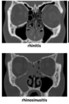
describe the condition in the photo below.

Describe the abnormal growth of tissue that arise from the nasal mucosa.

A

common pathologies of the nasal cavity

  1. deviated septum
    1. displacement of the nasal septum that typically occurs where the perpendicular plate of the ethmoid articulates with the vomer
  2. nasal polyps
    1. abnormal growths of tissue that arise from the nasal mucosa
  3. rhinitis
    1. inflammation of the nasal mucosa
  4. sinusitis
    1. inflammation of one or more papranasal sinuses
  5. rhinosinusitis
    1. when rhinitis and sinusitis occur together

loss of smell

  1. testing CN1
  2. how
    1. smell coffee with each nostril
    2. no use of noxious smells (vinegare, ammonia) that stimulate CNV
  3. what is tested
    1. impairment due to
      1. nasal obstruction
      2. inflammation of olfactory mucosa
      3. damaged olfactory nerves crossing fractured cribriform plate/ CSF rhinorrhea and meningitis
      4. abscess, intracranial lesions or subfrontal tumor affecting olfactory bulb where olfactory nerves synapse
      5. agining
46
Q

describe the condition in the following images.

what is inflammation in one or more paranasal sinuses?

A

common pathologies of the nasal cavity

  1. deviated septum
    1. displacement of the nasal septum that typically occurs where the perpendicular plate of the ethmoid articulates with the vomer
  2. nasal polyps
    1. abnormal growths of tissue that arise from the nasal mucosa
  3. rhinitis
    1. inflammation of the nasal mucosa
  4. sinusitis
    1. inflammation of one or more papranasal sinuses
  5. rhinosinusitis
    1. when rhinitis and sinusitis occur together

loss of smell

  1. testing CN1
  2. how
    1. smell coffee with each nostril
    2. no use of noxious smells (vinegare, ammonia) that stimulate CNV
  3. what is tested
    1. impairment due to
      1. nasal obstruction
      2. inflammation of olfactory mucosa
      3. damaged olfactory nerves crossing fractured cribriform plate/ CSF rhinorrhea and meningitis
      4. abscess, intracranial lesions or subfrontal tumor affecting olfactory bulb where olfactory nerves synapse
      5. agining
47
Q

What is used in loss of smell testing(what is not used)? What is being tested?

A

common pathologies of the nasal cavity

  1. deviated septum
    1. displacement of the nasal septum that typically occurs where the perpendicular plate of the ethmoid articulates with the vomer
  2. nasal polyps
    1. abnormal growths of tissue that arise from the nasal mucosa
  3. rhinitis
    1. inflammation of the nasal mucosa
  4. sinusitis
    1. inflammation of one or more papranasal sinuses
  5. rhinosinusitis
    1. when rhinitis and sinusitis occur together

loss of smell

  1. testing CN1
  2. how
    1. smell coffee with each nostril
    2. no use of noxious smells (vinegare, ammonia) that stimulate CNV
  3. what is tested
    1. impairment due to
      1. nasal obstruction
      2. inflammation of olfactory mucosa
      3. damaged olfactory nerves crossing fractured cribriform plate/ CSF rhinorrhea and meningitis
      4. abscess, intracranial lesions or subfrontal tumor affecting olfactory bulb where olfactory nerves synapse
      5. agining
48
Q

impairment of what structures leads to a loss of smell? Highlighted are most common.

A

common pathologies of the nasal cavity

  1. deviated septum
    1. displacement of the nasal septum that typically occurs where the perpendicular plate of the ethmoid articulates with the vomer
  2. nasal polyps
    1. abnormal growths of tissue that arise from the nasal mucosa
  3. rhinitis
    1. inflammation of the nasal mucosa
  4. sinusitis
    1. inflammation of one or more papranasal sinuses
  5. rhinosinusitis
    1. when rhinitis and sinusitis occur together

loss of smell

  1. testing CN1
  2. how
    1. smell coffee with each nostril
    2. no use of noxious smells (vinegare, ammonia) that stimulate CNV
  3. what is tested
    1. impairment due to
      1. nasal obstruction
      2. inflammation of olfactory mucosa
      3. damaged olfactory nerves crossing fractured cribriform plate/ CSF rhinorrhea and meningitis
      4. abscess, intracranial lesions or subfrontal tumor affecting olfactory bulb where olfactory nerves synapse
      5. aging
49
Q

the region posterior to the choanae.

describe the rooof and innervation.

A
  1. nasopharynx
    1. region of the pharynx posterior to the choanae and above the level of the soft palate.
    2. roof
      1. formed by
        1. body of the sphenoid
        2. basilar part of the occipital bones
      2. mucosal covering
        1. contains the pharyngeal tonsils
          1. largcollection of lymphoid tissue
          2. adenoids = enlarged pharyngeal tonsils
    3. tonsils
      1. tubual tonsils
      2. palatine tonsisls
      3. lingual tonsil
      4. pharyngeal tonsil
    4. sensory innervation
      1. pharyngeal nerve V2
  2. tonsils
    1. walderyer’s ring
      1. ttonsils form a stucture in the form of a ring around the nasal and oral openings of the pharynx.
    2. pharyngeal tonsils
    3. tubual tonsils
    4. palatine tonsisls
    5. lingual tonsil
  3. hparyngotympanic tube
    1. names
      1. pharyngotympanic (eustachian or auditory tube)
    2. location
      1. found on the lateral wall of the nasopharynx
    3. extension
      1. torus tubarius is an extension of its cartilage
    4. function
      1. connects the middle ear with thenasopharynx and equalizes pressure on both sides of the tympanic membran
      2. children below the age of 7
        1. have shorter and mor horizontal tube
        2. leaves them at increased susceptibility to infectious agents that enter the middle ear from the nasopharynx in conditions such as otitis media.
50
Q

what is the location of the pharyngeal tonsils? what are they called when enlarged?

A
  1. nasopharynx
    1. region of the pharynx posterior to the choanae and above the level of the soft palate.
    2. roof
      1. formed by
        1. body of the sphenoid
        2. basilar part of the occipital bones
      2. mucosal covering
        1. contains the pharyngeal tonsils
          1. largcollection of lymphoid tissue
          2. adenoids = enlarged pharyngeal tonsils
    3. tonsils
      1. tubual tonsils
      2. palatine tonsisls
      3. lingual tonsil
      4. pharyngeal tonsil
    4. sensory innervation
      1. pharyngeal nerve V2
  2. tonsils
    1. walderyer’s ring
      1. ttonsils form a stucture in the form of a ring around the nasal and oral openings of the pharynx.
    2. pharyngeal tonsils
    3. tubual tonsils
    4. palatine tonsisls
    5. lingual tonsil
  3. hparyngotympanic tube
    1. names
      1. pharyngotympanic (eustachian or auditory tube)
    2. location
      1. found on the lateral wall of the nasopharynx
    3. extension
      1. torus tubarius is an extension of its cartilage
    4. function
      1. connects the middle ear with thenasopharynx and equalizes pressure on both sides of the tympanic membran
      2. children below the age of 7
        1. have shorter and mor horizontal tube
        2. leaves them at increased susceptibility to infectious agents that enter the middle ear from the nasopharynx in conditions such as otitis media.
51
Q

list the tonsils in the nasopharnx and oropharynx.

Describe the walderyers ring.

A
  1. nasopharynx
    1. region of the pharynx posterior to the choanae and above the level of the soft palate.
    2. roof
      1. formed by
        1. body of the sphenoid
        2. basilar part of the occipital bones
      2. mucosal covering
        1. contains the pharyngeal tonsils
          1. largcollection of lymphoid tissue
          2. adenoids = enlarged pharyngeal tonsils
    3. tonsils
      1. tubual tonsils
      2. palatine tonsisls
      3. lingual tonsil
      4. pharyngeal tonsil
    4. sensory innervation
      1. pharyngeal nerve V2
  2. tonsils
    1. walderyer’s ring
      1. tonsils form a stucture in the form of a ring around the nasal and oral openings of the pharynx.
    2. pharyngeal tonsils
    3. tubual tonsils
    4. palatine tonsisls
    5. lingual tonsil
  3. pharyngotympanic tube
    1. names
      1. pharyngotympanic (eustachian or auditory tube)
    2. location
      1. found on the lateral wall of the nasopharynx
    3. extension
      1. torus tubarius is an extension of its cartilage
    4. function
      1. connects the middle ear with thenasopharynx and equalizes pressure on both sides of the tympanic membran
      2. children below the age of 7
        1. have shorter and mor horizontal tube
        2. leaves them at increased susceptibility to infectious agents that enter the middle ear from the nasopharynx in conditions such as otitis media.
52
Q

the pharyngeal opening found on the lateral wall of the nasopharynx. Several names

what is the extension of the cartilage?

A
  1. nasopharynx
    1. region of the pharynx posterior to the choanae and above the level of the soft palate.
    2. roof
      1. formed by
        1. body of the sphenoid
        2. basilar part of the occipital bones
      2. mucosal covering
        1. contains the pharyngeal tonsils
          1. largcollection of lymphoid tissue
          2. adenoids = enlarged pharyngeal tonsils
    3. tonsils
      1. tubual tonsils
      2. palatine tonsisls
      3. lingual tonsil
      4. pharyngeal tonsil
    4. sensory innervation
      1. pharyngeal nerve V2
  2. tonsils
    1. walderyer’s ring
      1. tonsils form a stucture in the form of a ring around the nasal and oral openings of the pharynx.
    2. pharyngeal tonsils
    3. tubual tonsils
    4. palatine tonsisls
    5. lingual tonsil
  3. pharyngotympanic tube
    1. names
      1. pharyngotympanic (eustachian or auditory tube)
    2. location
      1. found on the lateral wall of the nasopharynx
    3. extension
      1. torus tubarius is an extension of its cartilage
    4. function
      1. connects the middle ear with thenasopharynx and equalizes pressure on both sides of the tympanic membran
      2. children below the age of 7
        1. have shorter and mor horizontal tube
        2. leaves them at increased susceptibility to infectious agents that enter the middle ear from the nasopharynx in conditions such as otitis media.
53
Q

what does the phparyngotympanic tube connect?

describe the condition in children

A
  1. nasopharynx
    1. region of the pharynx posterior to the choanae and above the level of the soft palate.
    2. roof
      1. formed by
        1. body of the sphenoid
        2. basilar part of the occipital bones
      2. mucosal covering
        1. contains the pharyngeal tonsils
          1. largcollection of lymphoid tissue
          2. adenoids = enlarged pharyngeal tonsils
    3. tonsils
      1. tubual tonsils
      2. palatine tonsisls
      3. lingual tonsil
      4. pharyngeal tonsil
    4. sensory innervation
      1. pharyngeal nerve V2
  2. tonsils
    1. walderyer’s ring
      1. ttonsils form a stucture in the form of a ring around the nasal and oral openings of the pharynx.
    2. pharyngeal tonsils
    3. tubual tonsils
    4. palatine tonsisls
    5. lingual tonsil
  3. hparyngotympanic tube
    1. names
      1. pharyngotympanic (eustachian or auditory tube)
    2. location
      1. found on the lateral wall of the nasopharynx
    3. extension
      1. torus tubarius is an extension of its cartilage
    4. function
      1. connects the middle ear with thenasopharynx and equalizes pressure on both sides of the tympanic membran
      2. children below the age of 7
        1. have shorter and mor horizontal tube
        2. leaves them at increased susceptibility to infectious agents that enter the middle ear from the nasopharynx in conditions such as otitis media.
54
Q

equalizes pressure on both sides of the tympanic membrane. What conditions manifests in children and why?

A
  1. nasopharynx
    1. region of the pharynx posterior to the choanae and above the level of the soft palate.
    2. roof
      1. formed by
        1. body of the sphenoid
        2. basilar part of the occipital bones
      2. mucosal covering
        1. contains the pharyngeal tonsils
          1. largcollection of lymphoid tissue
          2. adenoids = enlarged pharyngeal tonsils
    3. tonsils
      1. tubual tonsils
      2. palatine tonsisls
      3. lingual tonsil
      4. pharyngeal tonsil
    4. sensory innervation
      1. pharyngeal nerve V2
  2. tonsils
    1. walderyer’s ring
      1. ttonsils form a stucture in the form of a ring around the nasal and oral openings of the pharynx.
    2. pharyngeal tonsils
    3. tubual tonsils
    4. palatine tonsisls
    5. lingual tonsil
  3. hparyngotympanic tube
    1. names
      1. pharyngotympanic (eustachian or auditory tube)
    2. location
      1. found on the lateral wall of the nasopharynx
    3. extension
      1. torus tubarius is an extension of its cartilage
    4. function
      1. connects the middle ear with the nasopharynx and equalizes pressure on both sides of the tympanic membran
      2. children below the age of 7
        1. have shorter and mor horizontal tube
        2. leaves them at increased susceptibility to infectious agents that enter the middle ear from the nasopharynx in conditions such as otitis media.