larynx, laryngopharynx and speech Flashcards
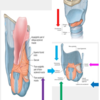
list the three parts of the pahrynx
- pharynx
- three parts
- nasopharynx
- lies above the soft palate
- oropharynx
- extends from the soft palate to the upper border of the epiglottis
- lies behind the oral cavity
- laryngopharynx
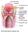
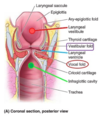
- extends from the upper border of the epiglottis to the lower border of the cricoid cartilage, where it is continuous with the esophagus.
- lies behind the aperture and posterior wall of the larynx
- nasopharynx
- three parts
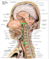
lies above the soft palate
- pharynx
- three parts
-
nasopharynx
- lies above the soft palate
- oropharynx
- extends from the soft palate to the upper border of the epiglottis
- lies behind the oral cavity
- laryngopharynx
- extends from the upper border of the epiglottis to the lower border of the cricoid cartilage, where it is continuous with the esophagus.
- lies behind the aperture and posterior wall of the larynx
-
nasopharynx
- three parts

lies behind the oral cavity
- pharynx
- three parts
- nasopharynx
- lies above the soft palate
-
oropharynx
- extends from the soft palate to the upper border of the epiglottis
- lies behind the oral cavity
- laryngopharynx
- extends from the upper border of the epiglottis to the lower border of the cricoid cartilage, where it is continuous with the esophagus.
- lies behind the aperture and posterior wall of the larynx
- nasopharynx
- three parts
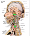
lies behind the aperture and posterior wall of the larynx
- pharynx
- three parts
- nasopharynx
- lies above the soft palate
- oropharynx
- extends from the soft palate to the upper border of the epiglottis
- lies behind the oral cavity
-
laryngopharynx
- extends from the upper border of the epiglottis to the lower border of the cricoid cartilage, where it is continuous with the esophagus.
- lies behind the aperture and posterior wall of the larynx
- nasopharynx
- three parts

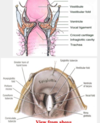
are depressions within the laryngopharynx on each side of the larngeal inlet and the lateral wall of the larynx
- piriform recesses (priform fossae)
- are depressions within the laryngopharynx on each side of the larngeal inlet and the lateral wall of the larynx
- are sites where food or foreign objectts may become lodged and may damage the internal laryngeal nerve lying just deep to the mucous membran
- attempts to remove foreign objects also may damage the nerve
- the nerve may be anestetized for endo tracheal intubation
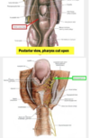
sites where food or foreign objectts may become lodged
What may be damaged deep to this location?
- piriform recesses (priform fossae)
- are depressions within the laryngopharynx on each side of the larngeal inlet and the lateral wall of the larynx
- are sites where food or foreign objectts may become lodged and may damage the internal laryngeal nerve lying just deep to the mucous membran
- attempts to remove foreign objects also may damage the nerve
- the nerve may be anestetized for endo tracheal intubation

describe the manipulatio nof the internal laryngeal nerve in the priform recesses
- piriform recesses (priform fossae)
- are depressions within the laryngopharynx on each side of the larngeal inlet and the lateral wall of the larynx
- are sites where food or foreign objectts may become lodged and may damage the internal laryngeal nerve lying just deep to the mucous membran
- attempts to remove foreign objects also may damage the nerve
- the nerve may be anestetized for endo tracheal intubation

between the pharynx and the trachea.
this complex organ of ____. What vertebrae align with this structure?
larynx
- part of the respiratory system between the pharynx and the trachea
- is a complex organ of voice production, lies in the anterior part of neck at the level of the bodies of C3-C6 vertebrae
- functions
- for the passage of air toward and from the lungs
- to prevent foreign objects from entering lower respiratory passages
- in phonation/voice production
- skeleton of the larynx
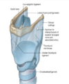
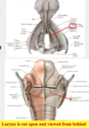
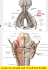
- consists of 9 cartilages, three unpaired and three pair
- paired
- cuneiform cartilage
- arytenoid
- corniculate
- unpaired
- thyroid
- cricoid
- epiglottis
- paired
- consists of 9 cartilages, three unpaired and three pair

what are the functitons of the larynx. 3
larynx
- part of the respiratory system between the pharynx and the trachea
- is a complex organ of voice production, lies in the anterior part of neck at the level of the bodies of C3-C6 vertebrae
-
functions
- for the passage of air toward and from the lungs
- to prevent foreign objects from entering lower respiratory passages
- in phonation/voice production
- skeleton of the larynx
- consists of 9 cartilages, three unpaired and three pair
- paired
- cuneiform cartilage
- arytenoid
- corniculate
- unpaired
- thyroid
- cricoid
- epiglottis
- paired
- consists of 9 cartilages, three unpaired and three pair

describe the skeleton of the larynx
larynx
- part of the respiratory system between the pharynx and the trachea
- is a complex organ of voice production, lies in the anterior part of neck at the level of the bodies of C3-C6 vertebrae
- functions
- for the passage of air toward and from the lungs
- to prevent foreign objects from entering lower respiratory passages
- in phonation/voice production
-
skeleton of the larynx
-
consists of 9 cartilages, three unpaired and three pair
-
paired
- cuneiform cartilage
- arytenoid
- corniculate
-
unpaired
- thyroid
- cricoid
- epiglottis
-
paired
-
consists of 9 cartilages, three unpaired and three pair

forms the laryngeal prominence.
what does it consist of two ____. Describe them
- laryngeal skeleton
- paired
- arytenoid
- cuneiform (wrisberg)
- corniculate (santorini)
- unpaired
- thyroid
- cricoid
- epiglottis
- paired
- thyroid cartilage
- forms the laryngeal prominence/ Adam’s appple in males
- consists of two laminae fused in the midline anteriorly
- forms bilateral cricothyroid jionts with the cricoid cartilage below, allowing the thyroid cartilage to tilt foward and backward on it
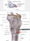
- cricoid cartilage
- is shaped like a signet ring with the broad lamina located posteriorly and the thinner arch in front
- articulates with the thyoid cartilage at bilateral cricothyroid joints
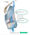
- is attached to it in the anterior midline at the cricothyroid ligament, where an emergency airway may be made
- cricothyrotomy
- is attached to it in the anterior midline at the cricothyroid ligament, where an emergency airway may be made
- is a useful landmark for locating Cv6 and its enlarged anterior tubercle,
- this is a compression site to control bleeding from the carotid arteries and their branches
- the vertebral artery enters the transverse foramen C6
- epiglottic cartilage
- is attached to the hyoid bone and to the thyroid cartilage by ligaments
- is covered by mucous membran to form the epiglottis
- is pushed back and down over the laryngeal inlet (adults ) during swallowing to help prevent the enterance of food and drink
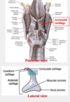
- artenoid cartilages
- are paired pyramidal-shaped carttilages that articulate with the superior border of cricoid cartilage lamina
- each has an apex
- muscular process and a coval process
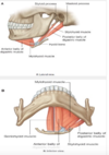
- has the vocal ligament attached to itts vocal process anteriorly
- therefore, the vocal ligament attaches anteriorly to the thyroid cartilage and posteriorly to the aryttenoid cartilage
- are capable of three basic movements
- sliding toward or away from each other
- tilting anteriorly or posteriorly around a horizontal axis
- rotating around a vertical axis
- corniculate and cuneiform cartilages
- aren’t of exceptional importance but ar landmarks during laryngoscopy

what does the adams apple form with the cricoid?
- laryngeal skeleton
- paired
- arytenoid
- cuneiform (wrisberg)
- corniculate (santorini)
- unpaired
- thyroid
- cricoid
- epiglottis
- paired
- thyroid cartilage
- forms the laryngeal prominence/ Adam’s appple in males
- consists of two laminae fused in the midline anteriorly
- forms bilateral cricothyroid jionts with the cricoid cartilage below, allowing the thyroid cartilage to tilt foward and backward on it
- cricoid cartilage
- is shaped like a signet ring with the broad lamina located posteriorly and the thinner arch in front
- articulates with the thyoid cartilage at bilateral cricothyroid joints
- is attached to it in the anterior midline at the cricothyroid ligament, where an emergency airway may be made
- cricothyrotomy
- is attached to it in the anterior midline at the cricothyroid ligament, where an emergency airway may be made
- is a useful landmark for locating Cv6 and its enlarged anterior tubercle,
- this is a compression site to control bleeding from the carotid arteries and their branches
- the vertebral artery enters the transverse foramen C6
- epiglottic cartilage
- is attached to the hyoid bone and to the thyroid cartilage by ligaments
- is covered by mucous membran to form the epiglottis
- is pushed back and down over the laryngeal inlet (adults ) during swallowing to help prevent the enterance of food and drink
- artenoid cartilages
- are paired pyramidal-shaped carttilages that articulate with the superior border of cricoid cartilage lamina
- each has an apex
- muscular process and a coval process
- has the vocal ligament attached to itts vocal process anteriorly
- therefore, the vocal ligament attaches anteriorly to the thyroid cartilage and posteriorly to the aryttenoid cartilage
- are capable of three basic movements
- sliding toward or away from each other
- tilting anteriorly or posteriorly around a horizontal axis
- rotating around a vertical axis
- corniculate and cuneiform cartilages
- aren’t of exceptional importance but ar landmarks during laryngoscopy

is shaped like a signet ring with the broad lamina located posteriorly and the thinner arch in front.
what does it articulate with?
- laryngeal skeleton
- paired
- arytenoid
- cuneiform (wrisberg)
- corniculate (santorini)
- unpaired
- thyroid
- cricoid
- epiglottis
- paired
- thyroid cartilage
- forms the laryngeal prominence/ Adam’s appple in males
- consists of two laminae fused in the midline anteriorly
- forms bilateral cricothyroid jionts with the cricoid cartilage below, allowing the thyroid cartilage to tilt foward and backward on it
-
cricoid cartilage
- is shaped like a signet ring with the broad lamina located posteriorly and the thinner arch in front
-
articulates with the thyoid cartilage at bilateral cricothyroid joints
-
is attached to it in the anterior midline at the cricothyroid ligament, where an emergency airway may be made
- cricothyrotomy
-
is attached to it in the anterior midline at the cricothyroid ligament, where an emergency airway may be made
- is a useful landmark for locating Cv6 and its enlarged anterior tubercle,
- this is a compression site to control bleeding from the carotid arteries and their branches
- the vertebral artery enters the transverse foramen C6
- epiglottic cartilage
- is attached to the hyoid bone and to the thyroid cartilage by ligaments
- is covered by mucous membran to form the epiglottis
- is pushed back and down over the laryngeal inlet (adults ) during swallowing to help prevent the enterance of food and drink
- artenoid cartilages
- are paired pyramidal-shaped carttilages that articulate with the superior border of cricoid cartilage lamina
- each has an apex
- muscular process and a coval process
- has the vocal ligament attached to itts vocal process anteriorly
- therefore, the vocal ligament attaches anteriorly to the thyroid cartilage and posteriorly to the aryttenoid cartilage
- are capable of three basic movements
- sliding toward or away from each other
- tilting anteriorly or posteriorly around a horizontal axis
- rotating around a vertical axis
- corniculate and cuneiform cartilages
- aren’t of exceptional importance but ar landmarks during laryngoscopy
articulates with the thyoid cartilage at bilateral cricothyroid joints. Describe the articulations and thte clincal applications
- laryngeal skeleton
- paired
- arytenoid
- cuneiform (wrisberg)
- corniculate (santorini)
- unpaired
- thyroid
- cricoid
- epiglottis
- paired
- thyroid cartilage
- forms the laryngeal prominence/ Adam’s appple in males
- consists of two laminae fused in the midline anteriorly
- forms bilateral cricothyroid jionts with the cricoid cartilage below, allowing the thyroid cartilage to tilt foward and backward on it
-
cricoid cartilage
- is shaped like a signet ring with the broad lamina located posteriorly and the thinner arch in front
-
articulates with the thyoid cartilage at bilateral cricothyroid joints
-
is attached to it in the anterior midline at the cricothyroid ligament, where an emergency airway may be made
- cricothyrotomy
-
is attached to it in the anterior midline at the cricothyroid ligament, where an emergency airway may be made
- is a useful landmark for locating Cv6 and its enlarged anterior tubercle,
- this is a compression site to control bleeding from the carotid arteries and their branches
- the vertebral artery enters the transverse foramen C6
- epiglottic cartilage
- is attached to the hyoid bone and to the thyroid cartilage by ligaments
- is covered by mucous membran to form the epiglottis
- is pushed back and down over the laryngeal inlet (adults ) during swallowing to help prevent the enterance of food and drink
- artenoid cartilages
- are paired pyramidal-shaped carttilages that articulate with the superior border of cricoid cartilage lamina
- each has an apex
- muscular process and a coval process
- has the vocal ligament attached to itts vocal process anteriorly
- therefore, the vocal ligament attaches anteriorly to the thyroid cartilage and posteriorly to the aryttenoid cartilage
- are capable of three basic movements
- sliding toward or away from each other
- tilting anteriorly or posteriorly around a horizontal axis
- rotating around a vertical axis
- corniculate and cuneiform cartilages
- aren’t of exceptional importance but ar landmarks during laryngoscopy

is a useful landmark for locating Cv6 and its enlarged anterior tubercle. what is this site used for during treatment of injury?
- laryngeal skeleton
- paired
- arytenoid
- cuneiform (wrisberg)
- corniculate (santorini)
- unpaired
- thyroid
- cricoid
- epiglottis
- paired
- thyroid cartilage
- forms the laryngeal prominence/ Adam’s appple in males
- consists of two laminae fused in the midline anteriorly
- forms bilateral cricothyroid jionts with the cricoid cartilage below, allowing the thyroid cartilage to tilt foward and backward on it
- cricoid cartilage
- is shaped like a signet ring with the broad lamina located posteriorly and the thinner arch in front
- articulates with the thyoid cartilage at bilateral cricothyroid joints
- is attached to it in the anterior midline at the cricothyroid ligament, where an emergency airway may be made
- cricothyrotomy
- is attached to it in the anterior midline at the cricothyroid ligament, where an emergency airway may be made
-
is a useful landmark for locating Cv6 and its enlarged anterior tubercle,
- this is a compression site to control bleeding from the carotid arteries and their branches
- the vertebral artery enters the transverse foramen C6
- epiglottic cartilage
- is attached to the hyoid bone and to the thyroid cartilage by ligaments
- is covered by mucous membran to form the epiglottis
- is pushed back and down over the laryngeal inlet (adults ) during swallowing to help prevent the enterance of food and drink
- artenoid cartilages
- are paired pyramidal-shaped carttilages that articulate with the superior border of cricoid cartilage lamina
- each has an apex
- muscular process and a coval process
- has the vocal ligament attached to itts vocal process anteriorly
- therefore, the vocal ligament attaches anteriorly to the thyroid cartilage and posteriorly to the aryttenoid cartilage
- are capable of three basic movements
- sliding toward or away from each other
- tilting anteriorly or posteriorly around a horizontal axis
- rotating around a vertical axis
- corniculate and cuneiform cartilages
- aren’t of exceptional importance but ar landmarks during laryngoscopy

Describe the important landmarks and route of the artery associated wit hthe cricoid cartilage.
- laryngeal skeleton
- paired
- arytenoid
- cuneiform (wrisberg)
- corniculate (santorini)
- unpaired
- thyroid
- cricoid
- epiglottis
- paired
- thyroid cartilage
- forms the laryngeal prominence/ Adam’s appple in males
- consists of two laminae fused in the midline anteriorly
- forms bilateral cricothyroid jionts with the cricoid cartilage below, allowing the thyroid cartilage to tilt foward and backward on it
-
cricoid cartilage
- is shaped like a signet ring with the broad lamina located posteriorly and the thinner arch in front
- articulates with the thyoid cartilage at bilateral cricothyroid joints
- is attached to it in the anterior midline at the cricothyroid ligament, where an emergency airway may be made
- cricothyrotomy
- is attached to it in the anterior midline at the cricothyroid ligament, where an emergency airway may be made
-
is a useful landmark for locating Cv6 and its enlarged anterior tubercle,
- this is a compression site to control bleeding from the carotid arteries and their branches
- the vertebral artery enters the transverse foramen C6
- epiglottic cartilage
- is attached to the hyoid bone and to the thyroid cartilage by ligaments
- is covered by mucous membran to form the epiglottis
- is pushed back and down over the laryngeal inlet (adults ) during swallowing to help prevent the enterance of food and drink
- artenoid cartilages
- are paired pyramidal-shaped carttilages that articulate with the superior border of cricoid cartilage lamina
- each has an apex
- muscular process and a coval process
- has the vocal ligament attached to itts vocal process anteriorly
- therefore, the vocal ligament attaches anteriorly to the thyroid cartilage and posteriorly to the aryttenoid cartilage
- are capable of three basic movements
- sliding toward or away from each other
- tilting anteriorly or posteriorly around a horizontal axis
- rotating around a vertical axis
- corniculate and cuneiform cartilages
- aren’t of exceptional importance but ar landmarks during laryngoscopy

what is the epiglottic cartilage attached to?
describe the mechanism
- laryngeal skeleton
- paired
- arytenoid
- cuneiform (wrisberg)
- corniculate (santorini)
- unpaired
- thyroid
- cricoid
- epiglottis
- paired
- thyroid cartilage
- forms the laryngeal prominence/ Adam’s appple in males
- consists of two laminae fused in the midline anteriorly
- forms bilateral cricothyroid jionts with the cricoid cartilage below, allowing the thyroid cartilage to tilt foward and backward on it
- cricoid cartilage
- is shaped like a signet ring with the broad lamina located posteriorly and the thinner arch in front
- articulates with the thyoid cartilage at bilateral cricothyroid joints
- is attached to it in the anterior midline at the cricothyroid ligament, where an emergency airway may be made
- cricothyrotomy
- is attached to it in the anterior midline at the cricothyroid ligament, where an emergency airway may be made
- is a useful landmark for locating Cv6 and its enlarged anterior tubercle,
- this is a compression site to control bleeding from the carotid arteries and their branches
- the vertebral artery enters the transverse foramen C6
-
epiglottic cartilage
- is attached to the hyoid bone and to the thyroid cartilage by ligaments
- is covered by mucous membran to form the epiglottis
- is pushed back and down over the laryngeal inlet (adults ) during swallowing to help prevent the enterance of food and drink
- artenoid cartilages
- are paired pyramidal-shaped carttilages that articulate with the superior border of cricoid cartilage lamina
- each has an apex
- muscular process and a coval process
- has the vocal ligament attached to itts vocal process anteriorly
- therefore, the vocal ligament attaches anteriorly to the thyroid cartilage and posteriorly to the aryttenoid cartilage
- are capable of three basic movements
- sliding toward or away from each other
- tilting anteriorly or posteriorly around a horizontal axis
- rotating around a vertical axis
- corniculate and cuneiform cartilages
- aren’t of exceptional importance but ar landmarks during laryngoscopy

is attached to the hyoid bone and to the thyroid cartilage by ligaments.
- laryngeal skeleton
- paired
- arytenoid
- cuneiform (wrisberg)
- corniculate (santorini)
- unpaired
- thyroid
- cricoid
- epiglottis
- paired
- thyroid cartilage
- forms the laryngeal prominence/ Adam’s appple in males
- consists of two laminae fused in the midline anteriorly
- forms bilateral cricothyroid jionts with the cricoid cartilage below, allowing the thyroid cartilage to tilt foward and backward on it
- cricoid cartilage
- is shaped like a signet ring with the broad lamina located posteriorly and the thinner arch in front
- articulates with the thyoid cartilage at bilateral cricothyroid joints
- is attached to it in the anterior midline at the cricothyroid ligament, where an emergency airway may be made
- cricothyrotomy
- is attached to it in the anterior midline at the cricothyroid ligament, where an emergency airway may be made
- is a useful landmark for locating Cv6 and its enlarged anterior tubercle,
- this is a compression site to control bleeding from the carotid arteries and their branches
- the vertebral artery enters the transverse foramen C6
-
epiglottic cartilage
- is attached to the hyoid bone and to the thyroid cartilage by ligaments
- is covered by mucous membran to form the epiglottis
- is pushed back and down over the laryngeal inlet (adults ) during swallowing to help prevent the enterance of food and drink
- artenoid cartilages
- are paired pyramidal-shaped carttilages that articulate with the superior border of cricoid cartilage lamina
- each has an apex
- muscular process and a coval process
- has the vocal ligament attached to itts vocal process anteriorly
- therefore, the vocal ligament attaches anteriorly to the thyroid cartilage and posteriorly to the aryttenoid cartilage
- are capable of three basic movements
- sliding toward or away from each other
- tilting anteriorly or posteriorly around a horizontal axis
- rotating around a vertical axis
- corniculate and cuneiform cartilages
- aren’t of exceptional importance but ar landmarks during laryngoscopy

is pushed back and down over the laryngeal inlet (adults ) during swallowing to help prevent the enterance of food and drink. Describe the attachment
- laryngeal skeleton
- paired
- arytenoid
- cuneiform (wrisberg)
- corniculate (santorini)
- unpaired
- thyroid
- cricoid
- epiglottis
- paired
- thyroid cartilage
- forms the laryngeal prominence/ Adam’s appple in males
- consists of two laminae fused in the midline anteriorly
- forms bilateral cricothyroid jionts with the cricoid cartilage below, allowing the thyroid cartilage to tilt foward and backward on it
- cricoid cartilage
- is shaped like a signet ring with the broad lamina located posteriorly and the thinner arch in front
- articulates with the thyoid cartilage at bilateral cricothyroid joints
- is attached to it in the anterior midline at the cricothyroid ligament, where an emergency airway may be made
- cricothyrotomy
- is attached to it in the anterior midline at the cricothyroid ligament, where an emergency airway may be made
- is a useful landmark for locating Cv6 and its enlarged anterior tubercle,
- this is a compression site to control bleeding from the carotid arteries and their branches
- the vertebral artery enters the transverse foramen C6
-
epiglottic cartilage
- is attached to the hyoid bone and to the thyroid cartilage by ligaments
- is covered by mucous membran to form the epiglottis
- is pushed back and down over the laryngeal inlet (adults ) during swallowing to help prevent the enterance of food and drink
- artenoid cartilages
- are paired pyramidal-shaped carttilages that articulate with the superior border of cricoid cartilage lamina
- each has an apex
- muscular process and a coval process
- has the vocal ligament attached to itts vocal process anteriorly
- therefore, the vocal ligament attaches anteriorly to the thyroid cartilage and posteriorly to the aryttenoid cartilage
- are capable of three basic movements
- sliding toward or away from each other
- tilting anteriorly or posteriorly around a horizontal axis
- rotating around a vertical axis
- corniculate and cuneiform cartilages
- aren’t of exceptional importance but ar landmarks during laryngoscopy

are paired pyramidal-shaped carttilages that articulate with the superior border of cricoid cartilage lamina. describe the apex
- laryngeal skeleton
- paired
- arytenoid
- cuneiform (wrisberg)
- corniculate (santorini)
- unpaired
- thyroid
- cricoid
- epiglottis
- paired
- thyroid cartilage
- forms the laryngeal prominence/ Adam’s appple in males
- consists of two laminae fused in the midline anteriorly
- forms bilateral cricothyroid jionts with the cricoid cartilage below, allowing the thyroid cartilage to tilt foward and backward on it
- cricoid cartilage
- is shaped like a signet ring with the broad lamina located posteriorly and the thinner arch in front
- articulates with the thyoid cartilage at bilateral cricothyroid joints
- is attached to it in the anterior midline at the cricothyroid ligament, where an emergency airway may be made
- cricothyrotomy
- is attached to it in the anterior midline at the cricothyroid ligament, where an emergency airway may be made
- is a useful landmark for locating Cv6 and its enlarged anterior tubercle,
- this is a compression site to control bleeding from the carotid arteries and their branches
- the vertebral artery enters the transverse foramen C6
- epiglottic cartilage
- is attached to the hyoid bone and to the thyroid cartilage by ligaments
- is covered by mucous membran to form the epiglottis
- is pushed back and down over the laryngeal inlet (adults ) during swallowing to help prevent the enterance of food and drink
-
artenoid cartilages
- are paired pyramidal-shaped carttilages that articulate with the superior border of cricoid cartilage lamina
-
each has an apex
- muscular process and a coval process
- has the vocal ligament attached to itts vocal process anteriorly
- therefore, the vocal ligament attaches anteriorly to the thyroid cartilage and posteriorly to the aryttenoid cartilage
- are capable of three basic movements
- sliding toward or away from each other
- tilting anteriorly or posteriorly around a horizontal axis
- rotating around a vertical axis
- corniculate and cuneiform cartilages
- aren’t of exceptional importance but ar landmarks during laryngoscopy
DESCRIBE the arytenoid attachments and their functions
- laryngeal skeleton
- paired
- arytenoid
- cuneiform (wrisberg)
- corniculate (santorini)
- unpaired
- thyroid
- cricoid
- epiglottis
- paired
- thyroid cartilage
- forms the laryngeal prominence/ Adam’s appple in males
- consists of two laminae fused in the midline anteriorly
- forms bilateral cricothyroid jionts with the cricoid cartilage below, allowing the thyroid cartilage to tilt foward and backward on it
- cricoid cartilage
- is shaped like a signet ring with the broad lamina located posteriorly and the thinner arch in front
- articulates with the thyoid cartilage at bilateral cricothyroid joints
- is attached to it in the anterior midline at the cricothyroid ligament, where an emergency airway may be made
- cricothyrotomy
- is attached to it in the anterior midline at the cricothyroid ligament, where an emergency airway may be made
- is a useful landmark for locating Cv6 and its enlarged anterior tubercle,
- this is a compression site to control bleeding from the carotid arteries and their branches
- the vertebral artery enters the transverse foramen C6
- epiglottic cartilage
- is attached to the hyoid bone and to the thyroid cartilage by ligaments
- is covered by mucous membran to form the epiglottis
- is pushed back and down over the laryngeal inlet (adults ) during swallowing to help prevent the enterance of food and drink
-
artenoid cartilages
- are paired pyramidal-shaped carttilages that articulate with the superior border of cricoid cartilage lamina
- each has an apex
- muscular process and a coval process
-
has the vocal ligament attached to itts vocal process anteriorly
- therefore, the vocal ligament attaches anteriorly to the thyroid cartilage and posteriorly to the aryttenoid cartilage
- are capable of three basic movements
- sliding toward or away from each other
- tilting anteriorly or posteriorly around a horizontal axis
- rotating around a vertical axis
- corniculate and cuneiform cartilages
- aren’t of exceptional importance but ar landmarks during laryngoscopy

these function are associated with what structure?
- sliding toward or away from each other
- tilting anteriorly or posteriorly around a horizontal axis
- rotating around a vertical axis
- laryngeal skeleton
- paired
- arytenoid
- cuneiform (wrisberg)
- corniculate (santorini)
- unpaired
- thyroid
- cricoid
- epiglottis
- paired
- thyroid cartilage
- forms the laryngeal prominence/ Adam’s appple in males
- consists of two laminae fused in the midline anteriorly
- forms bilateral cricothyroid jionts with the cricoid cartilage below, allowing the thyroid cartilage to tilt foward and backward on it
- cricoid cartilage
- is shaped like a signet ring with the broad lamina located posteriorly and the thinner arch in front
- articulates with the thyoid cartilage at bilateral cricothyroid joints
- is attached to it in the anterior midline at the cricothyroid ligament, where an emergency airway may be made
- cricothyrotomy
- is attached to it in the anterior midline at the cricothyroid ligament, where an emergency airway may be made
- is a useful landmark for locating Cv6 and its enlarged anterior tubercle,
- this is a compression site to control bleeding from the carotid arteries and their branches
- the vertebral artery enters the transverse foramen C6
- epiglottic cartilage
- is attached to the hyoid bone and to the thyroid cartilage by ligaments
- is covered by mucous membran to form the epiglottis
- is pushed back and down over the laryngeal inlet (adults ) during swallowing to help prevent the enterance of food and drink
-
artenoid cartilages
- are paired pyramidal-shaped carttilages that articulate with the superior border of cricoid cartilage lamina
- each has an apex
- muscular process and a coval process
- has the vocal ligament attached to itts vocal process anteriorly
- therefore, the vocal ligament attaches anteriorly to the thyroid cartilage and posteriorly to the aryttenoid cartilage
-
are capable of three basic movements
- sliding toward or away from each other
- tilting anteriorly or posteriorly around a horizontal axis
- rotating around a vertical axis
- corniculate and cuneiform cartilages
- aren’t of exceptional importance but ar landmarks during laryngoscopy


