Normal Histology "Pathology" of GI Flashcards

1
Q

how to tell it’s esophageal tissue

A

stratified squamous epithelium

2
Q
A

stomach

3
Q
A

small intestine

4
Q
A

large intestine, has smaller villi

5
Q
A

Appendix

6
Q
A

surface mucous cells of the stomach

7
Q

in barretts esophagus, ___ ___ epithelium (normal) becomes:

A

The process of cell change from flat, layered squamous to tall columnar epithelium is an example of metaplasia.

which the normal squamous cells lining the distal esophagus are replaced by intestinal-type columnar cells7. Barrett’s esophagus develops through the process of metaplasia, the replacement of one adult cell type by another.

8
Q

causes of this

A

this is esophagitis.

causded by

  1. GERD (can progress to barretts and then carcinoma)
  2. pills/medications wihtout sufficient fluids/food
  3. irradiation
  4. caustic/corrosive
  5. infectious (fungal or herpes)
  6. Graft vs Host
  7. Immunoallergic (esosinophilic esophagitis)
9
Q
A
10
Q
A

CMV esophagitis

11
Q
A
12
Q

mechanism for GERD aka Reflux esophagitis

A
13
Q

name these states

A
14
Q

endoscopal findings of eosinophilic esophagitis

A

Endoscopically characterized by esophageal rings and furrows

Common presentation of symptoms that include food impaction
and dysphagia in adults, feeding intolerance and GERD-like
symptoms in children • Many have atopy, including eczema, allergic rhinitis, asthma and
modest peripheral eosinophilia

(trachealization)

15
Q
A

eosinophilic esophagitis

16
Q

metaplasia

A

reversible change in which one differentiated cell type is replaced by another celltype. often an adaptive mechanism substition of cells tha are sensitive to stress.

The influences that predispose to metaplasia, if persistent, may initiate malignant
transformation in metaplastic epithelium

17
Q
A

Barrett Esophagus

18
Q
A

• Increased risk of esophageal adenocarcinoma
– Barrett Esophagus can develop dysplasia, considered premalignant
(0.2-2.0%/year)
• Risk of cancer progression: low grade (0.7%) or high grade (6%) depending on
how close to cancer it looks like (more later on dysplasia) • Endoscopic surveillance is required
– Vast majority of esophageal adenocarcinoma are associated with
Barrett Esophagus

19
Q
A
20
Q

benign or malignant?

A

benign peptic ulcer. often flat erosion through mucosa to deeper layer

21
Q
A

duodenal ulcer

22
Q

ulcers erode through the ___ and into the ____

A

erodes through the mucosa, goes through submucosa

23
Q

this ulcer bed is in a state of____ how?

A

inflammation. you can see neutrophils and fibrin

24
Q

features of benign vs peptic ulcers

A
25
Q
A

malignant (left) and benign (right) ulcer

26
Q

intestinal vs diffuse gastric adenocarcinoma?

A

intestinal: tumor cells form glands, and they also form masses and ulcers
diffuse: discohesive signet ring cells. Intense desmoplastic response creates markedly thickened, non-compliant stomach wall.

27
Q
A

diffuse gastric adenocarcinoma

28
Q
A

intestinal gastric adenocarcinoma

29
Q
A
30
Q
A

Gastric adenocarcinoma Diffuse type. there’s an obliteration of distinct stomach wall layers– reduced muscle layers.

31
Q

type of gastric adenocarcinoma and features?

A

desmoplastic stroma (really fibrous and dense) = reaction to the cancer cells.

  • discohesive malignant cells percolating through strome – correlates with absence of dicrete mass.
32
Q

two diseases of inflammatory bowel disease

A

idiopathic immune mediated diseases: crohn disease and ulcerative colitis.

33
Q

gross feature differences of corhns and ulcerative colitis

A

in crohsn you can develop skip lesions where various parts of the bowels become inflammed and ulcerated, whereas in ulcerative colitis, it mainly affects the descending colon – there’s continuous colonic involvement beginning in the rectum. In ulcerative colitis, pseudopoylps can also occur, whereas in chrohns disease, fissures are more likely.

34
Q
A

crohns disease. theres muscosa with obvious skip alternative lesions. Bit of a cobble stone appearance.

35
Q

fissure formation and erosion in crohns disease can lead to ____ foramtion

A

fistula formation: an abnormal connection between two epithelialized surfaces (ex/ colon-bladder, colon-colon)

36
Q
A

ulcerative colitis, continuous colonic inflammation

37
Q

differences in wall appearance in crohns and ulcerative colitis

A

crohns has a thick wall, UC is thin.

38
Q

duration, location, severity of COLITIS ASSOCIATED NEOPLASIA

A

duration- increased risk of neoplasia/malignancy 8-10 years after initiation.

location: pancolitis>LEFT SIDE DISEASES Pancolitis is a form of ulcerative colitis that affects the entire large intestine or bowel. It is a type of inflammatory bowel disease.

Severity; there’s an increased risk of malignancy if co-existing with primary sclerosing cholangitis (PSC)

39
Q

how does inflammation-dysplasia-cancer sequence differ from adenoma-carcinoma sequence.

A

IDC cancer sequence is depednent on chronic injury causing chronic inflammation (like barretts esophagus), where as AC sequence’s inciting factor is genetic (inherited or sporatic), causing polyp growths. (inflammation causes malfunctioning growth/neoplasia/metaplasia).

40
Q

4 types of polyps of the GI tract

A
  1. hyperplastic
  2. hamatomatous (abnormal growth of normal tissues such as colonic mucosa)
  3. inflammatory
  4. neoplasitc (conventional adenomas, serrated adenomas)
41
Q

two types of neoplastic polyps

A

conventional adenomas

serrated adenomas

42
Q

types of neoplasitc polyps/adenomas in the colon. outline the chances of dysplasia

A

conventional adenomas have lower chances of dysplasia compared to serated adenomas.

43
Q
A
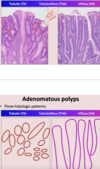
44
Q

types of polyp

A
45
Q

classify this polyp and outline features

A

tubular adenoma– looks PEDUNCULATED, low grade dysplasia only. The bigger. more dysplastic = more likely to precede carcinoma

46
Q

sessile serrated adenomas/polyps (SSA) are more common in the ___ colon

A

right colon. glands are saw-like

47
Q

T/F hyper plastic polyps have malignant potential

A

false. found in the sigmoid/rectum and there’s usually no malignant potential. There is serrated morphology, but only on the surface.

48
Q
A

hyperplastic polyp. There is serrated morphology, but only on the surface, unlike SSAs, which have saw like glands.

49
Q
A
50
Q

good slide to remembers

A