MSK 2 - Lower Limbs Flashcards

1
Q

what is the pelvic girdle and what makes it up?

A

The pelvic girdle is a bony ring consisting of the sacrum and right and left hip bones, joined anteriorly at the pubic symphysis and posteriorly by the sacroiliac joints.
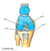
2
Q

a

A

Greater sciatic foramen

3
Q

b

A

Sacrotuberous ligament

4
Q

c

A

Sacrospinous ligament

5
Q

d

A

Lesser sciatic foramen

6
Q

Which sciatic foramen is the route for structures entering or leaving the pelvis?

A

Greater sciatic foramen

7
Q

Which sciatic foramen is a route for structures entering or leaving the perineum?

A

Lesser sciatic foramen

8
Q

Gluteal region - this posterior muscle group is organised into a _________ and ____ layer

A

superficial

deep

9
Q

what is muscle 1 and its innervation?

A

Gluteus maximus

inferior glutel nerve (L5, S1 and S2)

10
Q

what is muscle 2 and its innervation?

A

Gluteus medius

superior gluteal nerve (L4, L5 and S1)

11
Q

what is muscle 3 and its innervation?

A

Gluteus minimus

superior gluteal nerve (L4, L5 and S1)

12
Q

what is muscle 4 and its innervation?

A

Tensor Fascia Lata

superior gluteal nerve (L4, L5)

13
Q

The deep muscles of the hip ________ rotate and _______ the hip, and are covered in Lesson 1.

These are all supplied by branches of the ______ plexus

A

externally

stabilise

sacral

14
Q

What are the actions of gluteus maximus muscle?

A

Hip extensor and external rotator

15
Q

What is the action of gluteus medius, gluteus minimus and tensor fasciae latae?

A

They are hip abductors and internal rotators of hip joint

16
Q

The deep fascia of the thigh is called what?

A

the fascia lata

17
Q

Fascia lata - It extends posteriorly from the front of the thigh and is thickened laterally to form the iliotibial tract.

2 muscles attach to the iliotibial tract – these are what?

A

tensor fascia lata and gluteus maximus

18
Q

why is the iliotibial tract important?

A

The iliotibial tract is important, as it provides stabilisation to the lateral aspect of the knee joint

19
Q

The sacral plexus lies on which muscle?

A

Piriformis muscle

20
Q

The sacral plexus is formed by the union of the ventral rami of what spinal nerves?

A

L4, L5 and S 1 to S 4

21
Q

The supply from the lumbar ventral rami comes from the ________ trunk

A

lumbosacral

22
Q

The sarcal plexus supplies what?

A

the posterior aspect of the lower limb plus the perineum

23
Q

What are the two main branches of the sacral plexus?

A

Main branch to lower limb - Sciatic nerve

Main branch to perineum - Pedundal nerve

24
Q

The superior and the inferior gluteal nerves are smaller motor branches of what?

A

sacral plexus

25
The superior gluteal nerve has a root value of \_\_\_\_ The inferior gluteal nerve has a root value of \_\_\_\_\_
L4-S1 L5-S2
26
List the muscles supplied by the superior gluteal nerve below:
1. Gluteus medius 2. Glteus minimus 3. Tensor fasciae
27
The inferior gluteal muscle supplies only one muscle in the gluteal region. Which is it?
gluteus maximus
28
Other branches of the sacral plexus which innervate muscles in the lower limb are what?
1. Nerve to piriformis 2. Posterior Cutaneous Nerve to the Thigh 3. Nerve to quadratus femoris 4. Nerve to obturator internus A superficial knowledge of these nerve is all that is required.
29
The sciatic[](https://www.anatomy.tv/anatomytv/html5ui_2018/#/product/har_hip_2014/type/Text%20articles/id/43001/layer/18/angle/15/structureID/537728)nerve is the **\_\_\_\_\_\_ nerve** in the body and is the continuation of the main part of the _____ plexus
**largest** sacral
30
What are the root values of the sciatic nerve?
L 4 to S 3
31
Within the ______ region the sciatic nerve contributes very little motor or sensory innervation, aside from branches to the posterior aspect of the hip joint
gluteal
32
Sciatic nerve passes through the ______ \_\_\_\_\_\_ foramen, inferior to the ________ muscle, passing behind the deep abductors of the hip joint between the ischial tuberosity and greater trochanter of the femur to enter the _________ compartment of the thigh.
greater sciatic piriformis posterior
33
Sciatic nerve then passes distally It divides at the popliteal fossa into the what?
1. Tibial nerve 2. Common peroneal nerve
34
what is the muscle shown in green?
piriformis
35
Clinical – Sciatica: what is it?
Back pain is a pain!...Especially when it starts in your back and ends in your toes... As you get older the flexibility and elasticity of muscles, ligaments and joints diminish. Coupled with the forces that weight, gravity (plus 24 vertebrae, and a skull), place on the lower lumbar spine you can understand how vertebral foramen become narrowed and compress on the nerve roots. The L5 vertebrae is the most likely places where this happens. Compression of the nerve roots in this region can cause a syndrome called sciatica – where the proximal nerve compression can cause an ‘electric shock’ type pain passing from the back down to the toes (the dermatomal area supplied by the sciatic nerve).
36
Posterior compartment of the thigh: The _____ muscles in this region can be grouped together to be called the \_\_\_\_\_\_\_\_\_ These posterior thigh muscles act both on the ___ and the ____ joint \_\_\_\_\_\_ \_\_\_\_\_\_\_, is the most lateral of the 3 muscles, and is similar to its counterpart in the upper limb in that it has 2 heads (biceps)
three hamstrings hip knee Biceps femoris
37
1
1. Semimembranosus
38
2
2. Semitendinosus
39
3
3. Long head of Biceps femoris 4. Short head of biceps femoris
40
three muscles are collectively termed **hamstrings,** what are they?
1. Semimembranosus 2. Semitendinosus 3. Long head of Biceps femoris
41
What is the action of the hamstring muscles on the hip and knee joint?
Hip joint - Extension Knee joint - Flexion
42
Which nerve innervates the posterior compartment of the thigh?
Sciatic nerve
43
Which **branch** of the femoral artery contributes to the posterior muscle compartment blood supply?
Profunda femoris artery
44
what type of joint is the knee and what is it capable of?
The knee joint is a synovial hinge joint, capable of flexion, extension and a small amount of active and passive axial rotation
45
Bones: 1
Patella
46
Bones: 2
Fibula
47
Bones: 3
Tibia
48
Bones: 4
Femur
49
articular surfaces: a
Medial femoral condyle
50
articular surfaces: b
Lateral femoral condyle
51
articular surfaces: c
Lateral tibial condyle
52
articular surfaces: d
Medial tibial condyle
53
articular surfaces: e
Tibial tuberosity
54
The stability of the knee joints depends on what?
the strength and action of surrounding muscles and their tendons and ligaments connecting the femur and tibia
55
Which is the most important muscle which helps to stabilize the knee joint?
rectus femoris
56
ligaments of the knee - They are thickenings of the ____ \_\_\_\_\_\_\_
joint capsule
57
1
Fibular collateral ligament
58
2
Tibial collateral ligament
59
3
Anterior cruciate ligament
60
4
Posterior cruciate ligament
61
The fibular, or lateral collateral ligament (LCL) extends from the ______ epicondyle of the femur to the lateral surface of the ____ of the fibula.
lateral
62
The tendon of the _______ muscle passes deep to the LCL, separating it from the lateral meniscus.
popliteus
63
The tibial, or medial collateral ligament (MCL) extends from the ______ epicondyle of the femur to the medial surface of the tibia, with an attachment to the ______ epicondyle of the tibia.
medial medial
64
At its midpoint the MCL is attached onto the medial meniscus of the knee Why is the attachment of the MCL to the medial meniscus clinically important?
MCL injury will also injury MM and vice versa as they are joined
65
The cruciate ligaments cross each obliquely in the knee to form an ‘\_’ shape
‘X’ shape
66
The anterior cruciate ligament (ACL) prevents the femur from sliding ________ (anterior or posterior?) on the tibia. The posterior cruciate ligament (PCL) prevents the femur from sliding ________ (anterior or posterior?) on the tibia.
posterior anterior
67
**Clinical – Anterior Cruciate Ligament (ACL) Injuries:** **what is it?**
The name alone fills any sportsperson with dread... Whether is it caused playing rugby, skiing, or twisting your knee in a way it shouldn’t, an ACL injury or tear automatically means a long time out of playing sport. The reasons for this are simple. The ACL is a vital component in providing stability to the knee joint, preventing the femur from sliding on the tibia. After repair due to the poor blood supply to the ACL, it can take a long time to heal. Although both cruciate ligaments can be damaged, a PCL tear/rupture is less common due to the force required by a direct blow on the tibia to cause injury, compared to the twisting decelerating motions required to disrupt the ACL.
68
what is the popliteus muscle and its function?
The popliteus muscle is a small but, nevertheless important muscle in helping release the fully extended or “locked” knee. When flexing from a fully extended position, the muscle rotates the femur laterally on the tibia (or vice-versa), allowing for the unimpeded movement of the joint
69
**_Menisci of the Knee Joint:_** what are they?
The menisci are crescent shaped plates of fibrocartilage directly on the articular surface of tibia
70
1
Medial meniscus
71
2
Lateral meniscus
72
3
Anterior cruciate ligament
73
4
Posterior cruciate ligament
74
What are the main functions of the menisci?
Reduce friction and the stabilise knee joint
75
Which meniscus firmly adheres to the deep surface of the tibial or medial collateral ligament?
The medial meniscus
76
Clinical – Meniscal Tears: what are they?
One of the most common injuries around the knee joint are due disruptions to the cartilage or meniscal tears. Because of the shape of the menisci, any lateral impact to the knee joint risks both damaging the medial meniscus and disrupting both the ACL and medial collateral ligaments. If the cartilage becomes loose within joint space, there is a risk that the knee can become ‘locked in fixed flexion or extension
77
what is the patella, where is it and what is its function?
The patella is the largest sesamoid bone in the body, sitting at the anterior of the knee joint and acting as an anchor point for the quadceps[](https://www.anatomy.tv/anatomytv/html5ui_2018/#/product/har_knee_2014/type/Views/id/39303/layer/10/angle/33/structureID/555092)femorus[](https://www.anatomy.tv/anatomytv/html5ui_2018/#/product/har_knee_2014/type/Views/id/39303/layer/10/angle/33/structureID/555092)tendon superiorly and patellar[](https://www.anatomy.tv/anatomytv/html5ui_2018/#/product/har_knee_2014/type/Views/id/39303/layer/10/angle/33/structureID/550956)ligament inferiorly.
78
Which movement of the knee causes the patella to move?
Flexion and extension
79
what do birsae of the knee do?
Similar to the shoulder and elbow the knee has a number of bursae to act as shock absorbers
80
1
Prepatellar bursa
81
2
Deep infrapatellar bursa
82
3
Subcutaneous infrapatellar bursa
83
Which bursa communicates with the articular cavity of the knee joint? What is the clinical significance of this?
Suprapatellar bursa When bursa communicate with joint cavity, if injured or infection then it can spread it to the joint
84
What is housemaid’s knee (pre-patellar bursitis)?
Inflammation of the bursa in front of the knee cap
85
what are the muscles involved in knee flexion?
Biceps femoris, Semimembranosus, Semitendinosus, Gastrocnemius Sartorius and Gracilis
86
what are the muscles involved in knee extension?
Rectus femoris, Vastus medialis, Vastuls lateralis, Vastus intermedialis