More diseaess-Kerr, Dry mouth Flashcards
Erythroplakia
Treatment
○ Biopsy required for diagnosis
○ If a source of irritation can be identified and removed, biopsy may be delayed for 2 weeks to allow lesion to heal
○ Complete excision
What is this clinical presentation?

Erythroplakia.
Well-circumscribed red patch on the
posterior lateral hard and soft palate
What is this clinical presentation?

Erythroplakia.
Erythematous macule on the right
floor of the mouth.
Biopsy–
Turned out to be early invasive squamous cell
carcinoma.
What is this clinical presentation?

Smokeless tobacco keratosis/TOBACCO POUCH KERATOSIS
Smokeless Tobacco–related Gingival Recession.
Extensive recession of the anterior mandibular facial gingiv
What is this clinical presentation?

Smokeless tobacco keratosis/TOBACCO POUCH KERATOSIS
Tobacco Pouch Keratosis, Severe
What is this clinical presentation?

Smokeless tobacco keratosis/TOBACCO POUCH KERATOSIS
Tobacco Pouch Keratosis, Mild. A soft, fissured,
gray-white lesion of the lower labial mucosa located in the area of
chronic snuff placement.
Smokeless tobacco keratosis
Treatment:
typically resolves weeks after cessation
○ if persists 6+weeks -> biopsy to rule out dysplasia + SCC
What is this clinical presentation?

Pemphigus Vulgaris.
. Multiple erosions affecting the
marginal gingiva.
What is this clinical presentation?

Pemphigus Vulgaris.
Multiple erosions of the left
buccal mucosa and soft palate.
What is this clinical presentation?

Pemphigus Vulgaris.
Large, irregularly shaped ulcerations
involving the floor of the mouth and ventral tongue.
What is this clinical presentation?

Pemphigus Vulgaris.
What is this clinical presentation?

Pemphigus vulgaris
● Multiple, chronic, mucocutaneous ulcers
● Many patients also have
● Relatively non‐specific
● Very superficial, only in epithelium
● Occur on any mucosal surface: oral, ocular, nasal, GI, esophageal,
genital
What is this clinical presentation?

Pemphigus vulgaris
PV Lesions can affect
virtually any mucosal
surface (oral, nasal,
ocular, pharyngeal,
esophageal, genital)
What is this clinical presentation?

Pemphigus vulgaris
usually suffer from Desquamative
gingivitis (DG)
More superficial erosion of the marginal gingiva, typically with an
intense erythema and inflammation, and very often in the absence of
local factors that would typically cause a gingivitis
o Hurts to brush their teeth
Immediately look for areas where there are no local factors and look for
inflammation there
o To check the possibility of systemic factors causing local
gingivitis
What is this clinical presentation?

Pemphigus vulgaris
Combination of PV
inflammation and
gingival inflammation
accumulating local
factors can result in
advanced loss of
attachment and tooth
loss
Pemphigus vulgaris
Etiology
Pemphigus vulgaris is not fully understood.
Experts believe that it’s triggered when a person who has a genetic tendency to get this condition comes into contact with an environmental trigger, such as a chemical or a drug.
In some cases, pemphigus vulgaris will go away once the trigger is removed.
Pemphigus vulgaris
Treatment
Treatment has 3 stages:
● Stage 1: Control
○ Suppress inflammation / lesion activity with Systemic Corticosteroid: Remains initial / 1st‐line treatment…
○ Then quickly add steroid‐sparing agents (mycophenolate mofetil) to minimize dose and duration of corticosteroid treatment as well as improve disease control
● Stage 2: Consolidation
○ Reducing auto‐antibody production with the addition of Immunosuppressants
○ Assessed by the lack of development of NEW lesions
● Stage 3. Remission / Maintenance:
○ achieving complete remission of lesion activity OFF medication is the GOAL
○ When lesion activity OFF medications cannot be achieved, principle of MINIMALLY effective therapy is the goal, typically with combination of immunosuppressant medications
○ RITUXIMAB has become the FIRST CHOICE treatment after
○ the consolidation phase to achieve DISEASE REMISSION
● TOPICAL / INJECTABLE CORTICOSTEROID MEDICATIONS
○ o Can be used to help control limited number of lesions resistant to systemic therapy: it treats ONLY the disease
○ outcome (lesions) and not the systemic illness / pathologic antibody production
○ ex:clobetesol 0. 05% , halbetesol 0.05% (most potent)
What is this clinical presentation?

Mucous membrane pemphigoid
What is this clinical presentation?

Mucous membrane pemphigoid
SEVERE/HIGH RISK FORMS OF MMP
▪ Ocular
▪ Esophageal
can
result in functional
blindness
What is this clinical presentation?

Mucous membrane pemphigoid
Oral Hygiene: Plaque
related gingival
inflammation
contributing to
continued VB
desquamative
gingivitis
What is this clinical presentation?

Mucous membrane pemphigoid
REMEMBER:
▪ Plaque and calculus can be the consequence of painful MMP lesions
▪ When assessing MMP lesions/desquamative gingivitis, look for areas of intense inflammation WITHOUT local factors as evidence of VB disease
Mucous membrane pemphigoid
Etiology
Mucocutaneous autoimmune disease characterized by sub‐epithelial
blisters (bullae) which ruptures to form large, non‐healing ulcerations
Mucous membrane pemphigoid
Treatment
o Approach is similar to PV – but generally not as aggressive unless
hi‐risk areas ( ocular, esophageal ) where more intense immunosuppression indicated
▪ NON‐immunosuppressive treatments uniquely effective:
- *o** Dapsone
- *o Tetracycline + nicotinamide**
MMP & PV BIOPSY
take two different sites
○ For H&E, still must be perilesional
○ If you get only ulcer just because the clinician thinks
○ that is the pathology → there is no epithelium!
○ The sample is useless and no diagnosis can be made

What is this clinical presentation?

Actinic cheilitis
(Solar cheilosis)
Typical presentation of angular cheilitis with erythema, crusting and mild fissuring of the angles of the mouth bilaterally.
What is this clinical presentation?

Actinic cheilitis
(Solar cheilosis)
Early presetation:
Smooth, blotchy, pale, dry areas
Diffuse, irregular white plaque around line of the lip
Crusted, Scaly
Actinic cheilitis
malignant transformation
Actinic cheilitis has 2 times of risk for developing SCC of the lip.
SCC on the lips is 11 times as likely to metastasize compared to SCC found on other parts of the body
Actinic cheilitis
Etiology
due to chronic ultraviolet light exposure.
Actinic cheilitis
Treatment
- avoid sun exposure
- Laser ablation is preferred for severe actinic cheilitis
- surgical excision is recommended for severe actinic cheilitis with evidence of high-grade dysplasia
- Lip Shaving” (Vermilionectomy)
- can also use cryotherapy, electrodesiccation
It requires long term follow up and prognosis is good if caught early
What is this clinical presentation?

SCC
arising from Actinic Cheilitis
What is this clinical presentation?

Oral Melanoma
a highly malignant neoplasia, arising from melanocytes, the cells that produce the brownish pigment melanin.
What is this clinical presentation?

Oral Melanoma
an ulcerated, blue-black, slightly elevated lesion in the edentulous, posterior right maxilla. The lesion extends across the residual alveolar ridge onto the palate and onto the facial aspect of the ridge.
What is this clinical presentation?

Oral Melanoma
patient with extensive, black-pigmented and irregularly bordered macule in the maxillary labial mucosa and midline facial gingiva, (teeth 8 and 9). (The patient’s fingers are depicted.)
What is this clinical presentation?

Oral Melanoma
Large, blue-black, irregularly bordered lesion on the upper lip of a male Japanese patient. The diagnosis is oral melanoma.
What is this clinical presentation?

Amalgam tattoo
This image depicts two diffusely bordered, dark gray macules in the left posterior buccal mucosa adjacent to molar teeth that have been restored. .
What is this clinica presentation?

Oral melanoacanthoma.
the buccal mucosa of a middle-aged, black woman with a brown-black, irregularly bordered macule that arose suddenly. The patient was unaware of its presence.
What is this clinical presentation?
Oral melanotic macule
an irregularly shaped, tan-brown macule on the left hard palate in an edentulous patient.
Oral Melanoma
Etiology
Unknown. Ultraviolet radiation is an important causative factor for skin melanoma
Acute sun damage can cause it more than chronic exposure
Oral Melanoma
Risk Factors
Fair skin
A history of sunburn
Excessive ultraviolet (UV) light exposure.
Living closer to the equator or at a higher elevation
Having many moles or unusual moles
A family history of melanoma
Weakened immune system.
Oral Melanoma
Treatment
- Surgical excision
- Radiotherapy
- Chemotherapy
What is this clinical presentation

Oral Melanoma
What is this clinical presentation

Oral Melanoma
What is this clinical presentation

Oral Melanoma
What is this clinical presentation?

Traumatic ulcer
caused by sharp or puncturing food stuff
What is this clinical presentation?

Traumatic ulcer
a chronic ulcer on the left posterior lateral border of the tongue caused by lingually tilted mandibular 3rd molar. Note central ulceration with peripheral keratosis
What is this clinical presentation?

Traumatic ulcer
Post-anaesthesia traumatic ulcer on lower lip.
What is this clinical presentation?

Traumatic ulcer
Most often on tongue, lips, buccal mucosa
Any sites that may be injured by dentition
What is this clinical presentation?

Traumatic
Granuloma
What is this clinical presentation?

Traumatic
Granuloma
(traumatic ulcertaive granuloma)
What is this clinical presentation?

Traumatic Granuloma
( Traumatic Ulcerative Granuloma)
Traumatic ulcer/Traumatic ulcerative granluoma
Etiology
Etiology
- typically caused by trauma. In more than half the cases, the patient does not recall traumatizing the area although this may have occurred during sleep.
- Chronic mucosal trauma from adjacent teeth
- Some adjacent source of irritation
Traumatic ulcer/Traumatic ulcerative granluoma
Treatment
Remove cause of irritation
Topical anesthetic or film for pain relief
If there is no obvious cause then ► biopsy
What is this clinical presentation?

Squamous cell carcinoma
on the buccal mucosa)
What is this clinical presentation?

Erythroplakia and Squamous Cell Carcinoma
Erythroplakia is a general term for red, flat, or eroded velvety lesions that develop in the mouth. In this image, an exophytic squamous cell carcinoma on the tongue is surrounded by a margin of erythroplakia
What is this clinical presentation?

Leukoplakia and Squamous Cell Carcinoma
Leukoplakia is a general term for white hyperkeratotic plaques that develop in the mouth. About 80% are benign. However, in this image, squamous cell carcinoma is present in one of the leukoplakic lesions on the ventral surface of the tongue (arrow).
squamous cell carcinoma
Risk factors
HPV + SCC
Area affected: ( Oropharynx cancers largely involved tonsils, . Posterior 3rd of the Tongue)
Younger pts, 3:1 Males to females ratio, high socio-eco status
Incidence is decreasing
less aggressive → higher survival rates ( Better than HPV negative SCC)
HPV - SCC
The chief risk factors for oral squamous cell carcinoma are
Smoking (especially > 2 packs/day)
Alcohol use
Risk increases dramatically when alcohol use exceeds 6 oz of distilled liquor, 15 oz of wine, or 36 oz of beer/day. The combination of heavy smoking and alcohol abuse is estimated to raise the risk 100-fold in women and 38-fold in men.
( this affects these ares : the tongue, floor of mouth, buccal mucosa, or gingiva)
mostly men, low socio-economic factors
Incidence is decreasing
Very aggressive → lower survival rates
SCC treatment
Early stage: Radiation and/or Surgical removal
Late stage : combination of surgery, radiation therapy, or chemotherapy
What is this clinical presentation?

Graphite tattoo
Most common location on the palate and gingiva
Gray, black, or blue-ish macule
What is this clinical presentation?

Graphite tattoo
Gray, black, or blue-ish macule
Graphite tattoo
Treatment
If patient is concerned for cosmetic reasons ► then removal of lesion with autogenous graft
Graphite tattoo
Etiology
result from pencil lead that is traumatically implanted, usually during the elementary school years
What is this clinical presentation?

Traumatic ulcer of the tongue.
What is this clinical presentation?
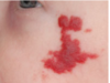
Hemangioma of Infancy
a relatively common benign proliferation of
blood vessels that primarily develops during childhood.
display a rapid growth phase with endothelial
cell proliferation, followed by gradual involution.
What is this clinical finding?

Hemangioma of Infancy
Hemangioma of Infancy
Treatment
○ Because most hemangiomas of infancy undergo involution, management often consists of “watchful neglect.”
What is this clinical finding?
Necrotizing Sialadenometaplasia
an uncommon, usually self-limiting, benign inflammatory disorder of the salivary glands.
Here it is on the palate
What is this clinical finding?

Necrotizing Sialadenometaplasia
we see two ulcers on the palate
Mostly
● Palatal salivary glands
○ Possible for parotid
● 75% of case on posterior palate
● Hard>Soft palate
● 2/3rd are unilateral
Necrotizing Sialadenometaplasia
Etiology
The cause is uncertain, although the hypothesis of ischemic
necrosis after vascular infarction seems acceptable.
Necrotizing Sialadenometaplasia
Treatment
No Treatment Needed
but we need to biopsy to rule out other diseases
What is this clinical finding?

Frictional Keratosis.
There is a rough, hyperkeratotic change to the posterior mandibular alveolar ridge (“alveolar ridge keratosis”),
because this area is now edentulous and becomes traumatized
from mastication.
Such frictional keratoses should resolve when the
source of irritation is eliminated and should not be mistaken for true
leukoplakia.
What is this clinical finding?

Frictional Keratosis
the white surrounding a a traumatic ulcer
Symptomatic traumatic ulceration of the left mid-ventral tongue associated with a sharp left lower molar. The ulcer has flat edges and is surrounded by an area of frictional keratosis.
Frictional Keratosis.
Differential Diagnosis

Leukoplakia
Linea alba
Chronic cheek chewing (bite injury)
Candidiasis
Oral Lichen planus
Squamous cell carcinoma
What is this clinical finding?

Frictional keratosis
on the tongue
Frictional Keratosis
Etiology

- Trauma from Sharp cusp & ortho appliance
- Chronic mechanical irritation (chronic biting)
- Masticatory function
- Normal hyperplastic response
- Dentures/missing teeth
Frictional Keratosis
Treatment

- Remove the cauative factor that caused the trauma
- observe large lesion regularly
excellent prognosis
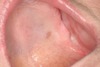
What is this clinical presentation?

dry‐mouth
patient
a classic example
• Classic fissuring
• depapillation of the tongue papilla
• some white changes on the tongue.
What is this clinical presentation?

dry‐mouth
from radiation
Note the Ropy, frothiness on the palate.
- The tissues are red and irritated due to candida infection as well.
What is this clinical presentation?

dry Mouth
Cervical caries related
to radiation.
The patient is a smoker and coffee drinker –> explains the staining
What is this clinical presentation?

dry Mouth
Incisal caries in a
radiation patient:
Incisal caries is a sure sign of severe dry mouth/ significant salivary gland hypofunction
What is this clinical finding?
Xerostomia-related Caries
Or
Dry Mouth
. Extensive cervical caries of
mandibular dentition secondary to radiation-related xerostomia.
Dry mouth
Subjective vs Objective
Xerostomia
The subjective experience of a dry mouth (ie a symptom)
Salivary Hypofunction
The objective measurement of a reduction in salivary flow (a sign)
What is the normal rate for Stimulated Saliva
Production
Stimulated Saliva
Production
▪ 200+ ml/day
▪ Flow rate: mean 1-2 ml/min, maximum 7 ml/min
o “Normal” range is very wide
What is the normal rate for Unstimulated Saliva
Production
300 ml/day
▪ Flow rate: mean 0.3 ml/min
What are Factors affecting unstimulated flow include?
- Dehydration
- Medical conditions
- Body posture
- Lighting conditions
- Circadian/circannual rhythm (lowest during)
- Medications
Age is an independent factor for whole saliva andsubmandibular/sublingual gland secretion (but notparotid).
What are Factors affecting stimulated flow include:
- Mechanical stimuli
- Vomiting
- Gustatory/olfactory stimuli (acid/smell)
- Gland size
Age is an independent factor for whole saliva (but not
for parotid and minor gland secretions)
What causes
dry mouth?
Central inhibition as a result of connections between the primary salivary centers and the
higher centers of the brain.
● What causes xerostomia in absence of measurable salivary hypofunction?
-
May be a reduction in baseline sialometry which is still above “normal.”
- If they get a decrease in the salivary flow, they still may be in the normal range, but for them the experience is that they have got dry mouth.
-
Saliva film thickness
- Palatal mucous gland secretions?
- Anterior dorsum of tongue?
-
Relative contributions by glands
- Mucins, proteins?
- Alterations in sensory perception?
- Mental status/central inhibition?
What cause
Salivary
Hypofunction?
● Dehydration
● Medications (Rx & OTC)
- Direct damage to glands
- Head and neck radiotherapy
- As a result of radiation it’s irreversible damage to the glands
- Chemotherapy (reversible)
- Autoimmune diseases
- Primary vs Secondary Sjögren’s Syndrome, GVHD
- HIV disease
● Decreased mastication (tooth loss, soft diet)
● Conditions affecting the CNS:
- Psychologic disorders (depression/anxiety?), Alzheimer’s, Parkinson’s, Cerebral palsy
To have dry mouth
xerostomia
what is the rate of Unstimulated and Stimulated Salivary flow
USFR
and
SFR
Abnormal unstimulated USFR= <0.1–0.2ml/min
Abnormal stiumated SFR = <0.5ml/min
Severity of patients
with xerostomia using
objective measures

How to manage with normal USFR and SFR?
- Salivary stimulation (OTC) to stimulate their glands
- Salivary lubrication ( to improve it)
- Humidification ( like a humidifier in the room at night)
- Hydration/prevent dehydration (ie avoid alcohol, caffeine both will act as a
- diuretic and lead to dehydration).
- Monitor closely to rule out emerging disease ( to see whether they are developing Sjogren’s or something else we want to follow them over time)
How to manage with abnormal USFR and normal SFR?
(respond to stimulated)
Abnormal unstimulated USFR= <0.1–0.2ml/min
- Look for possible causes (major cause will be medications & can dehydration or others)
- Restore chewing function (Masticatory issues)
- Reduce medication‐induced salivary hypofunction
- Prescribe Salivary stimulation OTC, Rx medications, others
- Prescribe Salivary lubrication
- Humidification ‐use humidifiers
- Hydration/prevent dehydration (ie avoid alcohol, caffeine)
- Treat oral consequences (such as candidiasis treated with an antifungal or caries with management of caries).
How to manage with abnormal USFR and abnormal SFR?
Abnormal unstimulated USFR= <0.1–0.2ml/min
Abnormal stiumated SFR = <0.5ml/min
- If dehydrated ► rehydrate or treat underlying condition
- People with uncontrolled diabetes, once you control the diabetes‐ their flow comes back.
- All we can do is offering Salivary substitutes (sprays, gels, rinses )
- For patients with high dose radiation treatment ► makes sure they get the INRT
- Minimizing damage to salivary glands ( there are other strategies for that)
- Prevention and treatment of oral complications
What are the
Prescription
Medications
for people with
low USFR and
some oral signs, but
responds to stimulation ?
(abnormal USFR, Normal/improved SFR)
(include dosage and usage)
– Muscarinic agonists:
– Pilocarpine 5‐7.5mg tid & qhs (can go as
high as 10mg qid)
– Cevimeline 30mg tid (can go as high as
60mg tid)
Contradicated for : CV disease, hepatic, renal or respiratory diseases or narrow angle glaucoma
Pilocarpine affects M1 & M3
side effects (sweating, flushing,
rhinitis, increased urination, weakness and
some experience the shakes. )
Cevimeline affects M3 only
fewer side effects
What is this clinical presentation?

SJÖGREN’S SYNDROME
Autoimmune exocrinopathy
Marked bilateral parotid gland enlargement in a patient with primary Sjögren syndrome
Dry mouth and eyes resulting from a chronic progressive loss of secretory function (Slowly but surely, the salivary glands and/or lacrimal glands (some cases are more lacrimal & less salivary or vice versa); slow & progressive
Patients with Sjogren’s can have bilateral salivary gland enlargement (parotid) (Sometimes we may see a unilateral enlargement of the salivary
glands due to retrograde infections.)
increased risk of lymphoma (MALT type)
What is this clinical presentation?

SJÖGREN’S SYNDROME
Dry Mouth
very severe
cervical disease & very dry lips
What is this clinical presentation?

Depapillated &
Fissured Tongue
SJÖGREN’S SYNDROME
What is this clinical presentation?

a patient
with a
bacterial sialadenitis
who has
SJÖGREN’S SYNDROME
When we examine such patients and ”milk” the gland ► you actually see a purulent drainage from the gland itself.
SJÖGREN’S SYNDROME
Management

These patients LOW USFR, no response to stimulation (aka abnormal USFR/SFR)
- Rehydrate if dehydrated
- Treat underlying conditions (i.e. DM)
- Salivary substitutes (glycerin)
- Minimize damage to glands from radiation
- Prevention of complications & palliative treatment
- Optimal hygiene
- Restore caries
- Smooth sharp edges in oral cavity
- Fluoride therapy
- Antifungals
- Chlorhexidine rinses w/o alcohol
- Sialendoscopy
- Salitron - salivary pacemaker
- ALTENS (acupuncture like transcutaneous electrical nerve stimulation)
What is this clinical presentation?

Sjögren Syndrome
- bilateral enlargement of the submandibular glands
- angular cheilitis, dry and cracked lips and fissured and despapilated tongue
- severe ocular lesions.
