Lecture 2: Histology of the Endocrine Glands Flashcards
What is produced by the thyroid gland?
T3: more potent, shorter half-life, less abundant
T4: less potent, longer half-life, more abundant
Calcitonin
What is the functional and structural unit of the thyroid gland; what is its structure?
- Thyroid follicle
- Single layer of follicular cells (simple cuboidal (inactive) to columnar (active) epithelium) surrounding a fluid called colloid
What is found within the colloid and what is its function?
- Thyroglobulin
- Storage form of T3 and T4
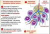
What is the arrow pointing to?

Follicle of the Thyroid Gland
What is the arrow pointing to; found where; produces?

- Parafollicular or ‘C’ cells
- Isolated clusters between follicles or within follicular epithelium
- Produce Calcitonin

Label A-C


Label the arrows from top to bottom!

Top: parathyroid gland
Middle: capsule
Bottom: thyroid gland
What are the two cell populations of the Parathyroid gland; function of each?
1) Chief (principle) Cells - secrete PTH
2) Oxyphil (acidophilic) Cells - function unknown
Label the 2 cells types A and B

A) Chief cells
B) Oxyphil glands - look more fluffy

Label the cell types A and B

A) Chief cells
B) Oxyphil cells
Why is the hypothalamus called the “master gland,” and what is its function?
- Connects nervous and endocrine systems
- Role in autonomic, endocrine, and limbic systems
- Helps maintain homeostasis
How is the Pituitary gland (hypophysis) connected to the hypothalamus?
By a thin stalk of tissue called the Infundibulum
What are the functional division of the anterior pituitary (Adenohypophysis)?
- Pars distalis
- Pars tuberalis
- Par intermedia

What are the functional divisions of the Posterior Pituitary (Neurohypophysis)?
- Pars nervosa
- Infundibular stalk (neural stalk)

Where does the adenohypophysis arise from; constitutes how much of the pituitary and is controlled by?
- Oral ectoderm
- 80% of the pituitary
- Neurohormones
Where does the neurohypophysis arise from; controlled by; function?
- Neural ectoderm
- Controlled by neurons
- Axons from hypothalamus carry ADH and oxytocin here for storage
What are the components of the Pars distalis?
- Glandular epithelial cells arranged in thick cords
- Connective tissue stroma
- Fenestrated capillaries (sinusoids): part of the secondary capillary plexus
What hormones are secreted by the Pars Distalis?
- FSH
- LH
- ACTH
- TSH
- Prolactin
- GH
What are the 2 cells classifications of the the Pars Distalis?
1) Chromophils (granules readily take up H/E stain - blue/pink)
2) Chromophobes (less affinitiy for stains)

What are the 2 categories of chromophils?
1) Acidophils = pink stain
2) Basophils = purple stain

Label the arrows from left to right?

Left: Basophil
Right: Acidophil
What are the major Acidophil cell types of the Pars Distalis; what does each secrete?
- Somatotrophs: GH
- Mammotrophs: Prolactin
What are the major Basophil cell types of the Pars Distalis; what does each secrete?
- Thyrotrophs: TSH
- Gonadotrophs: FSH and LH
- Corticotrophs: ACTH
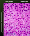
What is labeled by the arrows A and B?

A) Acidophils
B) Basophils
*Both of the Pars Distalis (Pars Anterior)