Lecture 12: Hip and Knee Joints Flashcards
(32 cards)
What makes up the hip joint?
Femoral Head and Acetabulum of Pelvis
Ball and Socket
What three bones fuse to make up the acetabulum?
Ilium
Ischium
Pubis

What is the femur mostly covered with?
Articular Cartilage
Calcar Femorale
Where is this found?
What does it do?
- Location: Vertical plane of bone on posterior aspect of femur and is deep to lesser trochanter
- Function: Helps determine if fractures are stable of not
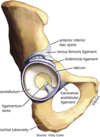
Iliofemoral Ligament
What are its attachments?
What does it do?
- Attachments: ASIS to the middle of greater and lesser trochanter of femur (B/t femur and ilium)
- Function: Prevents hyperextension of thigh
Strongest Hip Ligament

Pubofemoral Ligament
What are its attachments?
What does it do?
- Attachments: Superior pubic ramus to lesser trochanter of femur (B/t femur and pubis)
- Function: Prevents hyperabduction of thigh
Blends with Iliofemoral Ligament

Ischiofemoral Ligament
What are its attachments?
What does it do?
- Attachments: Lower part of Ischium to femoral neck
- Function: Limits internal rotation of hip

Weakest of all hip ligaments: why posterior hip injuries are most common
Ligamentum Teres (Ligament of Head of Femur)
What is its attachments?
What is its function?
- Attachments: Goes from the fovea of the head of the femur to the acetabulum
- Function: Holds head of the femur into the joint and provides blood supply

Acetabular Labrum
Where is it found?
What does it do?
- Location: Ring of cartilage that surrounds acetabulum on the rim
- Functoin: Deep pocket for the head of the femur

Anterior portion is most vulnerable when the labrum tears
Transverse Acetabular Ligament
What are its attachments?
What does it do?
- Location: Bridges Acetabular notch
- Function: Covers acetabular notch and prevents dislocation inferiorly
What artery supplies the acetabulum?
Anterior and Posterior Branch of Obturator Artery
What arteries supplies the head of the femur?
- Acetabular Branch of Obturator Artery
- Runs with Ligamentum Teres
-
Lateral Femoral Circumflex Artery (mostly anterior side)
- Ascending, Transverse, and Descending Branches
- Medial Femoral Circumflex Artery (mostly posterior side)
- Retinacular Arteries
What does Hilton’s Law state?
Nerves supplying muscles that diretly overly a joint also innervate that joint
What would you palpate on the hip?
Anterior
- Iliac Crest
- Greater Trochanter of Femur
- Pubic Tubercle
Posterior
- Iliac Crest
- PSIS
- Greater Trochanter
- Ischial Tuberosity
FADIR Test
How do you perform this test?
What does a positive test indicate?
- Flex hip to 90o
- Adduct
- Internally Rotate
Positive test (groin pain) indicates labral pathology/Femoroacetabular Impingement (FAI)

What is a Cam Impingement?
(Type of FAI Impingement)
- Femoral head is not perfectly round and cannot rotate smoothly inside the acetabulum.
- Results from a bump formed from excess bone growth at the end of the femur.
- During movement, the bump grinds the cartilage inside the acetabulum.

What is a Pincer Impingement?
(Type of FAI Impingement)
- Acetabulum is excessively deep or covers too much of the femoral head.
- Often results from excess bone growth that extends out over the normal rim.
- Overhang can impinge the neck of the femur and tear labrum

Patrick’s FABER Test
How do you perform this test?
What does a positive test indicate?
- Flex
- Abduct
- Externally rotate
Positive test (pain) indicates iliopsoas issues or SI pain

Log Roll Test
How do you perform this test?
What does a positive test indicate?
- Patient supine
- Internally and Externally rotate
Positive test (pain) helps indicates possible hip fracture

Thomas Test
How do you perform this test?
What does a positive test indicate?
- Patient supine
- Drops contralateral leg off table and pulls tested leg to chest
Positive test (contralateral thigh raises off thigh) indicates flexion contracture of hip.

Ober’s Test
How do you perform this test?
What does a positive test indicate?
- Patient lateral recumbent with affected side up
- Extend hip/flex knee and allow to drop
Positive test (affected leg not passing neutral adduction) indicates IT Band tightness.

Stinchfield Test
How do you perform this test?
What does a positive test indicate?
- Patient supine
- Resist hip flesion with straight leg
Positive test (pain) helps indicates intraarticular pathology.

Hip dislocations
What direction and position is it most likely?
Posterior (90% cases)
Flexed hip more susceptile to dislocation because of capsular laxity
Why do hip fractures need to be treated immediately?
The blood supply to femoral head will be compromised


