Lab Quiz 2: Labs 4, 5, 6 Flashcards
(136 cards)
1
Q
Identify the arteries
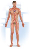
A

2
Q
Blood oxygen saturation level (SpO2)
- Describe
- What is used to measure it?
A
- Amount of oxygen present in blood compared to the maximum amount of oxygen the blood could contain
- Measured using a pulse oximeter
3
Q
Blood pressure
- Describe
- Where is it commonly measured?
- Normal range
A
- Amount of pressure exerted by the blood as it pushes against blood vessel walls
- Rises and falls as the heart contracts and relaxes
- Commonly measured in the brachial artery
- Normal range: 110-130 / 75-85 mm Hg
4
Q

A

5
Q

A

6
Q
A

7
Q
Identify the veins

A

8
Q

A

9
Q
Cardiac output formula
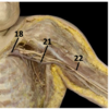
A
Stroke Volume (ml) * Heart Rate (beats/min) = Cardiac Output
10
Q
Which artery is used to measure the carotid pulse rate?
A
Common carotid artery
11
Q
Deoxyhemoglobin
- Describe
A
- Hemoglobin that is not bound to oxygen
- Less oxygenated blood appears dark red in color
12
Q
Describe how the pulse is generated
A
- As blood is forced out of the left ventricle, it expands the elastic arteries
- Blood moves through the arterial system
13
Q
Diastolic blood pressure
- Describe
A
The pressure measured when the ventricles relax
14
Q
Dubb sound
- Describe
- What creates the sound
A
- Second sound of the heart beat (S2)
- Sound is shorter and sharper than S1
- Associated with the closure of the semilunar valves
15
Q
What is used to listen (auscultate) for the S1 and S2 sounds?
A
Stethoscope
16
Q
What SpO2 level is considered to be that of hypoxemia?
A
- < 90% SpO2
17
Q
Identify #1

A
Pericardium
18
Q
Identify #1

A
Atrioventricular mitral valve
19
Q
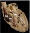
Identify #1

A
Brachiocephalic trunk
20
Q
Identify #1

A
P-wave
21
Q
Identify #10

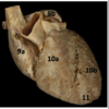
A
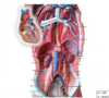
Superior mesenteric artery
22
Q
Identify #10

A
Interventricular septum
23
Q
Identify #10a
A
Right venticle
24
Q
Identify #10b

A
Left ventricle
25
Identify #11

Apex of heart
26
Identify #11
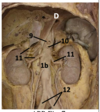
Renal arteries (right & left)
27
Identify #12

Right atrium
28
Identify #12

Common iliac arteries (right & left)
29
Identify #12a

Right atrium
30
Identify #12b

Left atrium
31
Identify #13

Interventricular septum
32
Identify #13

External iliac arteries (right & left)
33
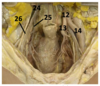
Identify #14

Apex of the heart
34
Identify #14

Internal iliac arteries (right & left)
35
Identify #14a

Right atrioventricular valve (tricuspid)
36
Identify #14b

Left atrioventricular valve (bicuspid)
37
Identify #15

Femoral arteries (right & left)
38
Identify #15

Papillary muscle
39
Identify #16
Chordae tendinae
40
Identify #16a

Superior vena cava
41
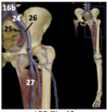
Identify #16b

Inferior vena cava
42
Identify #17

Brachiocephalic veins (right & left)
43
Identify #17b

Semilunar valve (aortic)
44
Identify #18
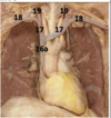
Subclavian veins (right & left)
45
Identify #18

Subclavian veins (right & left)
46
Identify #19

Internal jugular veins (right & left)
47
Identify #1b

Abdominal aorta
48
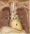
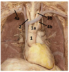
Identify #2

Ascending aorta
49
Identify #2
Heart
50
Identify #2
Brachiocephalic artery
51
Identify #2

Papillary muscles
52
Identify #2

T-wave
53
Identify #20

External jugular veins (right & left)
54
Identify #21

Axillary veins (right & left)
55
Identify #22
Brachial veins (right & left)
56
Identify #23
Renal veins (right & left)
57
Identify #24
Common iliac veins (right & left)
58
Identify #24

Common iliac veins (right & left)
59
Identify #24
Common iliac veins (right & left)
60
Identify #25

Internal iliac veins (right & left)
61
Identify #25

Internal iliac veins (right & left)
62
Identify #26

External iliac veins (right & left)
63
Identify #26

External iliac veins (right & left)
64
Identify #27

Femoral veins (right & left)
65
Identify #3

Left common carotid a.
66
Identify #3

Left auricle
67
Identify #3

Q-T interval
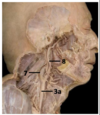
68
Identify #3a

Right common carotid artery
69
Identify #3a

Aortic arch
70
Identify #3b

Brachiocephalic artery
71
Identify #3b

Left common arotid artery
72
Identify #3c

Left common carotid artery
73
Identify #3d

Left subclavian artery
74
Identify #4

P-Q segment
75
Identify #4

Pulmonary trunk
76
Identify #4

Left brachiocephalic v.
77
Identify #4

Chordae tendinae
78
Identify #4a

Right subclavian artery
79
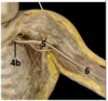
Identify #4b

Left subclavian artery
80
Identify #4b

Left subclavian artery
81
Identify #5

Left atrium
82
Identify #5

Axillary arteries (left & right)
83
Identify #5

Left subclavian a.
84
Identify #5a

Superior vena cava
85
Identify #5a & 5b

Superior and inferior vena cavae
86
Identify #6

Aortic arch
87
Identify #6
Brachial arteries (left & right)
88
Identify #6a

Right coronary artery
89
Identify #6b
Left coronary artery
90
Identify #7

Internal carotid arteries (right & left)
91
Identify #8

Right & left pulmonary vein
92
Identify #8
External carotid arteries (right & left)
93
Identify #9

Celiac trunk
94
Identify #9a

Right auricle
95
Identify #9b

Left auricle
96
Identify 1a

Aortic arch
97
Identify #1b

Thoracic aorta
98
Identify A

Finger sensor
99
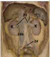
Identify K

Kidneys
100
Identify the following


101
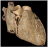
Identify the parts of the heart


102
Identify #12

Common iliac arteries (right & left)
103
Korotkoff sounds
* Describe
* How are they detected?
* Determines systolic and diastolic pressures
* Detected using a sphygmomanometer
104
Label the heart


105
List the artery & vein layers
1. Tunica intima/interna
2. Tunica media
3. Tunica adventitia/externa
106
Lubb
* First sound (S1)
* Produced when the _atrioventricular valves_ close
* Louder and more prolonged than S2 sound
107
Manual blood pressure procedure
* Wrap the cuff around the subject’s arm, above the elbow
* Inflatable portion of the cuff is on the _anterior medial side_ of the arm
* Clean the earpieces of the stethoscope with an alcohol swab before use
* Place the diaphragm of the stethoscope along the medial elbow
* Hold the rubber squeeze bulb so that the attached rubber tubing _leads away from you_. Turn the metal dial _clockwise_ until it is completely closed
* Pump the cuff up to about _150 mm Hg_ and listen carefully – you should **not** hear any sound at this point since the brachial artery is now closed, and there is no blood flowing through the blood vessel
* Gradually release the pressure so that the needle on the pressure gauge descends very slowly
* Listen very carefully for the **first** sound that you hear, and note the pressure at which this first sound occurs – this pressure corresponds to **systolic blood pressure.**
* Continue to slowly release the pressure
* Listen for the sounds to disappear
* Note the pressure at which this occurs – this pressure corresponds to **diastolic blood pressure.**
108
Name the test that examines the electrical activity of the heart through skin conductance
Electrocardiography
109
Oxyhemoglobin
* Hemoglobin _bound to four oxygen molecules_
* Well oxygenated blood appears bright red
110
Parts of a stethoscope
1. Earpieces
2. Diaphragm
*Note: When putting on the stethoscope, the earpieces should be angled in a **forward** direction*
111
Physiological splitting of the S2
* During _inspiration,_ S2 (dubb) sound splits into two separate sounds
* _Diaphragm muscle_ lowers, creating _negative pressure_ in the chest to bring in air
* Brings more _venous blood_ back to the _right atrium and right ventricle_
* Takes longer for the right ventricle to squeeze the extra blood into the _pulmonary arteries_, and it takes longer for the _pulmonary valve_ to close
* The closing of the _pulmonary valve_ is slightly later than the _aortic valve_, and that's called the *physiologic splitting of the S2*
112
Pulse oximeter
* Can detect both _pulse rate and SpO__2_
* Projects two different _wavelengths of light_ through the tissue
* Sensor picks up _color differences_ as well as color changes caused by varying amounts of _oxyhemoglobin_ and _deoxyhemoglobin_
* Color difference can be used to determine the % of oxygen saturated _hemoglobin_ (SpO2) present in the blood
113
Pulse pressure formula
Systolic BP (mmHg) – Diastolic BP (mmHg) = Pulse Pressure
114
Which artery is used to measure the pulse rate?
Radial artery
115
Sphygmomanometer
* Usually called a blood pressure cuff
* Most commonly used instrument to measure blood pressure
116
Sphygmomanometer procedure
1. Place cuff around the arm
2. Inflate the pressure high enough to completely block blood flow through the _brachial artery_
3. At this point, there are no sounds heard when listening with the _stethoscope_ because there is no blood flowing through the artery
4. As the pressure is gradually released and the blood vessel first opens, blood will spurt through the artery, even though it is still partially closed
5. The turbulence created by the spurting blood causes the first _Korotkoff_ sound heard
6. The pressure at which the first sound is heard corresponds to _systolic blood pressure_
7. As the pressure continues to decline, the sounds may become even louder because of greater blood turbulence, but eventually the sounds completely disappear as the artery fully opens, and the blood flows freely without turbulence
8. The pressure at which the sounds first stop represents the _diastolic blood pressure_
117
SpO2 ranges
* Normal = 95% - 100%
* \< 92% indicates inadequate oxygen levels in the blood typically caused by illness and can indicate _respiratory distress_
* \< 90% is hypoxemia
* Supplemental oxygen therapy may be prescribed \< 92%
118
Stroke volume formula
Pulse Pressure (mmHg) \* 1.7 ml/mmHg ∙ beat = Stroke Volume
119
Systolic blood pressure
The pressure measured at the moment the _ventricles contract_
120
Tunica adventitia/externa
* Describe
* Tissue type
* Most superficial layer
* _Dense irregular CT_ with _collagen fibers_ running in all directions for strength in many different directions (looks like squiggly lines)
121
Tunica intima/interna
* Describe
* Tissue types
* Innermost layer and lines the lumen of the blood vessels
* [_Simple squamous epithelium_](https://www2.victoriacollege.edu/dept/bio/Belltutorials/Histology%20Tutorial/Basic%20Tissues/Epithelial%20Tissues.html#Simple%20squamous%20epithelium) (provides a smooth surface for the blood to “slide past”) and a thin layer of [_areolar CT_](https://www2.victoriacollege.edu/dept/bio/Belltutorials/Histology%20Tutorial/Basic%20Tissues/Connective%20Tissue.html#Areolar%20CT) (basement membrane) to "stick it to the Tunica media"
* Continuous with the _endocardium_, makes-up the capillaries and is collectively referred to as the endothelium
122
Tunica media
* Describe
* What is it responsible for?
* Tissue types
* Middle layer
* Responsible for _vasodialation_ and _vasoconstriction_ of the blood vessels
* Made of [_smooth muscle_](https://www2.victoriacollege.edu/dept/bio/Belltutorials/Histology%20Tutorial/Basic%20Tissues/Muscle%20Tissue.html#Smooth%20muscle) and _elastic fibers_
123
What causes the S1 sound?
It is caused by the _atrioventricular valves closing_ at the _beginning of systole_
124
What causes the S2 sound?
* The _aortic and pulmonary valves closing_, at the beginning of _diastole_
125
What does a pulse oximeter measure and what do the measurements mean?
* Pulse/heart rate
* The number of times the heart beats per minute
* SpO2
* Measures the amount of oxygen in your RBCs
* The % saturation of hemoglobin in peripheral blood with oxygen as measured through the nail bed with a red light
* Maximum value is 100%
126
What is the avg pulse rate of an adult?
60-100 bpm
127
What is this?

Pulse oximeter
128
What procedure is shown here?

Pulse rate measurement
129
What vessel is being used here?

Left brachial a.
130
Where will you hear the mitral valve closing?
* Where the _mid clavicular line_ intersects with the _5th intercostal_ space
131
Where will you hear the right aortic valve closing?
2nd intercostal space (2nd & 3rd ribs)
132
Where will you hear the right pulmonary valve closing?
_Left 2nd intercostal space_ at the _left upper sternal_ border
133
Where will you hear the tricuspid valve closing?
_4th & 5th rib_ next to the _left lower border of the sternum_
134
Which EKG wave / segment / interval indicates the depolarization of the atria?
P-wave
135
Which EKG wave / segment / interval indicates the repolarization of the ventricles?
T-wave
136
Which EKG wave / segment / interval is the time between the end of the p-wave and the beginning of the Q-wave?
P-Q segment
