L9.3 Ear Flashcards
1
Q
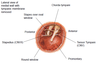
Auricle & Tragus
A
- Auricle - mostly cartilaginous
- Lobe does not have cartilage
- Tragus - little flap directly ANT to ear canal
2
Q
Features of the external acoustic meatus?
A
- Sound transmission
- Supported by:
- LAT: Cartilage (1/3)
- MED: bone
- Lines by cerumen glands → produces earwax
- Helps prevent masceration when it comes in contact with water

3
Q
NS of the ear
A
- POS/INF surface innervated by vagus
- ANT/SUP Surface innervated by auriculotemporal N (mandibular division of the trigeminal N)
- Also stimulates the tympanic membrane
- Refer pain to other areas of the mandibular N innervation (May be to the teeth)
4
Q
Tympanic membrane (LAT view)
A
- Indentation form by handle of malleus
- Concave LAT
- Concavity depends on pressure that exists in the middle ear
- Cone of light in ANT-INF quadrant
- Light shown due to concavity of the membrane
- If light changes position → may have problem with middle ear

5
Q
Middle ear
A
- Space b/w tympanic membrane and petrus part of temporal bone
6
Q
What does the middle ear split into?
A
- Tympanic cavity proper (MED to the tympanic membrane)
- Epitympanic recess (projects up above)
- Communicates with the mastoid air cells
7
Q
Communication of middle ear to the nasopharynx
A
- Via Auditory tube (aka: pharyngotympanic tube)
- Projects ANT and INF to nasopharynx
- Cartilaginous part towards the pharynx
- Embedded in bone in the tympanic part
8
Q
Ossicles of the ear
A
- Malleous - shaped like hammer
- Incus - shaped like a anvil
- Stapes - shaped like a steer
- Stapes sits on the oval window opening to the inner ear
- Transmit the energy to the inner ear

9
Q
What happens if the auditory tube is blocked?
A
- If auditory tube is blocked → impacts on the movement of the ossciles → impact energy into the inner ear → problems with hearing
10
Q
Difference in auditory tube of adults and infants? What is the significance?
A
- Vertical in adults → drains down → prevents infections
- More horizontal and shorter in infant (bacteria may move from nasal cavity through the nasal cavity and infect inner ear
- More difficult to drain infection as well
- Infection may interrupt movement of small bones which interrupts hearing and damage the tympanic membrane
11
Q
Tensor tympani
A
- ANT wall → malleolus
- Innervated by 5th cranial N
12
Q
Stapedius
A
- Attaches to stapes
- Innervated by the 7th cranial N
- Reduced reflex response when there is a problem with this facial N
- Increases hearing in these people
- Reduced reflex response when there is a problem with this facial N

13
Q
Why is there a reflex contraction of the ear muscles?
A
- When sound is too loud
- When the muscles contract the dampen the vibration amplitude of these bones → prevent damage
14
Q
LAT view of ear with tympanic membrane removed
A
- Muscles
- Round window
- Another opening of the inner ear
- Promontary (basal turn of the cochlear → makes an impression)
- Chorda tympani
- Runs through the middle ear (but doesn’t innervate the middle ear)
- Branch of the facial N
- Runs a tortous path to the ANT 2/3 of tongue
15
Q
Association of internal carotid artery with the middle ear
A
- Internal carotid artery
- Close association with the tympanic cavity and middle ear
- Infection dev in middle ear
- Energy from pulse → transmitted to the ossicles → able to hear your pulse

16
Q
Inner ear
A
- Inner ear is a system of spaces and membrane imbedded in the petrous part of the temporal bone
- Bony labyrinth filled with periplymph (white bits)
- Suspended inside → membranous labyrinth contains endolymph
- Also sensory for hearing and equilibrium
- Round window → provides escape of the transmitted energy

17
Q
Features of the bony labyrinth
A
- Cochlea
- Vestibule - connect cochlea to canals
- Opening of round and oval window relative to vestibule

18
Q
Membranous labyrinth
A
- Suspended in the bony labyrinth
- One twisted sac that approximates space that sits in the bony labyrinth
19
Q
Cochlear
A
- Contains sensory R for hearin
- Cochlear N (8th cranial N) connected to cochlear
20
Q
Semicircular ducts
A
- Sits in semicircular canals
- ANT/POS/horizontal
21
Q
Ampulla
A
- Has sensory R for dynamic equilibrium
- Information for our head MOVEMENT
22
Q
Vestibule
A
- Has Utricle & saccule for static equilibrium
- Information for our head POSITION
23
Q
What forms the 8th cranial N
A
- Vestibule + Ampulla gives information → forms the 8th cranial N
24
Q
Vibration of tympanic membrane relative to freq and loudness
A
- Tympanic membrane vibrates relative to freq and loudness → ossicles → oval window → perilymph → membranous labyrinth → endolymph → vibrates sensory R on membrane (along the organ of corti in the cochlea)
25
How does the brain code for high freq and low freq sound?
* Sensory neurons for:
* High freq = base of cochlear
* Low freq = apex of cochlear
26
Organ of corti
* Vibrates → pulls on hair → activate sensory R.
27
Dynamic equilibrium
* Rotate in 1 direction → endolymph in horizontal semi-circular canal is heavy → lags behind → moves in opposite head direction
* Cupula deflected and activates hair cells
* Brain decodes which ampulla is most active
28
Static equilibrium
* Otoconia (crystals) move with head by gravity → stays in one position → activates hair cells in that position
