L11.2 Regions of the head: Te orbit and eye Flashcards
1
Q
Blow out fracture of the orbit
A
- From trauma to the eye → Blood trauma pressure → breaks ethmoid and lacrimal bone in the orbit
- Extra-occular muscles gets trapped in the broken bone → develop double vision
- Surgery → replace with titanium plate
- But if have blood trauma again → may have irreversible damage to the eye as no structures are there to give way
2
Q
Supraorbital margin
A
- Formed by the Frontal bone
- Supraorbital notch → passage of BV and frontal N

3
Q
Infraorbital margin
A
- Zygomatic bone (LAT)
- Maxilla (MED)

4
Q
Boundaries of the bones in the orbit
A
- Apex is deep in the head, base is the front
- Roof
- Frontal bone (blue), lesser wing of sphenoid (yellow)
- Floor
- Maxilla (green), Zygomatic (orange), Palatine (Tiny bit of yellow)
- LAT wall
- Zygomatic, Greater wing of the sphenoid
- MED wall
- Maxilla, Lacrimal bone (purple - very thin), Ethmoid (also very thin), Body of sphenoid

5
Q
Fissures and foramina in the orbit
A
- Optic canal (optic N passes) next to SUP orbital fissure (most through here)
- Located at the apex
- Formed by lesser and greater wing of sphenoid

6
Q
3 layers of the orbit
A
- Outer coat
- Cornea & Sclera
- Tough coat for protection
- Middle layer
- Uvea
- Function for nutrition
- Inner coat
- Retina
- Vision

7
Q
Sclera
A
- White of the eye, forms 5/6 of eyeball
- Maintains shape and attachments of the muscles
- Made up of collagen → laid down series of whirl → extra strength
8
Q
Cornea
A
- ANT 1/6 eye
- Transparent and is continuous with sclera
- Layers:
- Corneal epithelial layer
- Stroma underneath → made up of collagen same as the sclera
- Endothelium
- Born with certain number
- Control water balance and thickness of cornea
9
Q
Why is the cornea transparent and the sclera not, despite being made up of the same substance
A
- Due to the way which collagen is laid down
- Collagen in sclera is of different size and laid down in whirl
- Collagen in cornea is of uniform size, evenly spaced, parallel bundles (lamellae)
- 200-300 lamellae in stroma

10
Q
What happens when different layers of the cornea is damaged
A
- If epithelium damaged → able to be repaired
- If stroma damaged → scarring occurs (collagen fibrils are disrupted)
- If endothelium damaged → cornea cannot be repaired and transplant is needed
11
Q
What is the ANT chamber angle
A
- B/w cornea and iris
- Where fluid (aqueous humour) drains out of the eye
- If blocked → disease like glaucoma
- Has a trabecular meshwork
- Drainage region
- Canal schlemm
- Drains to the venous system
12
Q
Uvea
A
- Ciliary body
- Iris
- Choroid
13
Q
2 key features of the ciliary body
A
- Ciliary processes and ciliary muscles
14
Q
Ciliary processes
A
- Formation of aqueous humour (like the CSF)
- Important for maintaining health of interior of eye
- Point which ligaments attach which attaches to the lens
15
Q
Ciliary muscles
A
- Accommodate and focusing of lens
- Look far = lens thin; close = lens fat
- Relaxation = look far; Contraction = read
- When contracting → ligaments are not under pressure and allows lens to become fat
16
Q
Iris
A
- Aperture of the eye
- 2 muscles:
- Sphincter pupillae: Constricts pupil: By PNS
- Dilator pupillae: Dilates pupil: By SNS
17
Q
Choroid
A
- Has 3 layers of BV and functions as providing the BS
- The small layer → sit beneath retina
- Providing vascular supply to outer part of retina (particularly the photoreceptors)
- Also supplies the structures of the front of the eye
18
Q
Retina features
A
- Fovea
- NO BV located
- Macula
- Crucial for central vision
- Fovea is within the area of macula
- POS pole
- Ora serrata
- Edge of retina
- Where retina joins the other structures of the eye (looks like a serrated edge)
19
Q
Specialised region: Fovea
A
- Fovea
- Best visual ability Anything impinging visual ability is removed (No Rods)
- ONLY photoreceptors are located here (Cones)
- Avascular
- Gets nutrients from choroid
20
Q
Specialised region: Optic nerve
A
- Formed by the axons of ganglion cells as they exit retina → pass info to higher coritcal areas
- Has to traverses sclera and choroid
- 2/3 of sclera fibres run down side of optic N → provides passage of optic N out
- 1/3 of the sclera fibres continues across optic N = Lamina cribosa
21
Q
Lamina cribrosa
A
- Network of collagen fibres that go across optic N to form a sieve
- Gives the optic N structure
- Pressure → may damage structure → damage optic N → glaucoma

22
Q
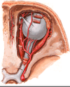
Orbital BS
A
- All originate from opthalmic A (from internal carotid A)
- Central Retinal A
- Ciliary A
23
Q
Central retinal A
A
- → pierece optic N → pass down mid optic N and fans out on the surface of the retina

24
Q
Ciliary A
A
- Long POS → goes around eyeball to the front
- Anastomose with the ANT ciliary A → inflam inside eye → may cause backflow and cause redness in the ANT ciliary A
- Supplies photoreceptors across retina and ANT structures (i.e. ciliary body and iris)
- Short POS → pierce around optic N into eyeball
- Both POS travel in the choroid
- ANT → Does not pierce the globe → supplies structure in the front

25
Which artery supplies which part of the retina?
* Dual BS to the retina
* Central retinal → inner retina (neurons that are not photoreceptors)
* POS ciliary A → outer retina (photoreceptors)
26
ANT BS of the eye
* Pass fwd along muscles of the eye to the front of the eye
* Conjuctiva, episclera, sclera, limbus
* Branch down to iris and ciliary body


