Inflammatory Bowel Disease (IBD) (Howden/Gupta) Flashcards
What is the major difference in the endoscopic appearances on the left and the right?

Left: Normal (you should see the veins throught the mucosa)
Right: Inflammatory (don’t know if its UC, CD, or other inflammatory process from single shot)
What histological features would you expect to see in someone with IBS?
• Remember IBS is NOT the same as inflammatory bowel disease (IBD).
IBS has a NL histological picture most of the time, this is in contrast to IBD that has abnormalities present
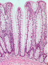
**Normal Intestinal epithelium shown below**

Is this appearance more consistent with Ulcerative Colitis or Crohn’s?

Seen here are lone islands of regenerating epithelium in ULCERATIVE COLITIS that resembles polyps => PSEUDOPOLYPS
***Histologic appearance of pseudopolyp below**

What is shown here?
• Key features? (Red, Tan, Black arrow)
• Are these features specific?

All you can say is that this is active colitis
• could be UC, Crohn’s, or other inflammatory process

What is the major difference in UC and Crohn’s disease if you could take a full section of colon?
Crohn’s causes full thickness lesions while UC only creates lesions that extend into the mucosa and submucosa

What is this arrow pointing to?

CMV that is causing Colitis (can do immunostaining to rule this out)
Surveillance biopsies are done in UC to evaluate for the complication shown below.
• what key features distinguish left and right sides of the frame?

Left Side: Neutrophils are attacking glands
Right Side: Dysplasia is seen with hyperchromatic cells and architectural changes

What pathology is seen in this abdominal X ray?

Alternating areas of blockage and pockets of air indicate that there is OBSTRUCTION (you can’t really say more than this)
What pathological features are seen in this section of small intestine?
• Is this more indicative of Crohn’s or IBD?

Thickening of the Small intestine wall with Submucosal Fibrosis and hypertrophy of mucularis propria has led to STRICTURE.
• Definitely crohn’s b/c UC doesn’t cause stricture, exist in small intestine, or involve muscularis propria

What abnormalities are seen in this tissue from a Crohn’s patient ?

Loss of Villi with inflammation of the Crypts and Architectural changes

What is this?

Non-caseating granuloma (ass’d with crohn’s if not infectious and in the colon)


