Inflammatory Bowel Disease Flashcards
What is Crohn’s Disease?
Chronic GRANULOMATOUS inflammatory disease than can affect any part of the GI tract. Can affect any segment of the GI tract from the mouth to anus. Characterised by transmural inflammation.
What is the aetiology of Crohn’s disease?
Unknown, though believed to be a mixture of environmental and genetic. Theories suggest that it is an inappropriate immune response against (?abnormal) gut flora in a genetically susceptible individual. Associated with altered neutrophil function and abnormality in epithelial cell integrity.
Where is the location of Crohn’s disease in the GI tract as a proportion of cases?
20% colic, 30% ileal, 50% ileocolic.
What are the risk factors of Crohn’s disease?
Mutation of NOD2 (aka CARD15) gene increases risk, smoking (x4 increased risk), altered cell mediated immunity, NSAIDs may exacerbate the disease, increased animal protein intake, contraception.
What is the epidemiology of Crohn’s disease: Age? Gender? Location? Prevalence?
Any age, but peak incidence 10-30 years, F>M, lower prevalence in Asian countries and populations. 50-80/100 000 prevalence in the UK.
What are the symptoms of Crohn’s disease? (x5)
- Crampy abdominal pain due to inflammation, fibrosis or obstruction
- Diarrhoea with urgency
- Blood/steatorrhea
- Fever and malaise
- Decreased weight and anorexia
What are the signs of Crohn’s disease? (x9)
- Abdominal tenderness/mass
- Abdominal distension
- Aphthous ulceration of mouth
- Perianal manifestations such as skin tags, anal strictures and signs of complications
- Signs of extra-intestinal complications
- Clubbing
- Signs of anaemia
- Weight loss
- Fever and tachycardia
What is anal stricture?
Narrowing of anal canal. Also called anal stenosis.
What are the investigations for Crohn’s disease? (x10) How is diagnosis made?
- BLOODS: decreased Hb, increased WCC, decreased Albumin (LFT), increased ESR/CRP, hypocholesterolaemia, hypocalcaemia.
- HAEMATINICS: look for deficiencies such as B12, folate (malabsorption) and ferritin (bleeding)
- STOOL: microscopy, culture and sensitivity and CDT (Clostridium difficile toxin) – exclude infective colitis. Can also look at FAECAL CALPROTECTIN as a simple test for GI inflammation with high sensitivity.
- AXR: look for complications, in particular, toxic megacolon
- ERECT CXR: if perforation risk
- CT/MRI: assess pelvic disease and fistulae, small bowel disease activity and strictures
- COLONSCOPY and BIOSPY: 70% effective in diagnosis. Not 100% as would miss disease when exclusively small bowel.
- Consider SMALL BOWEL BARIUM FOLLOW-THROUGH
- Consider CAPSULE ENDOSCOPY to visualise the small intestines
- Consider US: can provide small bowel imaging
- Diagnosis made by combination of investigations.
What does faecal calprotectin measure? Sensitivity?
Elevated levels is indicative of migration of neutrophils to the intestinal mucosa and is highly sensitive in IBD.
What is the purpose of small bowel barium follow-through in Crohn’s disease? (x3)
Look for fibrosis/strictures (you would see string sign of Kantor), deep ulceration (you would see rose thorn), or cobblestone mucosa in the SMALL INTESTINE.
What is the pathology of string sign?
Oedema or fibrosis associated with ulcerated mucosa
What does string sign look like in barium follow-through?
.

What does cobblestone mucosa look like in barium follow-through?
.
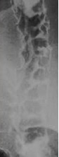
What is the clinical use of small bowel barium follow-through in Crohn’s disease?
Rarely used; useful when disease involves small intestine.
What is the purpose of colonoscopy in Crohn’s disease? (x4)
DEFINITIVE DIAGNOSIS, biopsy tissue, visualise mucosa to differentiate between UC and CD, monitor for disease progression or malignancy. Note that a biopsy is CONFIRMATORY rather than DIAGNOSTIC
!!! What do you look for in colonoscopy (or capsule endoscopy) for Crohn’s disease?
- Mucosal oedema
- Mucosal inflammation and discrete deep ulcers located transversely (penetrating deep; rose thorn fissures) and longitudinally (across sections of mucosal wall), creating a COBBLESTONE appearance
- Skip lesions: because of the transmural chronic inflammation
- Hyperaemia
- Fistulae and abscesses
- Involvement of the colon and ileum but NOT the rectum is suggestive of Crohn’s.
What are the histological findings in Crohn’s disease?
- Widening of submucosa
- Lymphoid aggregates in the submucosa associated with inflammation
- Cryptitis accompanied by abundant lymphatic and plasma cells
- Non-caseating granulomas (aggregation of macrophages from chronic inflammation) with epithelioid giant cells (aka epithelioid histiocyes; these describe macrophages and dendritic cells)
- Granulomas with epithelioid giant cells may be seen in blood vessels or lymphatics.
What does rose thorn fissures refer to?
Deep penetrating linear ulcers or fissuring typically seen within stenosed terminal ileum with a thickened wall.
What are the gastrointestinal complications of Crohn’s disease? (x10)
- Small bowel obstruction
- Toxic megacolon (rare complication relative to UC)
- Haemorrhage
- Bowel strictures
- Perforation
- Fistulae (between bowel, bladder and vagina)
- Perianal fistulae
- Abscesses (abdominal, pelvic or perianal)
- GI carcinoma
- Malabsorption
What is the risk of GI carcinoma in Crohn’s disease?
5% risk in 10 years.
What is toxic megacolon?
Bowel dilatation associated with inflammation and increased risk of stasis, perforation and haemorrhage. It is an acute presentation of Crohn’s with significant risk of death if left untreated.
What are the extraintestinal complications of Crohn’s disease? (x12)
- Uveitis: inflammation of uvea (middle pigmented layer of the eye comprising of iris, ciliary bodies and choroid)
- Episcleritis
- Gallstones
- Kidney stones
- Arthropathy (disease of joints) including arthritis
- Sacroiliitis (sacroiliac join inflammation)
- Ankylosing spondylitis
- Osteoporosis (from steroid treatment)
- Erythema nodosum (redness and swelling of skin arising from inflammation of subcutaneous adipose tissue)
- Pyoderma gangrenosum: immune system dysfunction leading to ulcer formation, typically on legs
- Amyloidosis
- DVT/PE from hypercoagulability associated with inflammation
What is amyloidosis?
Abnormal amyloid protein aggregates (fibrils) build-up in tissues leading to diarrhoea, weight loss, macroglossia, bleeding presenting as bruising, and more.
How is Crohn’s disease severity determined: Symptoms? Parameters?
- MILD/MODERATE: symptomatic but systemically well
- SEVERE: systemically unwell. High temp, high HR, high ESR, high WCC, high CRP, low albumin.
How is mild Crohn’s disease medically managed? (x3)
(1) Prednisolone 30mg OD/PO for a week, then taper by 5mg every week for next 7 weeks. (2) An alternative dietary approach based on ‘elemental’ or ‘polymeric’ diets is effective in children but less used for adults. (3) Plan maintenance therapy.
How is severe/acute exacerbation of Crohn’s disease medically managed? (x9)
- Fluid resuscitation
- IV/oral corticosteroids such as hydrocortisone
- 5-ASA analogues to induce remission (sulfasalazine, mesalazine – these are not immunosuppressants; they are in their own class)
- Metronidazole IV/PO for prophylaxis/treat active infection
- Analgesia as required
- Polymeric enteral diet is preferred, though consider total parenteral nutrition as a last resort.
- Consider need for transfusion if Hb below 80 g/L.
- Consider immunosuppression and biologics
- Monitor markers of activity (fluid balance, ESR, CRP, plt, stool freq, Hb, Alb).
How is Crohn’s disease medically managed long-term? (x5)
- Steroids for acute exacerbations
- 5-ASA analogues to reduce relapse
- Immunosuppression (steroid-sparing) e.g., azathioprine, methotrexate and 6-mercatopurine
- Biologics: Anti-TNF agents – immunosuppressants in the form of monoclonal antibodies e.g., infliximab and adalimumab. There is also anti-integrin and anti-IL12/23.
- DVT prophylaxis
When is Azathioprine (AZA) used in Crohn’s disease management? (x3)
Used if refractory to steroids, relapsing on steroids taper, or requiring more than 2 steroid courses in a year.
What are the problems with Azathioprine? (x2)
Takes 6-10 weeks to work. 30% will develop side effects requiring treatment cessation such as abdominal pain, nausea, pancreatitis, abnormal LFTs.
How must patients be monitored when on Azathioprine?
Monitor FBC, U&Es, LFTs weekly for 4 weeks, then every four weeks for three months, then every four months.
How does anti-TNFa work?
They counter neutrophil accumulation and granuloma formation and cause cytotoxicity to CD$+ T cells, thus clearing cells driving the immune response. In this way, they play a role in induction and maintenance therapy.
What are the complications of anti-TNFa? (x3)
Sepsis, active/latent TB (may reactivate), increased LFT.
How does anti-integrin work in the management of Crohn’s disease?
Monoclonal antibodies targeting adhesion molecules involved in gut lymphocyte trafficking, reduce disease activity, and have a more gut-specific mechanism of activity.
What kind of diets are recommended for Crohn’s disease management? (x2)
ELEMENTAL diets – contain amino acids and can give remission. LOW RESIDUE diets (low fibre) – help symptoms in those with active disease or strictures.
What are the indications for surgical management of Crohn’s disease? (x3)
Drug failure, complications (GI obstruction from stricture, perforation, fistulae, abscesses), and failure to thrive in children.
How is Crohn’s disease surgically managed? (x3 ways)
NOT CURATIVE = (1) resection, (2) to control perianal or fistulizing disease, (3) defunction (rest) distal disease e.g., with a temporary ileostomy.
What is a complication of resection in Crohn’s disease management?
Short bowel syndrome – malabsorption from lack of small intestine, when SI resected.
What is the prognosis of Crohn’s disease?
2/3 require surgery and chronically relapsing. Prognosis is poor.
How is small bowel follow-through performed?
Patient is fasted 6-hours prior to procedure, barium administered orally, and X-rays taken supine at 30 minute intervals.
What is ulcerative colitis?
Relapsing and remitting (suddenly stops) inflammatory disorder of the colonic mucosa.
As a proportion of cases, where does UC affect?
50% proctitis (affects just rectum), 30% left-sided colitis, 20% pancolitis (entire colon). NEVER spreads proximal to the ileocecal valve.
In what circumstances may there be some small intestine involvement in ulcerative colitis?
Backwash ileitis may present in patients with pancolitis – terminal ileum is oedematous.
What is the pathophysiology of ulcerative colitis?
Hyperaemic/haemorrhagic granular colonic mucosa +/- pseudo polyps formed by inflammation. Punctate ulcers extend deep into the lamina propria. STARTS at the rectum and moves upwards; anal sparing. Unlike CD, there are no skip lesions; the disease is continuous and rarely transmural.
What is the aetiology of ulcerative colitis? Genetics?
Unknown, though it has been suggested that, much the same as CD, there is an inappropriate immune response against (?abnormal) gut flora in a genetically susceptible individual. Genetic susceptibility is believed to lie in chromosomes 12 and 16. Associated with altered neutrophil function and abnormality in epithelial cell integrity.
What are the risk factors of ulcerative colitis? (x5) Protective?
Family history, high serum pANCA, HLA-B27, NSAIDs, primary sclerosing cholangitis. Smoking is protective.
What is the epidemiology of ulcerative colitis: Gender? Age? Ethnicity? Prevalence?
Gender equal up to 40, then more common in males. Mostly presents between 15 and 30. Higher prevalence in Ashkenazi Jews and Caucasians. 1/1500 in developed world.
What are the symptoms of ulcerative colitis? (x5)
- Episodic or chronic diarrhoea +/- blood or mucus
- Increased stool frequency – correlates with severity
- Crampy abdominal discomfort (less common than CD)
- Urgency and tenesmus (need to evacuate bowels with little or no stool passed)
- Systemic complications in ATTACKS: fever, malaise, weight loss.
What are the signs of ulcerative colitis? (x10)
- Abdominal tenderness/mass
- Abdominal distension
- PR – blood, mucus and tenderness
- Aphthous ulceration of mouth
- Perianal manifestations such as skin tags, anal strictures and signs of complications
- Signs of extra-intestinal complications
- Clubbing
- Signs of anaemia
- Weight loss
- Fever and tachycardia
What are the investigations for ulcerative colitis? (x8) How is diagnosis made?
- BLOODS: decreased Hb, increased WCC, decreased Albumin (LFT), increased ESR/CRP, low potassium, elevated sodium and urea. LFTs are IMPORTANT and used to screen for bile duct involvement (cholangitis risk factor AND complication!)
- HAEMATINICS: look for deficiencies such as B12, folate and ferritin
- STOOL: microscopy, culture and sensitivity and CDT (Clostridium difficile toxin) – exclude infective colitis. Can also look at FAECAL CALPROTECTIN as a simple test for GI inflammation with high sensitivity.
- BARIUM ENEMA – investigation to CONSIDER
- COLONSCOPY/FLEXIBLE SIGMOIDOSCOPY and BIOSPY
- MRI: assess pelvic disease and fistulae, small bowel disease activity and strictures
- AXR: no faecal shadows, colon dilatation and mucosal thickening – look at photo.
- ERECT CXR: if perforation risk
- Diagnosis made by combination of investigations.

What can be identified in barium enema in ulcerative colitis?
Fine granular appearance of the bowel wall from diffuse mucosal ulceration, thumbprinting due to mucosal oedema, featureless narrowed colon, and/or loss of haustral pattern (leadpipe or hosepipe appearance).
When is barium enema and colonoscopy contraindicated in ulcerative colitis?
In acute exacerbations as there is perforation risk. Note you can still perform limited flexible sigmoidoscopy.
What is the purpose of colonoscopy in ulcerative colitis? (x4)
Determine severity. Biopsy tissue for histological confirmation visualise mucosa to differentiate between UC and CD, monitor for disease progression or malignancy.
Why can severity of disease be assessed by colonoscopy in UC, but less accurately in CD?
UC is continuous and does not skip areas of the GI tract. Therefore, when you reach its proximal border, you know that the disease does not progress any further. In CD, this cannot be assessed, and there may be involvement beyond the colon.
What do you look for in ulcerative colitis in a colonoscopy/flex sig?
Continuous uniform distribution, loss of vascular marking, diffuse erythema, mucosal granularity, mucosal ulcers, fistulas (rarely seen), normal terminal ileum (or mild backwash ileitis).
What do you look for in histology (from biopsy) in ulcerative colitis? (x6)
Continuous distal disease, goblet cell depletion, crypt abscesses, basal plasmacytosis (presence of plasma B cells between the base of crypts and muscularis mucosae), no granuloma tissue, anal sparing, diffuse mucosal atrophy.
What are the gastrointestinal complications of ulcerative colitis? (x6)
- Haemorrhage
- Toxic megacolon (associated with fulminant colitis i.e. acute severe colitis)
- Perforation
- Colonic cancer
- Gallstones
- PSC (cholangitis)
What are the extra-intestinal complications of ulcerative colitis? (x11)
- Uveitis
- Renal calculi
- Arthropathy
- Sacroiliitis
- Ankylosing spondylitis
- Erythema nodosum
- Pyoderma gangrenosum
- Osteoporosis (from steroid treatment)
- Amyloidosis
- DVT/PE from hypercoagulability associated with inflammation
- Hypokalaemia from excess colonic secretion
How is toxic megacolon diagnosed?
AXR shows mucosal thickening, mucosal islands (pseudo-polyps) and diameter of larger than 6cm.

How is ulcerative colitis monitored? (x6)
Markers of activity are low Hb, low albumin, high ESR/CRP, diarrhoea frequency, bleeding and fever.
How does diarrhoea frequency relate to ulcerative colitis severity?
Less than 4 times per day is MILD, 4-6 MODERATE, and more than 6 is SEVERE.
How is toxic megacolon managed in ulcerative colitis and Crohn’s disease?
Low threshold for proctocolectomy and ileostomy as perforation has a mortality of 30%.
How is mild ulcerative colitis medically managed? (x2)
(1) 5-ASA e.g., sulphasalazine for as the mainstay for remission-induction and maintenance. PR for distal disease, or PO for more extensive disease. (2) And/or rectal steroids, given as topical steroid foams or retention enemas.
How is moderate ulcerative colitis medically managed? (x2)
Induce remission with oral prednisolone 40mg/d for 1wk, then taper by 5mg/week over 7wks, and maintain remission with oral 5-ASA.
What are the issues with 5-ASAs? (x5)
Side effects include rash, haemolysis, hepatitis, pancreatitis and paradoxical worsening of colitis.
How is severe/acute exacerbation of ulcerative colitis medically managed?
- IV rehydration
- IV corticosteroids e.g. hydrocortisone 100mg/6h
- Rectal steroids e.g. hydrocortisone 100mg in 100mL 0.9% saline/12hrs PR
- DVT prophylaxis regardless of rectal bleeding, because of high thromboembolism risk
- If, following 3-5 days of monitoring, CRP and stools/day remain high, rescue therapy with immunosuppressives such as cyclosporine, or infliximab.
- If improving, transfer to prednisolone PO and schedule maintenance infliximab if used for rescue, or azathioprine if cyclosporin rescue.
- If fails to improve, then urgent colectomy by day 7-10.
When is immunomodulation indicated in ulcerative colitis?
Patients flare on steroid tapering or require more than 2 courses of steroids in a year. This means that immunomodulation is not exclusively indicated in severe disease.
When are biologics indicated in ulcerative colitis? (x2)
Patients intolerant to immunomodulation or developing symptoms despite immunomodulation.
When is surgical management of ulcerative colitis indicated? (x3)
Failure of medical treatment, presence of complications, prevention of colonic carcinoma.
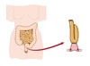
How is ulcerative colitis surgically managed? (x2)
CURATIVE. Removal of large intestine: completion proctocolectomy (resection of large intestine including rectum; permanent stoma), or ileo-anal pouch (see photo of ileum-anus anastomoses).
What are the advantages and disadvantages of ileo-anal pouch?
Pouches mean stoma reversal and the possibility of long-term continence, but pouch opening frequency may still be around 6 times a day and recurrent pouchitis can be troublesome (give antibiotics for two weeks).
What is the prognosis of ulcerative colitis? Poor prognostic factors? (x6)
Relapsing and remitting with NORMAL life expectancy. Poor prognostic factors (ABCDEF): low Albumin, blood PR, CRP raised, dilated loops of bowel, eight or more bowel movements per day, fever.
Ulcerative colitis and long-term colonoscopy?
Every 1-5 years to monitor for dysplasia as colon cancer risk is 5-10% with pancolitis for 20 years. Colonoscopy is done with multiple random biopsies or biopsies guided by differential uptake by abnormal mucosa of dye sprayed endoscopically.
SUMMARY: What are the diffierences between Crohn’s Disease and Ulcerative Colitis?
- LOCATION: anywhere in GI tract vs colon
- PATTERN OF INFLAMMATION: transmural vs continuous
- ULCERATION: ulcers penetrate entire thickness vs. penetrate submucosa
- BLEEDING: common vs. uncommon
- MANAGEMENT: induce and maintain remission vs. can be curative
!!! SUMMARY: What are the histopathological differences between UC and CD?
- UC: cobble stoning, rose-thorning, crypt abscesses, continuous, basal plasmacytosis
- CD: granuloma, transmural, skip lesions, cryptitis
- BOTH: mucosal ulcers, inflammatory infiltrate, goblet cell depletion
