Histiocyte, Spleen and Thymus Pathology Flashcards
Histiocyte Disorders
- Histiocyte pathology encompasses a heterogenous group of disorders ranging from benign, reactive disorders to highly aggressive malignant neoplasms.
- The most common of thes is the Langerhans cell histiocytoses (LCH).
Langerhans Cell
- LCs are produced in the bone marrow and are derived from the monocyte-macrophage lineage.
- LCs are a special type of dendritic cell that picks up antigen in the peripheral tissues and then migrates to lymph nodes where they are powerful activators of T-cells.
- Recent data has shown that the cell of origin for LCH is not a cutaneous LC, but rather a myeloid dendritic cell. For now, however, these disorders are still referred to as Langerhans cell histiocytoses.

Langerhans Cell Histiocytosis
- LCH is a clinicopathologic entity classified according the number of sites involved (single vs multiple) and their location (single organ or multiple organs).
- Older, historical names include Letterer-Siwe, Hand-Schuller-Christian, and eosinophilic granuloma
- These are RARE entities (~5 cases/million population/year) and most cases occur in childhood. The etiology is unknown in most cases.
LCH - Symptoms
•Preferential involvement of:
- Skin (80% of patients)
- Scalp, face, trunk, buttocks, intertriginous areas
- Closely spaced papules with scale and crust
- Bone
- Liver
- Spleen
- Bone marrow

LCH Entities
1) Multifocal System LCH (Letterer - Siwe Disease)
2) Unisystem (umifocal or multifocal)
3) Pulmonary LCH (eosinophilic granuloma of lung)
LCH - Multifocal multisystem (Letterer - Siwe Disease)
o Infants and children <2 years old, occasional adults
o Skin, bone, liver, spleen and bone marrow
o Aggressive course; most patients require chemotherapy

LCH - Unifocal (unifocal or multifocal)
o Unifocal disease:
§ Skeleton (skull, ribs and femur), older children/adults
§ Indolent, may regress spontaneously
o Multifocal disease:
§ Young children
§ Multiple erosive bone lesions
§ Exophthalmos
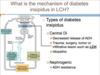
§ Involvement of the posterior pituitary stalk of the hypothalamus leads to diabetes insipidus (most common endocrine abnormality in LCH)
§ Prognosis depends on sites of involvement; may require systemic chemotherapy
LCH - Pulmonary
o Adult smokers (may regress upon smoking cessation)
§ Reactive, non-clonal process??
o Chronic interstitial lung disease with nodular and cystic lesions
o Nonproductive cough, dyspnea, fever, weight loss
o May be discovered incidentally on CXR

Diabetes in LCH
LCH Histology
- Langerhans cells have abundant cytoplasm and nuclei with linear grooves and minimal atypia; they may be admixed with eosinophils (often prominent), lymphocytes, plasma cells and neutrophils
- Nuclei
- kidney bean shaped
- linear grooves (coffee bean)
- minial atypia

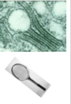
LCH - Electron Microscopy
• Birbeck granule
- Pentalaminar tubule with dilated terminal end
- Contains the protein Langerin
- Birbeck granules, CD1a and langerin play a role in antigen presentation to Tcells
LCH Genetics
- Phenotype: LC express HLA-DR, S100, and CD1a
- Genetic Profile: BRAF V600E mutation positive in ~50% of cases
White Pulp of Spleen
The white pulp is involved in generating immune responses to antigens in the blood, and consists of T and B cells and associated cells surrounding central arteries.

Red Pulp of Spleen
The red pulp consists of splenic sinusoids separated by splenic cords. Splenic sinusoids are made up of spindle-shaped endothelial cells and are the actual filter of the spleen. Blood cells that enter the red pulp must pass through the walls of the splenic sinusoids to re-enter circulation.

Spleen Functions
- Phagocytosis of blood cells (senescent and abnormal), particulate matter and microorganisms. Abnormally shaped or antibody-coated RBCs are phagocytosed by macrophages.
- Antibody production – dendritic cells in the periarteriolar lymphoid sheath (PALS) trap and present antigens to T-cells, leading to activation and proliferation of B and T lymphocytes and antibody-producing plasma cells.
- Hematopoiesis – normal prior to birth; can be seen after birth in response to marrow failure or in myeloproliferative neoplasms
- Sequestration of blood cells – especially RBCs and platelets
Splenomegaly
Splenic enlargement. The major manifestation of disorders of the spleen.
Splenomegaly Clinical Presentation
- Depends on the acuteness, nature of underlying illness and the size of the spleen.
- Possible symptoms include:
1) LUQ pain, fullness or discomfort (If pain is acute and accompanied by fever, consider splenitis or abscess)
2) Pain referred to the left shoulder
3) Early satiety
Spelnomegaly Causes
•The causes of splenomegaly are numerous; most are due to hepatic or hematologic disease, infection, or inflammation. In many instances, the spleen enlarges as it performs its normal functions.
Splenomegaly Classification
- by disease type
- by spleen compartment
Splenomegaly - White Pulp


Splenomegaly - Red Pulp


SPlenomegaly - Red and White Pulp


Congestive Splenomegaly
•Congestive splenomegaly is caused by chronic venous outflow obstruction, and may be seen in the following conditions:
1) Congestion (systemic or central venous) - causes moderate spleen enlargement
2) Cirrhosis of the liver - main cause of congestive splenomegaly
3) Obstruction of the extrahepatic portal vein or splenic vein
- Morphology: With long-standing congestion, the spleen may become markedly enlarged (1000-5000 gm) and firm.
- Microscopically, there is early congestion but eventually the red pulp becomes fibrotic.

Neoplasms of the Spleen
•Primary neoplasms
1) Vascular neoplasms
- hemangioma
- angiosarcoma
2) Lymphoid neoplasms
•Secondary Involvement
1) Metastases
2) Lymphoid neoplasm


