Autoimmmune Diseases Flashcards
Approach to Autoimmune Diseases
• Suspect the disease
– Patient history + family history + physical exam findings
• Perform serologic laboratory testing
– General anti-nuclear antibody (ANA) testing
– Specific autoantibody testing
- Refine the diagnosis, predict prognosis (consider tissue biopsy)
- Evaluate patient’s signs and symptoms according to American College of Rheumatology criteria
- Management/therapy
– Immune suppression: Corticosteroids, anti-B cell and anti-T cell therapies
Autoantibodies – Examples of Antigenic Targets Location
– Nuclear
- Anti-ds DNA
- Anti-nucleolar
- Anti-histone
– Cytoplasmic
- Anti-mitochondrial
- Anti-RBC (membrane)
– Non-cellular
- Anti-prothrombin
- Anti-immunoglobulin
Autoantibodies – Examples of Antigenic Targets Disease Specific
– Blistering skin diseases
- Anti-hemidesmosome
- Anti-desmosome
– Autoimmune hepatitis
• Anti-smooth muscle
– Primary biliary cirrhosis
• Anti-mitochondrial
– Hashimoto’s thyroiditis
• Anti-thyroid cytoplasmic antigen
ANA
• ANA = Autoantibodies commonly present in patients with autoimmune disease
– Directed against nuclear antigens
• Types:
– Antibodies to DNA, histones, non-histone proteins bound to RNA, nucleolar antigens
• Limitations of testing
– Up to 10% of population has positive ANA but no features of autoimmune disease
*Usually present in low titer
*May be detected following chronic inflammation, malignancy, viral illness
Antinuclear Antibody Detection Indirect IF test - Homogenous Pattern

Antinuclear Antibody Detection Indirect IF test - Speckled Pattern

Antinuclear Antibody Detection Indirect IF test - Rim Pattern

Antinuclear Antibody Detection Indirect IF test - Nucleolar Pattern

ELISA
•Enzyme Linked Immunosorbent Assay (ELISA)
– More specific: Will identify subtype of antinuclear antibody or other type of autoAb present
– Allows for evaluation of ab concentration (titer)

SLE Antibodies


Systemic Lupus Erythematosus Autoantibodies Continued…
•Anti-phospholipid antibodies
– Present in 30-50% patients with SLE
– Antibodies against proteins bound to phospholipids in cell membranes
• Examples: Anti-cardiolipin, anti-β2 glycoprotein, lupus anticoagulant
– In vivo (patient): Antibodies cause increased clotting
– Arterial and venous thrombosis
– “Antiphospholipid antibody syndrome”: DVT, pulmonary emboli, stroke, miscarriages (often multiple), bleeding (low platelets)
– Type II Hypersensitivity mechanism
– Paradox In vitro (test tube with patient serum): Lupus anticoagulant antibody causes delayed clotting of test blood
Systemic Lupus Erythematosus Etiology/Pathogenesis
• Genetic
– Runs in families
– Family members without SLE may have autoantibodies or other autoimmune diseases
– 20% concordance in monozygotic twins
– Some patients with inherited complement deficiencies
– Failure to clear immune complexes
• Non-genetic/Environmental
– Ultraviolet light
– Sex hormones, especially estrogen (pregnancy)
– Injury/trauma
– Drugs
– hydralazine, procainamide, D-penicillamine
– May develop anti-histone autoantibodies
• Immunologic
– Failure of tolerance: B-cells, CD4+ helper T cells
– Type I interferon (IFNα) chronically elevated
– Type III hypersensitivity (major)
• Antigen-antibody complexes form in circulation, deposit in tissue and cause inflammatory injury
– Kidney, skin, joints
– Type II hypersensitivity (minor)
• Anti-cellular component of SLE
– Autoantibodies directly target antigens on surface of RBC’s, WBC’s, platelets
Systemic Lupus Erythematosus Clinical Manifestations
•Clinical manifestations:
– Skin rash
– Malar “butterfly” rash
– Sun-exposed and non- exposed skin
– Photosensitivity
– Joints
– Non-erosive arthritis
– Cardiovascular
– Pericarditis
– Valve disease (Libman-sacks endocarditis )
– Lungs
– Pleuritis, pleural effusions
– Renal
– Immune complex glomerulonephritis
SLE Renal Manifestations
•Renal
- Immune complex mediated glomerulonephritis with “full house” staining by immunofluorescence (Type III Hypersensitivity):
i. Class I – Minimal lupus nephritis
ii. Class II – Mesangial lupus nephritis
iii. Class III – Focal lupus nephritis
1. Active lupus inflammatory lesions in 50% glomeruli
2. Most acutely severe and destructive renal lesion of lupus, may present as rapidly progressive glomerulonephritis (RPGN)
a. Requires prompt therapy
v. Class V – Membranous lupus nephritis
1. Clinical picture dominated by nephrotic syndrome (marked proteinuria, hypoabuminemia, edema, hyperlipidemia)
vi. Class VI – Advanced sclerosing lupus nephritis
1. 90% of greater glomeruli in the sample are completely sclerosed and obsolescent
2. Usually associated with extensive interstitial fibrosis, tubular atrophy and vascular sclerosis
3. Considered “end stage”
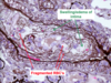
Skin Biopsy SLE


Skin Biopsy in SLE Immunofluorescence


Lupus Nephritis - Characteristic biopsy findings

– “Full house immunofluorescence”
• IgG, IgA, IgM, C3, C4, C1q
– “Wire loops”
- Thickened glomerular capillary loops due to large, continuous subendothelial deposits
- Seen in Focal LN (Class III) and Diffuse LN (Class IV)
– Membranous Lupus
- Numerous small subepithelial deposits (similar to idiopathic membranous) + mesangial deposits
- Patients with membranous lupus present with nephrotic syndrome, can have massive proteinuria

SLE Prognosis
• Survival
– >90% 10-year survival (40% in 1950’s)
• Increased mortality risk
– Severe disease activity, younger age, male gender, nonWhite
• Causes of death
– Short term:
- Severe inflammation (especially severe nephritis)
- Infection: May be opportunistic due to immune suppression
– Long term:
- Atherosclerotic cardiovascular disease, malignancy, infection
- Chronic kidney disease
– less common cause of death with better access to dialysis, transplantation
Discoid Lupus
• Similar rash as SLE
– Skin plaques with edema, erythema, scale
– Immune complex deposition in similar pattern as SLE (Type III hypersensitivity)
– Usually confined to sun exposed skin:
- Most severe on face, scalp
- Systemic manifestations rare/absent
– Multi-organ disease may develop late in 10% patients
– Better overall prognosis vs SLE
• Autoantibodies
– Only 35% pts with positive generic ANA
– Rare: anti-dsDNA, anti-Smith
Rheumatoid Arthritis
•Chronic inflammatory disease primarily affecting joints
a. Autoantibodies:
i. Rheumatoid factor (RF): an IgM antibody targets the Fc region of an IgG immunoglobulin
ii. Anti-cyclic citrullinated peptide (anti-CCP): Present in 60-70% of patients with RA, more specific for RA than rheumatoid factor.
b. Pathologic events likely orchestrated predominantly by cell-mediated immunity (lymphocytes and macrophages, Type IV hypersensitivity)
i. Unclear to what degree RF and/or CCP are actually pathogenic vs an epiphenomenon (i.e. helpful for diagnosis but not involved in causing the disease)
ii. A minor component of immune complex deposition (Type III Hypersensitivity) has been described, but not considered prominent. We do not assess for immune complexes in tissue.

RA Clinical Manifestations
a. Arthritis, primarily involving small joints of hands and feet
i. Synovium of joint is the principal immune target
ii. Chronic inflammation of synovium, with formation of germinal centers, recruitment of prominent plasma cells
iii. Reactive overgrowth/hyperplasia of synovium (pannus) onto articular cartilage, with irreversible cartilage destruction and bony erosion
iv. Secondary joint deformity
– Abnormal fusion/misalignment of bones
- Ulnar deviation of fingers
b. Non-joint manifestations: Rheumatoid nodules in soft tissue, inflammation of eye, and vasculitis

Scleroderma
• Multi-system disorder
– Systemic chronic inflammation
– Vascular abnormalities (small vessels)
– Progressive fibrosis of:
• Skin
– Starts with fingers, toes then progressively involves more proximal areas (upper arms, shoulders, face)
• Visceral organs:
– Gastrointestinal tract, kidneys, heart, and lungs
• Typical patient
– Female (3:1 ratio vs males), 50-60 yrs


Systemic Sclerosis Pathophysiology - Early
• Vascular injury
– Injury to endothelium + smooth muscle
– Narrowing of vessel lumen:
– Early: Intimal swelling due to edema
– Late: Fibrous proliferation with luminal occlusion
– Capillary dilation with leaking, capillary destruction
• Fibrosis
– Fibrogenic cytokines (like TGFβ) released from activated macrophages, T-cells
– Hyper-responsive fibroblasts
– Ischemia from vascular damage
• Not well classified into specific type of hypersensitivity (multiple mechanisms)