Final- Muscle Flashcards
(71 cards)
You are called out to a farm to investigate a sad scene…..
Clinical signs
Around 30% of 2 month old lambs in a mob of 40
Lying on their chest, roll on to their side
Weak, stiff
Die within a few days
What can we do to figure out what the pathological process was that resulted in these signs??
You are going to do a euthanize and necropsy an acute disease. You don’t want to wait for a natural death because you can get secondary infections etc that will hide the primary pathological process.
You are going to examine muscle bellies.
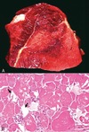
What is abnormal? There are muscles that are paleish white, swollen, firm, and dry to the touch(not shiny).

What is an addition pathological process for Skeletal Muscle?
Biochemical
What pathological process is responsible for this apperance(same as the sheep case)?

Degeneration/necrosis- results in a pale color because more water is coming into the muscle
What makes muscles pale other than degeneration/necrosis?
Fig. 15-9 Pathologic changes resulting in pale skeletal muscle.
A, Pale streaks, necrosis and mineralization, degenerative myopathy, canine X-linked muscular dystrophy, diaphragm (left side), dog.
B, Localized pallor, necrosis, injection site of an irritant substance, semitendinosus muscle, cow. The irritant was injected just under the perimysium and caused necrosis and disruption of the myofibers. Some irritant seeped down between the fascicles to cause necrosis, but the fascicles of myofibers are still in place.
C, Overall pale muscle with pale streaks from collagen and fat infiltration, denervation atrophy, equine motor neuron disease, horse. Equine motor neuron disease muscle (right) compared with normal muscle (left). Yellow tint; FAT WHERE YOU HAVE LOST SKELETAL MUSCLE, GREASY AND YELLOW
D, Enlargement and pallor, steatosis, longissimus muscles, neonatal calf. The majority of the muscles have been replaced by fat.
calicification can also do this
Fibrious connective tissue

Gross morphological diagnosis =

BEST ONE: skeletal muscle degeneration and necrosis
What is abnormal about this image?

Sacroplasm/cytoplasm is vacuolated, condensed, loss of cross striations
Things myofibers do when they are hurt:
They die:
- Vacuolation of sarcoplasm
- Condensation of sarcoplasm (looks hyper-eosinophilic, and lose striations)
- Nuclear pyknosis
- Calcification(dystropic)
They regenerate:
- Internalization of nuclei
- Macrophages infiltrate
What types of causes incite this pattern of degeneration and necrosis?

Pathology report: polyphasic skeletal muscle degeneration and necrosis( myocytes are in different stages of injury and regeneration vs in others where the myocytes are all in the same stage- monophasic)
This means that there is an ONGOING PROBLEM!
Causes of polyphasic lesions:
Ongoing insults
Nutritional deficiency – vitamin E/Se
Ongoing toxicities
Genetic defects in myocyte
structural/metabolic elements
How does nutritional deficiency cause this mess?

Pathogenesis: vitamin E/Se deficiency is needed for enzymes like glutathione peroxidase/reductase causeslack of ability to scavenge free radicals. This causes oxidative damage (lipoperoxidation of cell membranes) –> myocyte injury
Free Radicals cause necrosis
What would the path report say about this and what was the cause?

Monensin toxicity in a horse
path report- actue MONOPHASIC skeletal muscle degeneration and necrosis
Causes of monophasic lesions
Causes of Monophasic Lesions:
A single insult
Trauma (will be focal at the site of trauma)
Exertion, capture
Toxin – ionophores, plants
(coffee senna)
HORSES ARE REALLY SENSTIVE—–USUALLY FROM EATING A RUMINANT RATION

Edx

Monesin Toxicity
Our lambs with polyphasic muscle degeneration and necrosis…..
Follow up
You rule out access to toxic plants
Further research – they are from a Se deficient area
You arrange dietary vitamin E/Se supplementation
White Muscle Disease
Or Nutritional myopathy
Caused by Vitamin E / Se deficiency
See lesions in the most active muscles- because that is where the most free radicals are
Can see lesions in the heart with this condition and other metabolic/toxic myopathies of skeletal muscle
Mdx?

Heart from a calf with White Muscle Disease (nutritional myopathy from vitamin E/Se deficiency).
MDx: Cardiac myonecrosis.
Where else are you going to see white muscle disease?
- heart
- muscles of mastication
- tongue(espesically in suckling animals)
- diaphram
Single or multiple episodes
General signs of pain, anxiety, cramping most prominent after exercise
Multiple muscle groups look like this
In a horse

Exerional rhabdomyloysis(Tying up, azoteria)- necrosis/ lysis of skeletal muscle
ionic events of contraction can produce an adverse enviornment when extreme
may have an underlying metabolic conidtions which predispose such as polysaccharide storage disease
What types of causes typically incite this pattern of necrosis?

Acute Toxicity or Exertion
but NOT trauma because it’s mutifocal

“Exertional rhabdomyolysis” (Tying up, azoturia) – necrosis/lysis of skeletal muscle
During periods of exertion
Ionic events of contraction can produce an adverse environment when extreme
May have underlying metabolic conditions which predispose, such as polysaccharide storage disease
What other species is seen with foci of skeletal muscle necrosis?
Foci of skeletal muscle necrosis common in sled dogs, also observe in racing greyhounds
Exertional? Vit E/Se deficiency involvement? Underlying myocyte metabolism issue? lactosis, lesions in the heart, myglobin injures the kidneys
Sled dog myopathy – lethal, generalized lesions involving non-locomotory muscles
“Azoturia”
“Azoturia” = excess nitrogen in urine
This muscle is from a 1 year old deer with capture myopathy.
Why is the kidney and urine abnormal??
red urine from myoglobin from severe muscle necrosis

Capture myopathy
Zoo and wild birds
Exertion, stress during capture/handling/transport
• Anaerobic glycolysis —->hyperthermia, metabolic acidosis

“Malignant hyperthermia” – another example of a metabolic condition predisposing to necrosis
IN PIGS
muscle bellies are pale and dry
Pathogenesis: inherited defect in skeletal muscle ryanodine receptor==> excessive Ca release and contraction when stimulated —–>heat production and myocyte necrosis
stress can set off the receptor
MDx: focal muscle degeneration and necrosis
Are the pale streaks in this muscle due to degeneration and necrosis?
A. Yes B. No

NO because it’s fat!!!


