Final- Integument Flashcards

1
Q

Vesicle/Bulla

A

• Palpable elevation filled with clear fluid( fragile and they don’t stick around for long)

Degeneration and Necrosis or Inflammation and Repair

NAME DEPENDS ON SIZE:

– Vesicle < 1cm

– Bulla > 1cm

Causes:
– Auto‐immune dermatoses

– Viral infections
– Chemical irritants

– Burns

2
Q

What can lead to the formation of vesicle/bulba?

A
  • Intercellular edema ( histologically called “spongiosis”)- inflammatory process
  • Intracellular edema (“hydropic degeneration”- loose osmotic balance)
  • Disruption of intercellular junctions (“acantholysis”)
3
Q

How vesicles form

A

Fig. 17‐17 Schematic diagram of the sites of vesicle formation in the skin. A, In a subcorneal vesicle, the stratum corneum forms the roof of the vesicle (as in impetigo or pemphigus foliaceous). B, In a suprabasal vesicle, a portion of the epidermis (stratum spinosum) forms the roof (as in pemphigus vulgaris). C, In a subepidermal vesicle, the entire epidermis separates from the dermis and forms the roof (as in bullous pemphigoid).

Need to know– you can get a lot of information by knowning where the vesicle is forming- SAMPLE IT

4
Q

Pustule

A

Palpable elevation filled with pus

Cause – leukocyte infiltrate

Pathological process: Inflammation and Repair

I.e. Pimples

5
Q

Crust

A

can be when a pustule ruptures

• Dried exudate, serum, blood, and scale that is adhered to the skin surface

Causes:
– Severe disorders of keratinization

– Severe pustular dermatitis( when pustules burst)

– Secondary to ulcers

Pathological Processes: Degeneration/Necrosis, Inflammation/Repair, or Disorder of Growth

6
Q

Papules

A

• Palpable, solid (no fluid), elevated mass < 1cm diameter

i.e. mosquito bite

Causes:
– Infiltrate of inflammatory cells

– Infiltrate of neoplastic cells

– Epidermal hyperplasia
– Deposit of mineral

Termed Based on Size:

Nodules = >1cm diameter and deeper- usually thinking about tumors

Plaques = coalesced papules- has a flat surface

Pathological Process: Inflammation/Repair, Disorder of Growth, Deposits and Pigmentations

7
Q
A

Plaque- solid but with a flat top

8
Q
A

Nodule

9
Q

Ulcers

A

• Loss of epidermis with exposure of dermis- VERY NON-SPECIFIC

Cause: 2ndaryto…

– Epidermal necrosis

– Inflammation

– Infarction(cut off of blood supply)
– Neoplasia

Pathological processes: degeneration/necrosis, inflammation/repair, cirulatory disorders, or disorder of growth

Erosion— no dermis exposed

10
Q

Scale

A

• Accumulation of loose keratinized cells

  • dandruf

Causes:
– Disorders of keratinization

– Chronicdermatitis

Pathological Process: Inflammation/Repair or Disorders of Growth

11
Q

Epidermal collarette

A

a circular rim of scale that occurs secondary to rupture of a vesicle, pustule, or papule

FORM OF SCALE- vesicle, papule or pustule has ruptured

12
Q

Lichenification

A

• Thickening and hardening of the skin

  • becomes hyperpigmented

Causes:
– Chronic irritation/inflammation

Pathological Process: Inflammation and Repair

13
Q

What lesion terms can we use here?

A
  • ulcers
  • erthyema (skin term ONLY)
  • paules
14
Q

When collecting a skin biopsy…

A

• DO
– Biopsy early, before treatment
– Be gentle
– Collect multiple samples, range of changes – Include crusts

• DO NOT
– Surgically prep the site
– Grasp with forceps
– Biopsy the center of a lesion

– Hold out on history/DDx

15
Q

Pathological Processes of degeneration and necrosis

A

Can be difficult to grossly tell from other groups

  • Degenerative/Necrotic lesions tend to ulcerate, but other pathological processes lead to ulceration too
  • Degenerative/Necrotic lesions become inflammation&repair over time – normal response to injury, and secondary infections common
  • Primary circulatory disorders often lead to degeneration/necrosis

THINCK BURNS

16
Q

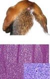
Flock of 300 merino ewes& lambs in the shade cuddling after a period of a lot of rainfall.

A

The skin lesions indicate something is injuring the epidermis because there are crusts and ulcers

The pathological process is likely epidermal degeneration&necrosis or inflammatory (infectious)

Differential Diagnoses

• Photosensitization

• Dermatophilus

• Viral infections – bluetongue, pox (orf), vesicular disease (FMD, VS

What can we do to determine the cause?

BIOPOSY, serum chem(NSF), bluetongue (negative), virus isolation(negative), plant id- st. john’s wort, histopath results- epidermal necrosis and ulceration(unknown etiology) comment- bacterial dermatitis which appears secondary

17
Q

Pathogenesis of Photosensitization

A

Uv light absorbed by photodynamic chemicals(shouldn’t be there) in skin causes free radical damage causes e_pidermal necrosis of lightly pigmented or sparsely haired skin_

18
Q

2 Types of Primary photosensitization

A

Primary photosensitization

_Type I (Exogenous)_
– Plants containing photosensitive chemicals

– St Johns Wort, lucerne, perennialryegrass

-TMS, quinolones, griseofulvin

Type II (Intrinsic)

– Porphyria

– Inherited deficiency of proporphyrinogen III cosynthetase leads to defect in heme synthesis leads to buildup of porphyrins(PINK TEETH)

19
Q

Secondary Photosenitization

A

Pathogenesis of Photosensitization

Light activates agents leads to free radical damage leads to epidermal necrosis of lightly pigmented or sparsely haired skin

Secondary (Type III, hepatogenous photosensitization)

– Poor hepatic clearance of phylloerythrin (product of rumenal chlorophyll transformation).

– Toxins causing biliary obstruction: nothing in these plants- phylloerythrin just can not clear the liver
• Lantadenes – Red Lantana
• Steroidal saponins – Tribulus, Pancium

  • Sporodesmin (Facial Eczema) ‐ Lolium perenne + Pithomyces chartarum—>sporodesmin toxicosis
  • Other hepatotoxins: Pyrrolizidine alkaloids, aflatoxin, phomopsin
20
Q
A

Liver from a sheep with photosensitization due to red lantana toxicosis

REMEMBER- THE LIVER CAN BE NORMAL

21
Q
A

Liver from a sheep with photosensitization due to Tribulus toxicosis

22
Q

A couple learning points

A
  • Important to use clinical clues to come up with differentials for likely pathological processes of skin pathology
  • Not always going to get definitive answers from your tests, including histology (gasp!)
  • How did we rule out type III photosensitization in this case?

We found the plant and we did a serum chemistry(if it was type III, then hyperbillirubia should show up)

• How do we know this simply wasn’t just too much sun exposure (i.e. not photosensitization)? History is different; photosensitization happens a lot faster

23
Q

Most Degeneration&Necrosis skin cases have these features…

A

pink storm= necrosis

24
Q

Skin healing by primary and seconary intention

A
25
Acute Solar injury
• Acute UV light exposure leads to “sunburn” – _erythema_ leads to _blistering/vesicles_ leads to _sloughing of necrotic skin_ – potentially due to uv‐mediated endothelial damage and cytokine production MDx: chronic locally extensive ulcers
26
Chronic Solar Injury
• Chronic (_years of_) UV light exposure leads to **“solar/actinic keratosis”** – Chronic injury leads to epidermal hyperplasia and dermal fibrosis and elastosis – Increased risk for neoplasia due to direct DNA injury and subsequent mutations- SQUAMOUS CELL CARCINOMA MDX: epidermal hyperplasia, dermal fibrosis and elastosis increased risk for neoplasia due to direct DNA injury and subsequent mutations
27
dermal fibrosis and comedones ## Footnote The epidermis is thickened by acanthosis, and three comedones (follicular distention and hyperkeratosis) are present. If comedones rupture, a large amount of endogenous foreign material (stratum corneum, hair shafts, and sebum) is released into the dermis, causing a foreign‐body inflammatory response. Bacteria are also released and cause a secondary bacterial infection.
28
29
Thermal burns
• Caused by exposure to excessive heat – hot liquids, flames, friction, electricity, heating pads, blow dryers, drying cages, and lightning _1st degree_ – epidermis – Reddened/darkened necrotic epidermis – Complete healing _2nd degree_ – epidermis and dermis – Vesicle formation – Some adnexa are preserved allowing epidermal _3rd degree_ – full thickness epidermis and dermis +/‐ subcutis – Sloughing of necrotic tissue, followed by granulation tissue – Scar; life threatening (fluid/protein loss and portal for sepsis) Examples: 1st degree burn from my savvy cooking skills- Mdx: focal acute epidermal degeneration and necrosiss regeneration with some scarring
30
Thermal burn, full thickness (third‐degree) skin, dog. A, There is necrosis of the epidermis (arrow), follicular epithelium, and dermis. Because of increased capillary permeability, fluid has accumulated between the dermis and epidermis, forming vesicles (V). H&E stain. B, The dry necrotic skin is the site of the burn (arrow).
31
Chemical burns
• Caused by body or wound secretions, application of drugs, exposure to acids, alkalies, soaps, detergents, or irritant plants Most cases mild (“irritant”) Exposure via direct application – Must penetrate hair and protective epidermal layers – Adsorptive medium may facilitate • Main issue differentiating vs contact hypersensitivity dermatitis
32
Erythema Multiforme & Toxic Epidermal Necrolysis
**THIS TYPE IS HISTOLOGICALLY AND GROSSLY DISTINCT!** • Pathogenesis thought to involve _type IV hypersensitivity_ towards antigens on the surface of keratinocytes inducing apoptosis – induced via antigenic mimicry from drug administration, underlying infection, neoplasia, dyes/preservatives in pet foods? – Histologically distinct – Widespread coalescing erythematous macules(spots that are not elevated or depressed, but they are a different color) that leads to vesicles and ulcers _EM_ – Milder macules leads to vesicles and ulcers – Single cell apoptosis +/‐ lymphocyte satellitosis _TEN_ – More severe – Sheets of apoptotic/necrotic cells resembling a burn
33
Is Degeneration&necrosis the primary pathological process in our unknown case?
Erythema Multiforme & Toxic Epidermal Necrolysi
34
Ways to tell the difference between Inflammation & Repair
Tips for differentiating - Look for ‘cardinal signs of inflammation’ – erythema, edema, exudate, heat, pain; + itching! - If there are pustules/crust, there is an inflammatory component to the lesion - Depigmentation – immune mediated inflammatory lesions - If inflammatory infiltrate is severe enough, will see papules, plaques, nodules, but disorders of growth (neoplasia) can also result in these changes
35
Gross Features of Dermatitis
_Early_ Gross Features of Dermatitis – Edema – Erythema – +/‐ pustules, crust, vesicles _Later_ – Scaling – Ulceration – Alopecia – Lichenification – Pigmentary change – Fibrosis/scarring
36
Inflammation & Repair
Inflammation and Repair- DERMATITIS – Suppurative/Pustular/Exudative/Neutrophilic – bacterial, auto‐immunity – Proliferative – viral – Vesicular – viral, auto‐immunity – Granulomatous – ‘higher’ bacteria, mycobacteria, fungal, foreign substance – Eosinophilic – allergy, parasitic – Interface – auto‐immunity- HISTOLOGICAL DX
37
Pyoderma
• “pus in the skin” – usually bacterial infection involved
38
Superficial vs deep pyoderma
**_• Superficial – epidermis and hair follicles_** – Examples * Canine superficial spreading pyoderma / Bacterial folliculitis * Impetigo (superficial pustular dermatitis) Greasy pig disease Dermatophilosis **_• Deep – deep dermis_** – Examples • Bacterial furunculosis • Abscesses
39
Dog with superficial spreading pyoderma, a superficial pyoderma with bacterial folliculitis
40
Dog with chin acne, a deep pyoderma with bacterial furunculosis hair explodes and bacteria then forms chin acne
41
Bacterial infection of skin Portals for:
Pores (follicular openings) Hematogenous spread- less common Direct entry through damaged skin
42
Predisposing factors for bacterial infection of the skin
Allergy Disorders of keratinization (seborrhea) Immunodeficiency Anatomic predisposition
43
Why are bacterial infections extremely common in dogs?
- Thin stratum corneum, - lack of lipid seal of hair follicles, - high skin pH
44
Skin infections often involve
**Often involve Staphylococcus spp.** Exception: opportunistic gram negatives, and cases of dermatophilosis Produce _exfoliative toxins_ that cause intraepidermal splitting
45
Impetigo
(Superficial pustular dermatitis)- bacteria colonize WHEREVER in the epidermis * Nonfollicular pustules which develop into crusts * Prepubescent puppies – healthy otherwise * Adults – look for underlying disease can be from poor over all condition
46
Puppy with pustular dermatitis “impetigo”
47
Intertrigo
• Skin fold pyoderma Intertrigo • Pathogenesis: closely apposed skin surfaces → frictional trauma → moisture → opportunistic bacterial infections You need histo to confirm dx
48
Intertrigo
49
Pyotraumatic Dermatitis
Pyotraumatic Dermatitis (“Hot Spots”) • Very common in dogs Pathogenesis • **_Self trauma_** ----\>bacterial infection Gross: moist, alopecic, slightly raised, red well‐ circumscribed lesions -----\> ulceration/crusting • Underlying pruritis – especially flea allergy dermatitis! **Usually Staph!**
50
crusts and papules
51
Papule or if it gets bigger nodule
52
Pyotraumatic Dermatitis (“Hot Spots”)
53
Pyotraumatic Dermatitis (“Hot Spots”)
54
Greasy Pig Disease (Exudative Epidermitis)
Fatal in neonatal pigs Caused by **Staphylococcus hyicus** Gross: erythema--\>pustules---\>crusts • Predisposing factors – other skin lesions, poor nutrition/husbandry, lacerations
55
Greasy Pig Disease (Exudative Epidermitis)
56
Dermatophilosis
* Caused by **_Dermatophilus congolensis_**(Train tracks!) – gram positive filamentous bacteria which subdivide longitudinally and transversely * Lesions on back or distal extremities * Stimulate neutrophilic exocytosis: pustules--\>exudate--\>matting of hair/wool ---\> alopecia _Predisposing factors:_ – **Wet weather** in humid climates (“rain rot”) – prolonged wetting of skin/hair/wool allows penetration of epidermis by “zoospores”
57
Dermatophilosis
58
Also called ‘lumpy wool’ in sheep Dermatophilius
59
Canine superficial spreading pyoderma
Usually secondary condition **Bacterial infection of superficial follicles and adjacent skin- Follicultis- when we have inflammation of the hair follicle before the hair follicle ruptures** **furunculosis- when the hair follicle ruptures** _Gross:_ scaling, erythematous macules (often circular--\>ring shaped), papules, pustules, crusts, epidermal collarettes, alopecia _DDx:_ – Dermatophytosis (‘ring worm’) – Demodicosis _Diagnostics:_ – Cytology of pustule/crust -------------neutrophils with cocci – Woods lamp – Fungal culture – Skin scraping
60
Canine superficial spreading pyoderma
61
Fig. 17‐27 Folliculitis and furunculosis, skin, haired, dog. A, The wall of the follicle has become disrupted, resulting in the release of follicular contents (a hair shaft, stratum corneum of the follicle, and exudate) into the dermis. The follicular lumen also contains numerous coccoid bacteria (arrow). The hair shaft has variably sized and shaped melanin pigment granules, indicating that this dog also has coat color dilution. H&E stain. B, A circular area of erythema, scaling, and crusting is caused by follicular inflammation and rupture (furunculosis). The inflammation in the perifollicular dermis has extended into the surrounding dermis and formed a draining sinus to the epidermal surface. The exudate on the surface has dried to form a crust.
62
Demodicosis
Two Conditions Grossly Indistinguishable from Canine Superficial Spreading Pyoderma (because they also have folliculitis; erythematous macules---\>alopecia and scaling) **Demodicosis** Cause: Demodex spp. mite Lesion vary by host/mite species – Distribution on body – Neutrophilic to granulomatous (exudative vs nodular grossly) • In dogs – Localized form – face, forelegs – Generalized form – Adult‐onset demodicosis is often associated with systemic disease such as neoplasia, endocrinopathy, or immunosuppressive therapy
63
Dermatophytosis
Two Conditions Grossly Indistinguishable from Canine Superficial Spreading Pyoderma (because they also have folliculitis; erythematous macules--\> alopecia and scaling) Dermatophytosis * Cause: Epidermophyton, Microsporum, and Trichophyton spp. (keratinophilic fungi) * **Contagious(only one that is)** – acquired by contact with scales shed from infected animals * Colonize keratin, do not need to invade tissue to cause disease * Self‐limiting in healthy animals, can become chronic/generalized in immunocompromised animals _Predisposing factors:_ – Youngorimmunosuppressed – Hot/humid environments Cattle have a different lesion!
64
5 yo FS dog has acutely developed some nasty skin lesions... How do we describe these changes? What can we do to determine the pathological process and cause?
There is a leukocyte infiltrate of the epidermis... The pathological process is likely _inflammatory_ Differential Diagnoses?? What can we do to determine the cause? Skin scraping: • No mites or fungus seen Wood’s lamp: • No fluorescence Fungal culture: no growth Cytology of a pustule: • Eosinophils **• Some bacteria, but not within cytoplasm of leukocytes, and not many neutrophils- NOT A LOT OF BACTERIA THOUGH!**
65
Acantholytic keratinocytes consistent with Pemphigus foliaceous MDx: Eosinophilic pustular dermatitis with intralesional acatholytic keratinocytes
66
Pemphigus foliaceous
* “Pemphigus” – group of auto‐immune diseases involving type II hypersensitivity against cell adhesion proteins (desmosomes) * PF is the most common and milder form of pemphigus (reported in horses, dogs, cats and goats) – others are rare but covered in text * Often involves face, ears, footpads, clawbeds * Vesicles, pustules, crusts, ulcers * _Can be spontaneous, drug‐induced, or associated with allergic skin disease_
67
Piglets at a zoo display have come down with skin lesions...
Something is being added to the epidermis... The pathological process is likely a disturbance of growth or inflammatory- MDx: papular dermatisis **_Proliferative- Viral etiology_** Differential Diagnoses?? What can we do to determine the cause? Skin scraping: • No mites or fungus seen Cytology (aspirate a papule): * A few lymphocytes, some keratin * Staph. sp
68
Thickening of the epidermis(2-3x normal) Histopathology report: Mdx: Proliferative dermatitis with “ballooning degeneration” & intracytoplasmic inclusion bodies Comment: _Consistent with swine-pox infection_
69
Pox Viruses * Sequence of lesions: macule → papule → vesicle → umbilicated pustule → crust → scar * Poxviruses have a gene whose product is similar to _epidermal growth factor → epidermal hyperplasia_ * Many cutaneous lesions only, some systemic and fatal * Some are foreign animal diseases
70
Lamb with contagious exanthema, early papular to vesicular phase pox virus
71
Lambs with contagious exanthema, late crusty phase
72
Seeing lameness, anorexia, ptyalism in some backyard livestock...
Something is injuring keratinocytes... The pathological process is likely degeneration&necrosis or inflammatory Differential Diagnoses?? * Early herpesvirus * Early poxvirus Viruses!! • Vesicular diseases – FMD, VSV, SVD, VES What is the next best step in our diagnostic process?? This was VSV. * The only sure way to distinguish these diseases is by laboratory testing * Producers and private practitioners, are the first line of defense against these economically devastating diseases. * WHENEVER YOU SEE ULCERS – LOOK FOR VESICLES!
73
What else causes cutaneous vesicles?
Herpesviruses can cause cutaneous vesicles!
74
herpes virus in cows
75
4 year old mixed breed with multiple skin lesions... Papules, plaques, and nodules in multiple foci. What is the most likely pathological process?
Something is being added to the epidermis... The pathological process is likely a **disturbance of growth or inflammatory** Differential Diagnoses?? What can we do to determine the cause? Skin scraping: • No mites or fungus seen – how helpful is this with nodular disease? NOT VERY HELPFUL! Cytology (aspirate a papule): * Macrophages; inflammatory, cause not apparent * Staph. sp
76
Histopathology report Multifocal granulomatous dermatitis No infectious agents or foreign material seen with special stain
77
Examples of Granulomatous Dermatitis w/o Agents **(“sterile”)**
Pathogenesis is not understood- treat with immunosupressives * Juvenile sterile granulomatous dermatitis and lymphadenitis * Sterile pyogranuloma syndrome (idiopathic sterile granuloma and pyogranuloma) Canine reactive histiocytosis Canine Langerhans’ cell histiocytosis Feline dendritic cell histiocytosis Idiopathic sterile nodular panniculitis Xanthoma (xanthogranuloma) * Equine generalized granulomatous disease (see the section on Vetch Toxicosis and Vetch‐Like Diseases) * Feline nutritional pansteatitis (see the section on Vitamin E Deficiency Don't need to memorize all of these! Just know that we don't quite know why this happens
78
Idiopathic Sterile Granuloma and Pyogranuloma Syndrome
* Rare * Esp. dogs Pathogenesis • Pathogenesis – cause unknown – Caused by fastidious unrecognized organisms? – Abnormal immune response to unidentified microbial antigen? – Variant of histiocytic neoplasia? **• Diagnosis of exclusion**
79
Fungal dermatitis
• Uncommon Fungal dermatitis * Pythium, Lagenidium spp. * **“Swamp Cancer”** ‐ clinically resembles neoplasia...invasive lesions, involvement of regional lymph nodes * Dx with Culture + PCR – why?
80
Actinomycete Mycetomas
* Bacteria introduced by _traumatic injury_ * Nocardia and Actinomyces sp. * Can form _large clumps_ (grossly evident as ‘sulfur granules’) * Gross ‐ Nodules, ulceration, draining sinuses, involvement of underlying bone(Actinomyces bovis- lumpy jaw)
81
Mycobacterial Dermatitis Feline
Mycobacterial Dermatitis Feline leprosy * Cause ‐ Mycobacterium lepraemurium, obligate intracellular organism (potentially other strains) * Cats in cold, wet areas * Transmission unknown – biting insects? * FIV/debilitation may be predisposing factors * Numbers of mycobacteria in lesions can be low – might not see on histo * DOES NOT GROW IN CULTURE – Must use PCR – Can not get sensitivity results
82
Mycobacterial Dermatitis Opportunistic mycobacteriosis
Mycobacterial Dermatitis Opportunistic mycobacteriosis • Cause – **“atypical mycobacteria”, facultative saprophytes (inhabitants of soil, water, and decomposing vegetation)** – Rapid‐growing (more common) – M. fortuitum, M. smegmatis, M. chelonae, M. abscessus, M. thermoresistible Dermatology for Animals – Slow‐growing – M. avium‐intracellulare complex, M. kansasii, M. ulcerans • Infection tends to occur via wound contamination or traumatic implantation Usually localized, but can disseminate Can do culture and sensitivity
83
Mycobacterial Dermatitis Canine Leproid Granuloma
* Cause – saprophytic mycobacteria * Transmission – fly bites? * Nodules involving dorsal pinna, less commonly other distal extremities- EARS IN DOGS Short coated breeds, esp **_Boxer_** Self‐limiting in immunocompetent dogs but may last months • Difficult to culture, may need PCR; but usually can find organisms with histo
84
What is this and species it is common in?
Mycobacterial Dermatitis Canine Leproid Granuloma BOXER!
85
Puppy Strangles
Puppy Strangles (Juvenile sterile granulomatous dermatitis) Day 1 – pustules, enlarged LN * Pups\<4mosold * One or more in litter * _Pathogenesis unknown_ * Gross – pustules, nodules, swelling of face, ears, mucocutaneous junctions * Histo – (pyo)granulomatous dermatitis and panniculitis +/‐ lymphadenitis * Fever, joint pain STARTS WITH PUSTULES ON THE FACE!
86
Puppy Strangles
87
“Lick Granuloma”
“Lick Granuloma” Acral lick dermatitis Common in dogs Cause – persistent licking/chewing * Underlying factors – boredom? Sensory polyneuropathy? * Gross – extremities; circumscribed, hairless, and ulcerated * Histo – **Not really a granuloma!** – Epidermal hyperplasia, granulation tissue, fibrosis
88
“Lick Granuloma” Acral lick dermatitis
89
year old mixed breed itching constantly, especially in the middle of the night, and driving everyone crazy...
eryethema on the ventrum and inter-digitally The pathological process is likely inflammatory BECAUSE IT'S ITCHY! Skin scraping: • No mites or fungus seen Bacterial Culture: • Staph. sp Fungal culture: • negative
90
Histo report: Eosinophilic dermatitis with epidermal hyperplasia; no cause evident. Consistent with ALLERGIC SKIN DISEASE. Must distinguish clinically.
91
Atropy in a dog: MDx: chronic dermatitis
92
Web Fig. 17‐2 Atopic dermatitis, skin, dog. A, This Golden retriever has erythema, alopecia, and erosions in the skin around the eye and muzzle. The lesions are caused by self‐trauma from rubbing and scratching as a result of pruritus. DX WITH INTRADERMAL SKIN DISEASE TYPE I HYPERSENSITIVITY TO ENVIRONMENTAL ALLERGENS
93
Contact Dermatitis • Pathogenesis: Type IV hypersensitivity reaction; exposure via direct contact – Plastics in food bowls – Dyes in carpets – Plant residues in bedding * Low molecular weight haptens present in chemicals require binding to cell‐associated proteins prior to being recognized by CD8+ (cytotoxic) T lymphocytes * Distribution ‐ depends on site of contact, often in poorly haired areas
94
Insect Bite Hypersensitivity • Pathogenesis: Type I and/or IV hypersensitivity reaction • Examples – Culicoides saliva ‐ horses – Flea saliva ‐ dogs and cats – Mosquito saliva ‐ cats • Distribution: depends on areas favored by insect, but can become generalized – Culicoides – tail base, withers, head – Fleas – tail base (dog), neck (cats) – Mosquito – nose and face * Gross: often includes papules * Histo: may have eosinophilic pustules, folliculitis, or granulomas IN the picture:Horse with culicoides hypersensitivity (above); Cat with mosquito bite hypersensitivity (below)
95
Dog with flea bite hypersensitivity; Remember: pyotraumatic dermatitis (secondary self‐trauma associated pyoderma) very common with this condition.
96
“Miliary dermatitis” in Cats- _Clinical Term_ not patholgical term Not a disease, a pattern of lesions. Small crusty erythematous papules. Commonly seen in cats with allergic skin disease.
97
“Eosinophilic granuloma complex” in Cats
ALSO not a disease, a pattern of lesions. Indolent ulcer – ulcers on upper lips. Eosinophilic plaque – discrete red to ulcerated plaques on abdomen or medial thighs. Eosinophilic granuloma – nodules (may be ulcerated) on thighs, face or mouth. Commonly seen in cats with allergic skin disease.
98
“Eosinophilic granuloma complex” in Cats
99
“Eosinophilic granuloma complex” in Cats
100
“Eosinophilic granuloma complex” in Cats
101
Eosinophilic Granuloma
* Grossly similar to non‐eosinophilic granulomas * Often see collagenolysis – due to proteolytic enzymes of eosinophil granules? _• Causes:_ – Parasite reaction – Insect bite hypersenstivity – Foreign body reaction – Underlying mast cell neoplasia
102
Degenerate collagen fibers within an eosinophilic granuloma
103
Case of cutaneous habronemiasis (**“Summer sores**”): Cutaneous eosinophilic granulomas caused by larval migration of _Habronema or Draschia sp. deposited into a wound by house or stable flies_ Fig. 17‐59 Cutaneous habronemiasis, Habronema sp., skin, horse. A, Face. Multiple coalescing nodular granulomatous and ulcerated areas are present on the skin of the medial canthus, skin immediately ventral to the eye, and the skin of the lateral surface of the face. Lesions of cutaneous habronemiasis develop in areas of the skin that are traumatized (often the legs) or in soft moist skin (around the genitalia or eyes). In this case, moisture from ocular secretion (tears) may have predisposed to bites of house or stable flies, with subsequent emergence and migration of Habronema sp. larvae into the dermis. B, Note sections of larvae within necrotic debris bordered by macrophages and mixed inflammation including eosinophils. H&E stain.
104
Discoid Lupus Erythematosis * 2nd most common autoimmune skin disease * Induction/exacerbation by UV light * Dorsal **nose** and nasal planum \>\>\> pinnae, lips, periocular region \>\>\> oral mucosa * Gross: depigmentation, erythema, scaling, erosion, ulceration, and crusting Dermatology for Animals * Histo: interface dermatitis * DDx: Uveodermatologic syndrome – auto‐ immune disease against dermal and uveal melanocytes; esp arctic breeds Uveodermatological Syndrome in an Akita
105
Mononuclear leukocytes infiltrate at the epidermal junction and obscure the basal lamina Basal keratinocyte apoptosis There are several other immune-mediated dermatoses with similar interface dermatitis.
106
Pigmentary incontinence
107
Tips for differentiating Circulatory Disorders
- Discrete reddened areas (hemorrhage)! - Lesions follow a linear pattern - Lesions are in geometrical shapes (indicating mapping to a vascular supply) - Vascular lesions which result in ischemia -----\>see gross features of degeneration/necrosis
108
What are the 4 main mechanisms of subcutaneous edema?
1. increased vascular permeability 2. decreased oconotic pressure(loss of protein) 3. increased hydrostatic pressure(hypertension or chronic congestion) 4. lymphatic obstruction
109
MDx: Multifocal cutaneous ecchymotic hemorrhages Cause = Primary hemostasis defect (Vasculitis vs thrombocytopenia) hemorrage WILL NOT BLANCH OUT
110
Infarcts
No blood supply to the area- this is how you get the pigmentation _Gross:_ – Sharply demarcated geometrical shaped dark red to blue area – As time goes by becomes firm, dry, sunken, darkened (features of necrosis begin to predominate over hemorrhage) _Causes:_ – Vasculitis _Gram negative septicemias_ - Erysipelothrix rhusiopathiae, Salmonella, Pasteurella, E. coli E. coli shiga toxin 2e (damages endothelium) “Edema Disease” _Spider/snake bite_ hypersensitivity Drug reaction Secondary to ulceration – Frost bite- distial extremidities – Toxins causing extreme vasoconstriction (ergot)
111
Pig with “diamond skin disease”. MDx: Cutaneous infarcts Cause: E. rhusiopathiae bacteremia, associated thrombosis & vasculitis.
112
MDx: Multifocal cutaneous infarcts. Disease: Diamond skin disease Cause: Erysipelothrix rhusiopathiae DDx: Salmonellosis (+ African swine fever, hog cholera viruses) Sharp demarkcation on the ears
113
MDx: Vasculitis and thrombosis (with cutaneous hemorrhage and necrosis) see a lot of hemmorrage
114
Erysipelothrix rhusiopathiae infection, skin, haired, pig. B, The epidermis and dermis are necrotic from infarction. The only normal dermis and epidermis are at the extreme left. H&E stain.
115
pig with “diamond skin disease”. MDx: Cutaneous _ecchymotic hemorrhages_ Cause: E. rhusiopathiae bacteremia and associated vasculitis.
116
Purpura hemorrhagica
Pathogenesis: Type III hypersensitivity immune-mediated vasculitis Cause: Streptococcus equi infection (rarely with other infections or vaccinations) Gross: Red or purple macules or patches (hemorrhage or infarct) in the skin or mucous membranes
117
Purpura hemorrhagica
118
Horses with purpura hemorrhagica. Ecchymotic hemorrhages (above) and subcutaneous edema (below).
119
Frost bite
Cause: exposure to cold temperature Pathogenesis: – Formation of ice crystals which physically disrupt cells – Vasoconstriction and endothelial damage---\> reduced blood flow---\> thrombosis ---\>infarction Lesions in cold exposed extremities Fig. 20-30 Auricular infarction, frostbite, goat. Note that the distal (upper) half of the ear has **_dry gangrene_** from frost-bite.
120
Fig. 20-30 Auricular infarction, frostbite, goat. Note that the distal (upper) half of the ear has dry gangrene from frost-bite.
121
12yo TB gelding with acute onset of ‘welts’. These are transicent.
Something is being added to the dermis... The pathological process is a vascular disorder What can we do to determine the cause? These are called WHEALS. These are trascient. T_hese are from a vascular disorder from edema._ These are a Hyper I and III hypersentivity. These are itchy. Wheal = Elevated, irregular-shaped area of cutaneous edema; solid, transient
122
Wheal = Elevated, irregular-shaped area of cutaneous edema; solid, transient (arrows)
123
Urticaria (“Hives”)
Dogs, horses Localized areas of edema – Urticaria – superficial dermis – Angioedema – dermis + subcutis- when head is swelling Pathogenesis: Type I and III hypersensitivity; mast cell degranulation  focal edema, congestion, pruritis Triggers – foods, drugs, antisera, insect stings Histo – subtle (don’t waste your time!)
124
MDx
MDx: multifocal cutaneous edema and congestion
125
My dog “Jessie” with hives in St Kitts (in the middle of the night) due to suspected centipede bite reaction
126
Allergic Skin Disease
Food Allergy Contact Hypersensitivity Insect Bite Hypersensitivity * Distribution of lesions vary * Gross – Lesions due to self‐inflicted trauma: erythema, alopecia, excoriation – Lesions due to secondary pyoderma: papules, pustules, crusts – Lesions due to chronicity: lichenification, hyperpigmentation, scaling Dog with atopy. • Histo – Lymphocytic and eosinophilic dermatitis
127
Atopy
* Affects 10‐15% of dogs * Pathogenesis: Type I hypersensitivity to environmental allergens (“inhalant allergy”) Diagnose with intradermal skin testing Distribution ‐ ventrum, face, distal extremities
128
129
what's the MDx?
MDx: Generalized subcutaneous edema.
130
Causes of developmental anomalies
Things that injure cells at a critical stage of development. The defect depends on tissue tropism and timing. Genetic defect – inherited mutation, spontaneous somatic mutation In utero infection – BVD, bluetongue, border disease, akabane, panleukopenia, classical swine fever In utero exposure to teratogens – toxic plants, griseofulvin in cats
131
Hypotrichosis
Less than the normal amount of hair Hereditary most common More susceptible to environmental extremes and infections
132
Calf with hypotrichosis (“Singy calf”) Due to in utero BVD infection born without hair
133
Collagen Dysplasia (Cutaneous Asthenia, Dermatosparaxis, Ehlers-Danlos) Fig. 17-36 Collagen dysplasia, skin, dog. A, The skin is hyperextensible. In this dog, the skin can be stretched more than the skin of a normal dog. B, The collagen bundles are irregular in size and shape and are arranged haphazardly. The abnormally formed collagen is responsible for the hyperextensibility of the skin, which predisposes to tearing with normal handling and activity. H&E stain. C, Deeper level of the sample shown in B. Collagen bundles are stained blue. The variation in diameter and shape of the collagen bundles and their haphazard arrangement is accentuated with this stain. Masson's trichrome stain.
134
Epitheliogenesis Imperfecta **fatal due to 2ndary infections** skin has not developed
135
Ichthyosis calf extra thick stratum corneum
136
Disorders with Alopecia
_Endocrine disorders_ – Hypothyroidism – Hyperadrenocorticism – Hyperestrogenism (sertoli cell tumor) – Alopecia X _Hair cycle abnormalities_ – Postclipping alopecia – Telogen effluvium – Antimitotic drugs - chemotherapeutics, immunosuppressants Excessive grooming – feline psychogenic alopecia **_Self-trauma (pruritis_**- most common) – ectoparasites, dermatitis of any cause, but particularly allergic skin disease Autoimmune - alopecia areata (antibodies made against hair follicle)- only one that is HISTOLOGICALLY DISTINCT General poor nutrition - hairs are produced weak Hyperkeratosis - interferes with the hair follicle decreasing hair production Cicatricial alopecia (scar)
137
Fig. 17-65 Truncal alopecia, hyperadrenocorticism, skin, dog. A, Note the alopecia, distended abdomen, and thin skin in which blood vessels are faintly visible (arrow). The distended abdomen and visibility of blood vessels are a result of protein catabolism and loss of muscle and dermal collagen, respectively. The distended abdomen and thin skin with greater visibility of blood vessels in conjunction with symmetric alopecia suggest that a catabolic endocrine disease, such as hyperadrenocorticism, is likely. B, Atrophy of dermal collagen fibers is so severe that the collagen has almost disappeared, leaving the adnexa and arrector pili muscles readily visible. Hair follicles are in the telogen (resting) stage of the hair cycle. H&E stain.
138
Fig. 17-67 Symmetric alopecia and hyperpigmentation, hyperestrogenism (iatrogenic from diethylstilbestrol therapy), skin, dog. A, Note the symmetric alopecia and hyperpigmentation over the caudal dorsal trunk and caudolateral hind legs. In male dogs, the symmetric alopecia in conjunction with enlargement of nipples, pendulous prepuce, and attraction of other male dogs suggest the possibility of hyperestrogenism. B, Note epidermal orthokeratotic hyperkeratosis (arrowhead), follicles dilated with keratin (F), and small atrophic follicles (arrows) in the telogen (resting) stage of the hair cycle. H&E stain.
139
**_Epidermal Hyperplasia_** _Acanthosis –_ increased thickness of stratums basale and spinosum ------ very active areas which is why they thickenss _Hyperkeratosis –_ increased thickness of stratum corneum – Parakeratotic - nuclei retained – Orthokeratotic - anuclear
140
**Hyperkeratosis** Gross – scaling “seborrhea” _Primary (Idiopathic seborrhea) -_ especially Cockers, Springers, Westies _Secondary_ – Endocrine imbalances – hyperadrenocorticism, hypothyroidism, hyperestrogenism – Chronic dermatitis – especially allergy and ectoparasitism – Zinc responsive
141
Primary Idiopathic Seborrhea
Inherited disorder of keratinization or cornification Pathogenesis: thought to involve hyperproliferation of the epidermis, hair follicle infundibulum, and sebaceous glands _Clinical presentations:_ – Dry form (seborrhea sicca) → dry skin and white-to-gray scales that exfoliate – Greasy form (seborrhea oleosa) →excessive brown to yellow lipids
142
Primary Idiopathic Seborrhea
143
Zinc responsive dermatosis
• Types – Arctic breeds – inherited defect in zinc absorption – Rapidly growing large-breed dogs fed low zinc diet • Gross: scaling around mouth, chin, eyes, pressure points, pawpads
144
Fig. 17-70 Zinc-responsive dermatosis, skin, dog. A, Siberian husky. Periocular skin is thickened, alopecic, pigmented, and covered by tightly adherent scale. In Siberian huskies and Alaskan malamutes in particular, scaling and crusting develop in the skin around the mouth, chin, eyes, external ears, pressure points, and pawpads. B, Note the papillary epidermal hyperplasia (H) with marked parakeratosis (P). The parakeratotic hyperkeratosis and acanthosis form the thickened adherent scale. Although epidermal hyperplasia and parakeratosis are features of zinc-responsive dermatosis, they also occur in other conditions (such as superficial necrolytic dermatitis, chronic surface trauma, and nasal parakeratosis). Therefore breed, lesion distribution, and other features in the clinical history or clinical chemistry analysis are important in differential diagnosis. H&E stain.
145
Sarcoptic Mange
Caused by Sarcoptes scabiei Highly contagious and zoonotic Pathogenesis: _burrow into stratum corneum_ ----\>intense pruritis through hypersensitivity mechanism----\>self trauma, chronic irritation----\>hyperkeratosis, lichenification, alopecia **Characteristic of a chronic dermatitis that ends in hyperkeratosis** **VERY ITCHY**
146
Normal wombat (above) Wombat with sarcotic mange (below) MDx: chronic dermatitis
147
MDx: chronic dermatitis
148
Sarcoptes mites burrowed beneath the stratum corneum, and associated hyperkeratosis.
149
Callus Raised, irregular patch of thickened skin developing from chronic friction, usually over pressure points Histo: epidermal hyperplasia May develop secondary pyoderma
150
Are many skin tumors neoplasms?
Many skin tumors are not true neoplasms... They are other benign disorders of growth such as: – Nodular hyperplasia – Hamartoma–redundant disorganized tissue normally present at the site – Cysts
151
Follicular cyst
152
Nodular sebaceous hyperplasia
153
Collagenous hamartoma skin tags
154
8 yo MN lab “Harley”...
How do we describe these changes? What can we do to determine the pathological process and cause? Something is being added to the epidermis... papules The pathological process is likely a disorder of growth or inflammatory Differential Diagnoses?? What can we do to determine the cause? _Cytology:_ • Low numbers of spindle cells, some mixed leukocytes. – Probably not a round cell tumor or carcinoma. What next? Excisional vs incisional biopsy? How big of margins? Histopathology or not? Surgical margin assessment? want 1 fasical plane under the tumor and about 1 cm for margins Culture?
155
Pathologist report: MDx: Grade II soft tissue sarcoma Comment: excision does not appear complete
156
Cutaneous Soft Tissue Sarcomas (spindle cell tumors)
Very common in dogs Types-- cells that have several different possible origins that can not be distinguished pathologically – Fibrosarcoma – Nerve sheath tumor – Malignant fibrous histiocytoma – Liposarcoma – Myxosarcoma Have overlapping histological features and similar prognosis --\>treat the same Prognosis predicted by grade and margins **Locally invasive, slow to metastasize**
157
Papillomas
Caused by papilloma virus _Pathogenesis:_ viral genes inactivate host tumor-suppressor proteins Benign, spontaneously regress Gross: horny cauliflower-like masses
158
Fig. 17-45 Cutaneous papillomas, skin. C, The papillary projections (arrows), often called fronds, are composed of hyperkeratotic epidermis covering a collagenous core. H&E stain.
159
Equine Sarcoid
Very common in young adult horses **Thought to be caused by bovine papilloma virus** Frequently involve sites of previous wounds Invasive, high rate of recurrence, but do not metastasize Gross: variable range from nodular to plaque-like to wart-like **_Histo: composed of both epithelial and dermal components – need a biopsy that is not ulcerated to diagnose!_** Remember that it looks like proud flesh, so you have to do a biospy to tell the difference.
160
Equine Sarcoid
161
Squamous cell carcinoma
Pathogenesis: solar radiation, chronic injury commonly involved Gross: – Verrucous and ulcerated _– Poorly pigmented_,sparsely haired, sun-exposed areas Histo: keratinizing squamous cells gone wild
162
Squamous cell carcinoma
163
Bilateral squamous cell carcinoma in ear tips of sheep, site of chronic fly strike (R);
164
Eyelid squamous cell carcinoma in a cow due to UV exposure.
165
Sebaceous adenoma
* Benign growth of dogs * Gross: – White-yellow , greasy, cauliflower- like
166
Sebaceous adenoma
167
Lipoma
* Benign growth of dogs \>\>\> cats * Gross: – Looks and feels like fat,only forming a nodule • Histo: – Looks like normal fat
168
Lipoma
169
Mast Cell Tumor
Dog - behavior varies with grade but all considered potentially malignant Cats, horses - benign---- NO GRADING SYSTEM DEVELOPED! • Gross: – Can look like anything – Often resembles inflammation
170
Mast Cell Tumor
171
Histiocytoma
Dogs, mostly **young**, but any age Head, ears, neck, distal forelegs Gross: dome-shaped Cyto: round cells Langerhans’ cell origin (epidermal dendritic antigen-presenting cell) **Benign, often spontaneously regress**
172
Histiocytoma
173
Melanoma
Dog, horse, angora goat Gross: usually dark brown Histo: characteristic “junctional change” (in nonulcerated biopsies!) Location, size, mitotic index, and cell morphology may help predict behavior _– Canine:_ oral, mucocutaneous, subungual,- often malignant haired skin often benign _– Gray horses_ • usually progressive and multicentric WANT EPIDERMIS IN THE BX
174
Hemangioma / Hemangiosarcoma
Pathogenesis: solar radiation Hemorrages everywhere Young adult dogs • Hemangiosarcoma – Not as malignant as visceral counterparts – Primary to skin or metastatic from visceral tumor – Metastatic potential may vary with depth of tissue invasion these bleed a lot
175
junctional change in melanomas
176
melanoma
177
Hemangioma / Hemangiosarcoma
Pathogenesis: solar radiation Young adult dogs • Hemangiosarcoma – Not as malignant as visceral counterparts – Primary to skin or metastatic from visceral tumor – Metastatic potential may vary with depth of tissue invasion
178
Hemangioma
179
Hemangiosarcoma.
180
Cutaneous Lymphoma
Many species, esp. dogs Poor prognosis * Epitheliotropic – T-cells * Nonepitheliotropic – T or B cells BAD PROGNOSIS
181
Epitheliotropic
182
CD3 (T-cell) immunohistochemistry
183
Non-epitheliotropic
184
185
Deposits & Pigmentations Tips for differentiating
Tips for differentiating - Look for color change - Melanin – darker color (added), lighter color (lost) - Calcium – chalky white, gritty to hard texture
186
Melanin
The pigment which imparts skin color Factors that influence production: – Hormones – Genes – Age – Inflammation
187
Melanin Synthesis
tyrosainse requires copper
188
Hyperpigmentation (Hypermelanosis)
**_• Usually increase in amount of melanin rather than number of melanocytes_** • Generalized – Endocrine dermatosis (Cushing’s, Hypothyroidism, Hyperestrogenism) – **Acanthosis nigricans**– genetically determined disease of young dachshunds • Localized – Chronic inflammation or physical irritation (rubbing/friction) – Congenital (freckles)
189
Dog with Cushing’s disease (hyperadrenocorticism). MDx:cutaneoushyperpigmentation
190
Dog with chronic flea allergy dermatitis. MDx: Chronic dermatitis and cutaneous hyperpigmentation. Regional------\> dermatitis
191
Hypopigmentation (Hypomelanosis) • Causes
Numbers can decrease or amount of melanin can decrease **_Congenital_** – Inherited lack of melanocytes – various conditions of horses, dogs, cats; associated with deafness – _Piebaldism_ – foci of lack of melanocytes – _Albinism_ – melanocytes are present but defect to synthesize melanin (usually tyrosinase gene); color dilution is a mild form **_Acquired_** – Copper deficiency – Destruction of melanocytes or melanin-containing keratinocytes (“pigmentary incontinence”) – scars, autoimmune dermatoses, vitiligo
192
MDx: Leukoderma. Dog with discoid lupus (below).
193
MDx: Focal leukoderma/leukotrichia Horse acquired hypomelanosis (depigmentation) due to freeze branding Cow with acquired hypomelanosis (depigmentation) due to copper deficiency
194
vitiligo
Horses with vitiligo, an idiopathic acquired melanocytopenic hypomelanosis (depigmentation). Gradually expanding pale macules, often symmetrical. Genetic inheritance. Pathogenesis of melanocyte death? not understood but genetic predisposition Micheal Jackson
195
Calcification
The most common forms observed in the skin are both classified as dystrophic calcification, but pathogenesis not understood for both! _Calcinosis cutis_ – Associated with hyperadrenocorticism(Cushing's Disease) – Erythematous to white gritty plaques and nodules _Calcinosis circumscripta_ – Young, rapidly-growing, large-breed dogs – Single hard subcutaneous nodule, usually over pressure points or at previous site of trauma/injection
196
Fig. 17-66 Calcinosis cutis, **hyperadrenocorticism**( CUSHING"S DISEASE- PATHONUERMOTIC LESIONS) skin, dog. A, Dorsal neck. The skin is partially alopecic, ulcerated, and crusted. It is also palpably thickened. B, Subcutis. Mineral deposits are visible as white nodular foci.. chalky foci in the skin
197
Dog with **_calcinosis circumscripta._** Mild granulomatous inflammation surrounds deposits of calcium within the dermal and subcutaneous connective tissue. young, large bred dogs often over pressure points MDx: Cutaneous calcification
198
Mucinosis
• Mucin is normally in the dermis (protein bound to hyaluronic acid) • Gross: – Thickened/puffy gelatinous skin – If severe can exude viscous fluid when pricked with needle – Prone to injury _Seen with:_ – Inherited mucinosis in the **_Chinese Shar-Pei_** – Myxedema with hypothyroidism When you squeeze out fluid, it's still a nodule(opposite of a papule)
199
MDx?
Excessive mucin deposition (mucinosis) in the shar pei. MDx: cutaneous micinosis or cutaneous mucin
200
201
Looking at the organism in a crust in ___ will be benefical
DERMATOPHILIUS