Eye Stuff (56b - 65b) Flashcards
Includes eye pathologies
Between which two layers of the eye is the retinal pigment epithelium located?
Between photoreceptos and the choroid

What are some of the common causes fo cataract?
- Lens fibers lose transparency
- Usually age related
- May be caused by steroids
- Results in posterior subcapsular cataract
What are the symptoms of cataract?
- Improved near vision
- Worse:
- Glare
- Far vision
- Night vision
- Changes in color
Describe the symptoms/signs of retinal vein occlusion
- Sudden, painless, visual field defect + loss of vision
- Will see hemorrhages (not blanching) on eye exam

What are the consequences of damage to burst neurons?
Cannot generate saccadic eye movements
Visual acuity of _____ or worse is classified as legal blindness
Best corrected visual acuity of 20/200 or worse is classified as legal blindness
- Someone with 20/200 vision that can be corrected (with glasses) is not legally blind (in terms of disability elligibility)
How would you inhibit accomodation to get an accurate measurement of a patient’s refractive power?
Cycloplegic refraction
Important especially in younger patients who are really good at accomodating (suspect especially if pupils are always constricted and/or they look a bit cross-eyed; due to synkinetic refelex during accomodation)
A patient’s fundus exam looks like this
What lifesytle interventions would you suggest?

These are drusen, indicative of age-related macular degeneration
- Stop smoking
- Take vitamins! AREDS 1 or AREDS 2
- Eat green leafy vegetables
Note: Anti-VEGF treatment begins when AMD has progresssed to exudative stage
When retinal ganglion cells are damaged, will rod vision, cone vision, or both be affected?
Both!
- The rod and cone pathways converge on the RGCs
- The rod pathway “piggybacks” onto the cone pathway
(RGCs may be damaged in glaucoma or neurodegenerative disease)
What is “disparity” as it applies to vision?
Slightly different views seen by the right eye and the left eye
- The brain uses these differences to make calculations about depth - this is stereopsis
What two features of primate vision does the midget system support?
High accuity
Red/green color opponency
Until what age is eye patching useful to treat amblyopia?
9-10 years old
Scotomas that are “homonymous but noncongruent” indicates what kind of damage to the lateral geniculate nucleus?
Damage to some but not all layers
Which area of the brain is involved in visual memory, learning, and recognition?
Temporal lobe
Vs. parietal lobe, which is responsible for attention/awareness of objects in the visual field
A patient presents with ptosis and diplopia that is worse in the evening, and not always present.
They note that sometimes they have trouble getting words out, and they are choking on food more often when eating.
When you apply ice to the patient’s face the ptosis improves
What is your leading diagnosis? How would you confirm?
Myasthenia gravis
Confirm using tensilon test
What is the differential for leukocoria in an infant?
- Cataract
- Coloboma
- Retinoblastoma
- Most concerning
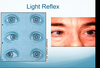
What test is used to see if a patinet’s eyes are in alignment?
Light reflex

How would damage to the 6th nerve nucleus present?
Bilateral loss of abduction
CN VI only has one nucleus for both sides?
What changes to the retina occur with diabetic retinopathy?
How will this affect vision?
- Loss of pericytes and endothelial cells
- Basement membrane thickening
- Decompensated endothelial function
- Leakage and microvascular occlusion
- -> Retinal hypoxia
- -> Expresssion of molecules
- -> Breakdown of retina/blood barrier
- Can lead to neovascularizaiton (in proliferative DR)

If a rod cell catches some light, does it become hyperpolarized or depolarized?
How does this fit into the visual pathway?
If a rod cell catches some light, it becomes hyperpolarized
- Rod cells are very sensitive to single photons, even in really dim light
- This means they can help support the cone pathway / allow us to deduce what is going on when there isn’t a lot of light
- The hyperpolarized rod cell is turned OFF
- Allows amacrine (A2) cells to send activating signal to ON bipolar cells
- “Helloooo we see some light!”
- Signal that there is some light to RGCs
- Allows us to make some sense of what is going on
- But no color vision and decreased acutiy because the cone cells aren’t signaling as much/providing as much invo to ON bioplar cells
Although somehow cones are insensitive to light? If the above is WAY off base please lmk so I can attempt to pass this exam :o
A patient presents with flashes and floaters
What are the most likely causes?
How do you differentiate?
Posterior vitreous detachment vs. Retinal detachment
- Retinal detachment will be accompanied by a curtain/veil loss in vision
- This is an emergency! Call the ophthamologist!
- PVD is less serious and typically doesn’t present with vision loss
- Normal as we age
What systemic conditions are associated with retinal artery occlusion?
- Hypertension
- Diabetes
- Hypercholosterolemia
These, plus hypercoagulable state are the conditions also associated with retinal vein occlusion
How might a posterior communicating aneurysm present?
3rd nerve palsy - eye dilated, in down and out position
MUST evaluate for Pcomm aneurysm to prevent subarachnoid hemorrhage
In myopia:
- Light is focused [anteroir/posterior] to the retina
- The eye is [over/under] powered
- May be because the eye is too [long/short] or the cornea is too [steep/flat]
- Correct with a [converging/diverging] lens
In myopia:
- Light is focused anterior to the retina
- The eye is over powered
- May be because the eye is too long or the cornea is too steep
- Correct with a diverging lens
What defines wet/exudative age-related macular degeneration/
Neovascularization
- Irrecularities caused by drusen promote angiogenesis
- Leads to bleeding (toxic to the retina) and fibrosis
- Wet AMD = end-stage
Picture = drusen

Describe the pathway of choroidal circulation, staring with the internal carotid artery
- Internal carotic
- Short posterior ciliary artery
- Long posterior ciliary artery
- Anterior ciliary artery
Anastomoses exist in the choroidal circulation :)
[But not in the retinal circulation :(]

Flattening the cornea would fix [myopia/hyperopia]
Steepening the corena would fix [myopia/hyperopia]
Flattening the cornea would fix myopia
Steepening the corena would fix hyperopia
What is Adie’s puil?
How does it present?
Acute, idiopathic, postganglionic ciliary nerve dysfunction
-> pupil cannot constrict

What is the definition of glaucoma?
Visual field defects due to optic nerve damage
Typically associated with high intraocular pressure
Postganglionic neurons that control pupillary dilation are located in the [location]
Postganglionic neurons that control pupillary dilation are located in the superior cervical ganglion
- Arousal
- -> IML: Sends preganglionic sympathetics
- -> Superior cervical ganglion: sends postganglionic sympathetics
- -> Pupil dilator
The choroidal circulation is under [autonomic/metabolic] control
The retinal circulation is under [autonomic/metabolic] control
The choroidal circulation is under autonomic control
The retinal circulation is under metabolic control

What prevents vascular growth in the retina?
Soluble VEGF receptors
- Bind VEGF and prevent it from initiating angiogenesis
- Basically, these receptors sequester VEGF o prevent it from having any effect
A right “pie in the sky” visual field defect results from damage to what structure?
Left Meyer’s loop (temporal) lesion
List the steps in the phototransduction cascade
(Staring with light hitting a rod cell)
*Remember, there is amplification at each step*
- Photon absorption converts 11-cis-retinal to all-trans retinal
- Rhodopsin is activated
- Activates transducin via GPCR
- PDE synthesized
- Converts cGMP to 5’-GMP
- Decreased cGMP -> closing of Na+ channel
- Cell hyperpolarization
- Rod cell is turned off
- Allows amacrine cells to activate ON bipolar cells, which signal to RGCs
- Basically, this pathway is more sensitive in low light. It helps signal to RGCs so we put an image together, even when we’re not getting a lot from the cone pathway
Preganglionic neurons that control pupillary dilation are located in the [location]
Preganglionic neurons that control pupillary dilation are located in the interomediolateral cell column (IML)
- Arousal
- -> IML: Sends preganglionic sympathetics
- -> Superior cervical ganglion: sends postganglionic sympathetics
- -> Pupil dilator
What is the limiting factor in our visual acuity?
Cone spacing in the fovea
Which veins collect uveal blood and carry it away from the eye?
Vortex veins
Basically, these are the veins of the choroidal circulation, but they are NOT paired with the arteries
What isthe function of the omnipause neurons in the retiular formation?
Inhibit burst neurons
Response to movement in the visual world is associated with the [temporal/parietal] stream, and the [parvocellular/magnocellular] pathway
Response to movement in the visual world is associated with the parietal aka dorsal stream, and the magnocellular pathway
This is the “where” pathway, associated with the medial LGN
(layers 1-2)
Damage -> pizza on the floor, neglect of objects
*Movement = magnocellular (but parietal stream) Perception = parvocellular (temporal stream)*
Perception of the visual world is associated with the [temporal/parietal] stream, and the [parvocellular/magnocellular] pathway
Perception of the visual world is associated with the temporal aka ventral stream, and the parvocellular pathway
This is the “what” pathway, associated with the lateral LGN
(layers 3-6)
Damage -> pie in the sky
*Movement = magnocellular (but parietal stream) Perception = parvocellular (temporal stream)*
Partial damage to the right lateral geniculate nucleus results in what visual field defet?
Nearly homonymous (but not identical) left visual field defects
What is the treatment for retinopathy of prematurity/
- Diode laser retinal photocoagulation
- Anti-VEGF therapy
Treat if stage 3 with plus disease (idk what this means)
What is the most common cause of blindness in the working-age population in developed countries?
Diabetic retinopathy
A 72 year old woman presents with acute, painless, severe vision loss. She is having trouble seeing things above her.
Eye exam shows chalky white swelling
What additional symptoms would make you concerned for temporal (giant cell) arteritis?
This patient has AION
MUST rule out temporal arteritis
Concerning symptoms include:
- Headache
- Tender scalp
- Jaw claudication
- Fever, weight loss, malaise
Treat with steroids if there is any suspicion
Postganglionic neurons that control pupillary constriction are located in the [location]
Postganglionic neurons that control pupillary constriction are located in the ciliary body
- Light
- -> Melanopisn contianing RGC
- -> Olivary pretectal nucleus
- -> Edinger-Westphal nucleus - start of the afferent pathway
- Generates signals to constrict to both pupils
- Sends preganglionic neurons
- -> Ciliary ganglion
- Sends postganglionic neurons with CN III parasympathetics
- Postganglionic neurons go to pupillary constrictor muscle
Which photoreceptors do we use for high-acuity vision?
Red and green cones
- Red and green light are better concentrated in the fovea, so these cones are most active in high acutity vision
- Blue light does not focus well in the fovea
- We have way fewer blue cones
What are the signs/symptoms of 4th nerve palsy?
Eye on legion side is higher (hypertropia), more prominent when that eye looks medially
Head tilts away from lesion side
Ex: Right 4th nerve palsy:
- Right eye higher than left, more prominent when eye looks to the left
- Head tilts to the left
What tumor is associated with horner syndrome?
Does this affect preganglionic or postganglionic fibers?
Pancost lung tumor
Affects preganglionics (coming from IML to superior cervical ganglion
Preganglionic neurons that control pupillary constriction are located in the [location]
Preganglionic neurons that control pupillary constriction are located in the Edinger-Westphal** **nucleus
- Light
- -> Melanopisn contianing RGC
- -> Olivary pretectal nucleus
-
-> Edinger-Westphal nucleus - start of the afferent pathway
- Generates signals to constrict to both pupils
- Sends preganglionic neurons
- -> Ciliary ganglion
- Sends postganglionic neurons with CN III parasympathetics
- Postganglionic neurons go to pupillary constrictor muscle
Describe the pathophysiology of retinopathy of prematurity
- Phase 1
- Extrauterine hyperoxic environment
- Baby is introduced to oxygen too early -> decreased VEGF
- Leads to vaso-obliteration
- At this stage, VEGF is good and oxygen is bad
- Phase 2
- Not enough oxygen
- Too much VEGF
- Results in neovascularization
- At this stage, VEGF is bad and oxygen is good
- -> Retinal traction
- -> Detachment
Which structure in the eye makes aqueous humor?
Ciliary epithelium
What does a contact prescription of +3.00 +1.50 x 90° mean?
+3.00 power to one axis, and an extra 1.50 at 90°
This would correct a hyperopic astygmatism
These spots are associated with which eye disease?

Age-related macular degeneration
- These are drusen
- Appear early in AMD
- Increase as the disease progresses
- Cause irregularities that can lead to neovascularization (indicates wet/exudative AMD; this is end-stage)
- Tissue loss over the drusen = permanent eye damage
A 3 year old healthy boy presents with crossed eyes, noticed by his caretaker over the past few months.
The patinet does not complain of vision loss or diplopia.
How would you treat this patient?
- Evaluate with cycloplegic refraction
- Possible that eyes are crossing becaues the patient is constantly accomodating to correct his hyperopia
- This is caused by the synkinetic reflex
- Convergence and pupillary constriction both occur with accomodation
- If the child has been accomodating
- Prescribe glasses so the patient can see without self-accomodating
- If the child has not been accomodating
- Surgery to straighten the eyes
If untreated, risk of permanent amblyopia
What are the roles of the frontal eye field and the parietal cortex in generating saccades?
They both transform a sensory signal into a motor command
Frontal eye field = “oh I want to look out the window”
Parietal cortex = “WTF just went by the window” (and then you reflexively look over there)
- Frontal eye field or parietal cortex - sensory signal to motor command
- -> Superior colliculus - Controls size and direction
- -> Lateral geniculate
- -> Reticular formation - Generate burst and tonic command components
- -> Final saccade command
What are the symptoms of acute angle closure glaucoma?
Sudden onset:
- Pain, headache, nausea, vomiting
- Redness
- Photophobia
- Blurred vision/halos
If it is LIGHT, are each of the following hyperpolarized or depolarized?
- Cones
- OFF bioplar cells
- ON bipolar cells
In the LIGHT
- Cones - hyperpolarized
- OFF bioplar cells - hyperpolarized
- ON bipolar cells - depolarized
- Signaling to RGC: I have seen the light!!
Note: in very dim light, the signals from the ON bipolar cells need some help. This is where the rod pathway comes in (I think). Due to the phototransduction cascade, rod cells are hyperpolarized and turned off in the light, allowing amacrine cells to send activating signals to ON bipolar cells, and tell the RGCs “I have seen the light!!”

Describe the activity of burst neurons…
- When our eyes our still
- During a saccade
- When our eyes our still
- Burst neurons are inhibited by omnipause neurons
- During a saccade (ex: to the right)
- Omnipause neurons “pause” their inhibition of burst neurons
- Burst neurons signal to ocular motor and tonic neurons
- Ocular motor neuron sends signal extraocular muscle (ex: abduct right eye)
- Tonic neuron helps us hold our gaze there
- Burst neuron also sens signal to contrlateral brainstem (ex: to the left MLF)
- Results in congugate gaze shift (ex: left eye looks right)
The retinal circulation supplies the [inner/outer] retina
The choroidal circulation supplies the [inner/outer] retina
The retinal circulation supplies the inner retina
The choroidal circulation supplies the outer retina
^ includes photorecepotors

What is the role of the superior colliculus in generating saccades?
Cotnrols the size and direction of the saccade (contralateral)
This is where the initial motor command is generated
- L superior colliculus controls saccade to the right
- Frontal eye field or parietal cortex - sensory signal to motor command
- -> Superior colliculus - Controls size and direction
- -> Lateral geniculate
- -> PPRF - Generate burst and tonic command components
- -> Final saccade command
What method is used to assess refractive error in patinets that are pre-verbal?
Retinoscopy
What is the efffect of a lesion in the calcarine cortex?
Blindness in the affected regions of the visual field
What is papilledema?
How does it present?
What causes it?
Papilledema = disc swelling due to elevated intracranial pressure
- Headache
- Tinnitus
- Grey outs of vision when standing up
- Nausea, vomiting
Most concerning cause: tumor
May also be
- Venous sinus thrombosis
- Severe HTN
- Idiopathic (pseudotumor cerebri)
- Classic patient is a young, obese woman
What tools can be used to meausre corneal astigmatism?
Keratometry
Corneal topography
Slit lamp examination (look for scar causing irregular astigmatism)
What horizontal eye movement can still be made after right side brainstem damage produces one-and-a-half syndrome?
Which structures specifically are damaged?
Left eye abduction is still possible
- Right eye cannot look left (adduct) due to right MLF damage
- Neither eye can look right (saccade to the right) due to right PPRF damage
- Right eye abduction imparied due to PPRF damage
- No signal for right eye to look right = no signal to left MLF to adduct, even if the left MLF is intact
Damage to the medial right LGN results in loss of which visual field?
Left lower quadrant
Medial LGN = layers 1-2 = parietal stream
(vs damage to the lateral right LGN leads to loss of the left upper quadrant)
(Pizza on the floor)
Homonymous hemianopsia with macular sparing result in damage to which area of the brain?
Contralateral calcarine cortex
Which cells are damaged in glaucoma, leading to vision loss?
What is the mechanism of damage?
Retinal ganglion cells
Damaged due to high intraocular pressure that either:
- Compresses axons, resulting in reduced axonal transport
- Compresses blood flow to the axons, causing RCGs to die
What is the most common cause of untreatable visual loss in the elderly?
Age-related macular degeneration
[Retinal/choroidal] circulation supplies the photoreceptors
Choroidal circulation supplies the photoreceptors

What is the leading cause of vision loss among children/
Amblyopia
- Strabismic (misaligned eyes)
- Brain ignoes eye that isn’t straight to prevent diplopia
- Refractive
- Brain ignores the eye with larger refractive error
- Deprivation
- Ptosis, cataract, corneal scar
- Brain ignores the deprived eye
At what age should congenital cataract surgery be performed?
6 weeks
- Early enough to correct vision without long-term consequences
- Late enough to avoid risk of serious complications of surgery/anesthesia in an infant
What are the consequences of damage to tonic neurons?
Gaze nystagmus
- Can make the saccade to lood at an object, but cannot hold gaze there
- Results in a “jump then drift” eye movement pattern
What symptoms are suggestive of a reitnal detachment?
- Flashes and floaters in one eye
- Visual field cut / loss of vision
Floaters and flashes only may indicate vitreous detachment
Floaters, flashes, loss of vision = suggestive of retinal detachment
Which structure generates vertical saccade burst and tonic commands?
Which structure generates horizontal saccade burst and tonic commands?
- Vertical saccades - rostral interstitial nucleus of the MLF
- Horizontal saccades - PPRF
Note: the burst and tonic commands are like the “final go ahead” that communicates with the extraocular muscles
For horizontal saccades:
- Frontal eye field or parietal cortex - sensory signal to motor command
- -> Superior colliculus - Controls size and direction
- -> Lateral geniculate
- -> PPRF - Generate burst and tonic command components
- -> Final saccade command
What are the functions of the RPE? (6)
- Blood-retinal barrier
- Light absorption - anything not caught by photorecptors, prevents scatter
- Epithelial transport
- Visual cycle - regenerates the visual pigment
- Phagocytosis - of photorecptors that are worn out
-
Secretion - of growth factors PEDF, VEGF
- PEDF = anti angiogenic, to the retinal side
- VEGF = angiogenic, to the basal side; bad if upregulated
Damage to the lateral right LGN results in loss of which visual field?
Left upper quadrant
LateralLGN = layers 3-6 = temporal aka ventral stream
(vs damage to the medial right LGN leads to loss of the left lower quadrant)
(Pie in the sky)
What refractive error can be corrected with a limbal relaxing incision?
Severe corneal astigmatism
- Tiny cuts through the limbus can help relax one axis of the cornea, reducing the difference in refractive power
In hyperopia:
- Light is focused [anteroir/posterior] to the retina
- The eye is [over/under] powered
- May be because the eye is too [long/short] or the cornea is too [steep/flat]
- Correct with a [converging/diverging] lens
In hyperopia:
- Light is focused posterior to the retina
- The eye is under powered
- May be because the eye is too short or the cornea is too flat
- Correct with a converging lens
What causes coloboma?
Failure of the embryonic fissure to close
- May be anterior or posterior
- Anterior: iris, lens
- Posterior: retina, choroid, optic nerve

What eye movement cannot be made after the left MLF is damaged?
Left eye cannot adduct
(Left eye cannot look right)
Results in unilateral internuclear ophthalmoplegia
Which premature infants are at risk for retinopathy of prematurity?
Low birth weight, earlier gestational age
Also: Anemia and need for supplemental O2
-
Screen for retinopathy of prematurity if:
- Weighs <1500 grams
- Born at <=30 wks
What are the consequences of damage to the right PPRF?
Saccades to the right cannot be generated
(Cannot look toward the legion)
- Right eye cannot look right
- No signal to left MLF to look right
- => Neither eye can look at an object to the right
If it is DARK, are each of the following hyperpolarized or depolarized?
- Cones
- OFF bioplar cells
- ON bipolar cells
In the DARK
- Cones - depolarized
- OFF bioplar cells - depolarized
- Signaling to RGC: it is dark!!
- ON bipolar cells - hyperpolarized
Which layer of tissue regenerates visual pigment?
Retinal pigment epithelium
In what pathologies wil you see cotton wool spots?
- Nonproliferative diabetic retinopathy
- Still present in proliferative, but will see more hemorrhage
- Hypertensive retinopathy
- Temporal arteritis
Which gene is mutated in retinoblastoma?
RB1
- Very concerning if retinoblastoma in both eyes - indicates germline mutation
- => All cells are affected :(
What is the main function of the ventral stream pathways?
Visual perception
What are feature detectors in the eye?
Detectors of visual patterns that are more complex than those that excite hypercomplex neurons