Differentiation of the endoderm and mesoderm Flashcards
What does the endoderm form?
The digest tube that extends the entire length of the body
What membrane have only two tissue types? What tissue types are they?
Buccopharyngeal membrane and cloacal membrane They only have the ectoderm and endoderm
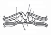
Label the diagram
What developmental stage is it in at the moment?

Day 20 just before longitudinal bending and transverse folding

Label the diagram
Where on the embryo is this found?

The Buccopharyngeal membrane

Label the diagram
Where is this found on the embryo?

The neural plate

Label the diagram
Where does this cross section pass through?
The intermediate mesoderm is connected onto two other mesoderm tissue types, what are they called and where are they found?
Throught the somites
The parietal lateral plate mesoderm, adjactent to the ectoderm (top)
The visceral lateral plate mesoderm, adjacent to the endoderm (bottom)

Label the diagram
Where is this cut from?
Has this section undergone full gastulation? What indicates this?

Cut through the primitive streak at the tail end
No, because there is still mesenchyme (mesoderm that is still loosely packed)

Label the diagram
What is the developmental stage of the embryo at this point?
What is happening at this point?

21 days
The embryo is beginning to undergo longitudinal bending and transverse folding

Label this diagram
What is the embryos development stage?
What are the 3 vesicles that form the brain?

23 days
Forebrain vesicle, midbrain vesicle and hind brain vesicle

What happens to the buccopharyngeal membrane and cloacal membrane during longitudinal bending? What happens to these as the embryo develops? What do they form?
They block the ends of the tube These membranes disappear Forms the mouth and the anus
What forms on the ‘embryonic tube’ once folding and bending is complete? What does this develop into?
Budding begins, this leads to the development of all of the digestive glands and the lungs
How does the somitomere tissue vary during development based on body location?
In the head it remains as somitomeres (FYI a pseudo segmeneted tissue) while from the head lower (trunk) it develops into somites
What is the process of forming somites from somitomere?
Somitogenesis
How does the formation of somites progress during embryonic development (eg regular/irregular)? What does this indicate?
Somites are formed at very regular intervals
Inidcates that there is some kind of clock device within the embryo
How does the clock within the embryo work? What is a developmental sequence that is linked closely with this clock?
The levels of mRNA production fluctuate with a regular time interval creating a genetic clock
The formation of somites is syncronised with the peaks of mRNA production of this gene
What is the clock and wave form model? What does this control and how do they work together?
The clock is the genetic clock determined by the mRNA production
The wave is a chemical signal that determines the competency of a cell (if it has passed a cell it become competent, if not then it is incompetent) that travels from the rostral to caudal end (head to tail)
Only when the wave is competent (chemical signal has gone by the somitomere) and the gene expression is at its maximum will a somite form
Once the clock and wave form model has passed through the entire embryo, what is formed?
Somites that are lined along the length of the notohord
What segments are the somites divided into?
The medial and lateral axis (left and right side) and the dorsal and ventral surface (top and bottom)
How are the dorsal and ventral surface distinguished? Where does it come from?
Through the concentration differences from the chemical sonic hedgehog produced by the notochord
How does the concentration of sonic hedgehog influence what surface phenotypes the somites develop?
The side which gets a higher concentration will develop ventral phenotypes while the side which gets a lower concentraiton will develop dorsal phentypes
What kind of chemical is sonic hedgehog? What does this make the notochord?
It is a morphogen however the notochord is not regarded as an orgnaiser, it only has orngaiser properties
What does sonic hedgehog do to the somites?
It causes the dorsal side to express the Pax3 gene and the ventral side to express the Pax1 gene
Label the diagram
What have the dorsal and ventral somites developed into?

Dorsal = dermatome adn myotome, Ventral = syndetome and sclerotome

What is the differentiation of the dorsal and ventral somites determied by? Explain the conditions for each of the four types formed
It is determied by what gene is expressed and the position of the cells
Dermatome: Pax3 expressed and furtherest away from notochord/closest to epithelium
Myotome: Pax3 expressed adjacent to somite cavity
Syndetome: Pax1 expressed adjacent to somite cavity
Sclerotome: Pax1 expressed and closest to notochord/furthest from epithelium
What part of the embryo will form the the epidermis and dermis?
The outside layer forms the epidermis and the dermatome forms the dermis layer (as it is just below the epithelial tissue of the embryo which will form the epidermis FYI)
What do each of the layers from the somites form in a fully develped organism?
Dermatome: dermis
Myotome: skeletal muscle
Syndetome: tendons
Sclerotome: skeletal structures
What does myotome do before forming skeletal muscle?
It migrates out of the somites first
Do sclerotome form bones? Explain how
Eventually it does but first is becomes mesenchymal and migreates towards the midline where it then normally first forms cartilage which then transforms into bones
What kind of skeletal structures do the sclerotome form?
Forms the ribs and verterbrate
How are limbs formed?
Cells from the lateral plate mesoderm migrate and form limb buds, these then develop into skeletal structures (yes, limb bones are formed from lateral plate mesoderm NOT sclerotome)
What two main embryonic tissue types are limbs formed from? Why not all three types?
Ectoderm and mesoderm, not endoderm becasue that is used for forming organs which are not presnet in the limbs
What are the differences between the embryonic formation of the trunk vs the limbs?
The trunk has derivatives of the endoderm, the limbs don’t
Skeletal structure of the limbs is from parietal lateral plate mesoderm (lateral plate mesoderm closest to the ectoderm) while the trunk’s is derived from sclerotome (somatic mesoderm)
When and how is mesoderm first formed? What does it initiatlly form? What does it develop into?
First formed during gastrulation as the cells migrate through the primitive streak and placing themeselves between teh ectoderm adn endoderm
First forms mesenchyme (loosely packed mesenchmal cells)
Forms chordamesoderm (notochord), Paraxial mesoderm (somites), intermediate mesoderm and lateral plate mesoderm
Where is the notochord formed? Is it a permanent structure? What does it do?
Along the midline up to the level of the hindbrain (about the level where the ear will form)
No it is not, it is important in the development of the nervous system but eventually disappears
Where is paraxial mesoderm found? What does it form?
It is formed at both sides of the embryo near the mid line
Form somitomeres (loosely packed mesodermal cells) that soon become compacted and form somites
Are all somites the same? How do we know this?
No
Each somite will form a different structure
How does the somitomeres develop differently in the cranial region?
Somitomeres do not become compacted to fomr well defined somites, instead it combines with chordamesoderm (aka notochord) which then gives rise to connective tissues and muscles of the head and neck
What does the intermediate mesoderm form?
Forms the urogenital system (aka the organs associated with the reproductive and urinary system, eg kidneys, gonads etc.)
What does the lateral plate mesoderm split into? At what developmental stage does it do this?
The visceral lateral plate mesoderm and the parietal lateral plate mesoderm
At week three
What does each layer of the lateral plate mesoderm develop into?
Visceral: heart, smooth muscle and connective tissue of the gut
Parietal: forms inner lining of the body wall and the skeletal structues of the limb
