Chapter 16: Oral and Salivary Glands Flashcards
___________ is one of the most common diseases in the world and a major cause of tooth loss before 35YO.
Dental carries (tooth decay)
What are dental carries?
Reversible?
-
Tooth decay that occurs when there is focal demineralization of a tooth – enamel and dentin—by acid metabolites of fermenting sugar that is made by bacteria.
- Reversible up until cavitation (hole is formed)
Where are rates of dental carries highest and lowest?
- Dropping in countries like US where oral hygeine is improving and fluoridation of water occurs.
- Increase rate in developing countries (eating more processed food)
What are symptoms of dental carries?
- Pain to the point where it affects activities of daily living
- WL/nutritional problems
- Loss of self-esteem/confidence
- Potentially life threatening infections
What is gingivitis?
Who is it most common in?
a reversible inflammation of the gums due to poor oral hygeine.
Most common in adolescence.
What causes gingivitis?
Gingivitis is caused by dental plaques that build up under gumline, which mineralized to form calculus (tarter).
What are dental plaques?
- sticky, clear, biofilm that collects between and on the surface of the teeth.
- it contains a mixture of [bacteria, salivary proteins and desquamative epithelial cells]. If plaque is not removed => mineralized => forms calculus (tarter).
What changes charcterize gingivitis?
- Redness of gums
- edema,
- bleeding,
- changes in countor,
- less of soft tissue adaptation of the teeth
_________ is always preceeded by gingivitis, however gingivitis does not always progress into it.
Periodonitis (gum disease)
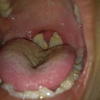
What is Periodontitis?
Chronic inflammatory process affecting the supporting structures of the teeth (peridontal lig.), alveolar bone, and cementum, that causes teeth loss.
Periodontitis can lead to what?
gums can pull away from the tooth, bone can be lost, and the teeth may loosen or fall out.
Periodontitis is caused by _______________ in the mouth that affect surrounding tissue. What bacteria is normally found in our mouth?
- - Anaerobic and microaerophilic gram (-) bacteria
- - Facultative gram (+)
Which systemic diseases increase the risk of Periodontitis?
- AIDS
- Leukemia
- Chron disease
- DM
- Down syndrome (high risk for leukemia)
- Sarcoidosis
- Dz asso. w/ defect in neutrophils (Chediak-Higashi, agranulocytosis, and cyclic neutropenia)
Adult periodontitis is primarily associated with which bacteria?
- Aggregatibacter (actinobaccilus) actinomycetemcomitans
- Prophyromonas gingivalis
- Prevotella intermedia
Which diseases can periodontal infections be the origin for?
- Infective endocarditis
- Pulmonary and Brain abscesses
Common, often recurrent, painful superficial oral mucosal ulcers whose cause is not known.
Aphthous ulcers (canker sores)
- D/t stress and gray/blue base surrounded by erythema.

Reactive lesions in the mouth (fibrous proliferations) include ___________ and are ALL __________.
- Traumatic fibroma/irritation fibroma
- Pyrogenic granuloma
- all BENIGN
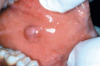
Which inflammatory lesion is typically found on the gingiva of children, young adults, and pregnant woman (pregnancy tumor)?
Pyogenic granuloma

Trauma fibroma (irritation fibroma)
Trauma fibroma (irritation fibroma)
Submucosal nodule of CT that formed due to trauma, most common along bite line and ginigiva.
What are the 3 important inflammatory/reactive lesions?
- 1. Aphthous ulcers (canker sores)
-
Fibrous proliferative lesions
* Irritation fibroma/traumatic fibroma
* Pyogenic granuloma
-
Fibrous proliferative lesions
Canker sores (apthous ulcers) are most common when?

0-20 YO
Canker sores are commonly seen in what diseases?
- 1. Celiacs disease
- 2. IBD
- 3. Behcets disease
CIB
What is an ulcer?
-
Breach in the surface of a tissue or organ that is made by the shedding of inflamed necrotic tissue.
- ONLY occurs when tissue necrosis and inflammation exist on or near a surface.
Where do ulcers most commonly occur?
- Mucosa of mouth, stomach, intestines, GU tract
- Skin or subQ tissue of LE in older people who have circulation problems that predispose to ischemic necrosis.
Describe the bacterial infiltrates in canker sores.
- At first, largely mononuclear.
- Secondary bacterial infection may be d/t neutrophilic infiltrate.
Describe the features of pyogenic granuloma and its course.

- Red/purple lesion that is ulcerated.
- Highly vascular proliferation of organzing granulation tissue.
Course:
- Regress into dense fibrous mass.
- Develop into a peripheral ossifying fibroma.
What infections can occur in the oral cavity?
- HSV 1 ** and oral HSV 2 (genital herpes)
- Candida
- Deep fungal infections
What are other viral infections that can involve the oral cavity?
- Herpes zoster
- EBV (mononucleosis, nasopharyngeal carcinoma, lymphoma)
- CMV
- Enterovirus (herpangina, hand-foot-mouth disease, acutre lymphonodular pharyngitis)
- Rubeola (meales)
HSV occurs most commonly in _________ and are often…
- 2-4 YO
- Asymptomatic (& do not cause significant morbidity)
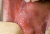
10-20% of primary infections of HSV can present as _____________, with abrupt onset of vesicles and ulcers in the mouth, particularly the gums. These lesions are also accompanied by what symptoms?
acute herpetic gingivostomatitis
Symptoms:
- lymphadenopathy
- fever
- anorexia
- irritability
Which test is diagnostic for Acute Herpetic Gingivostomatitis?
What are you looking for?
- Tzanck test (microscopic examination of the vesicle fluid)
- Multinucleate polykaryons (giant cells)
- Eosinophilic intranuclear viral inclusions
What is the course of vesicles and ulcers seen in Acute Herpetic Gingivostomatitis?
- First: vesicles filled with a clear, serous fluid
- Rupture quickly and create painful, red-rimmed shallow ulcers.
- Spontaneously go away in 3-4 weeks, but the virus then travels along regional nerves and becomes latent in local ganglion (trigeminal/semilunar ganglion)
- Reactivated during stress or sunlight, leading to vesicles (cold sores) on the lips
With HSV, infection is most common and most adults have ________.
latent HSV-1
Viral reactivation of HSV (recurrent herpectic stomatitis) occurs where?
- Site of primary inoculation or adjacent mucosa assx with the same ganglion & go away in 7-10 days.
- Groups of small vesicles on the lips, nasal orifices, buccal mucosa, gum and hard palate.
What is the most common fungal infection of the oral cavity and a NL part of the oral flora in 50% of the population?
Candidiasis (thrush)
Which form of Oral Candidiasis is the most common?
How does it appear in the oral cavity?
- Pseudomembranous (thrush)
- Superficial, gray to white inflammatory membrane, that can be readily scraped off, revealing red inflammation.
Oral candidiasis remains ________, except in people with immunosupression.
superficial
Which infection produces a characteristic dirty white, fibrinosuppurative, tough, inflammatory membrane over the tonsils & retropharynx?
Diptheria
What oral findings do we see in patients with measles?
1. Spotty enanthema (ulcers or eruptions in the buccal mucosa) that occur before a skin rash
- => Koplik spots via Stenson Ducts (small red lesions with blue/white centers)
Pt presenting w/ a fiery red tongue w/ prominent papillae (raspberry tongue); white-coated tongue through which hyperemic papillae project (strawberry tongue) should raise suspicion of which infection and organism?

- Scarlet Fever
- Strep pyogenes –> Gram (+)
What oral findings do you see in a patient with infectious mono (EBV= a dsDNA virus)?

- Acute pharyngitis and tonsilitis with a grey/white exudutaive membrane
- Petechiae on the palate
- Large LN in neck
Which infection
- HIV predisposes ppl to oral infections ________, _______ and ________
- OR is a common diagnosis in people with ___________ and _________
- Herpes
- Candida
- Other fungi
Karposi sarcoma and hairy luekoplakia
In addition to superficial funal infections, which occur at sites of infection, certain deep fungal infections have a predilection for the oral cavity, head and neck. What are they?
- Aspergillosis
- Cryptococcosis
- Murcomycosis
Other:
- Histoplasmosis, Blastomycosis, Coccidiodomycosis, Zygmocosis
What is the key predisposing factor for deep fungal infections (infections that have a predilection for oral cavity/head and neck?
Immunosupression
- Hairy leukoplakia is caused by what virus?
- Found where in oral cavity?
- Oral manifestation of what systemic diseases?
- EBV => squamous cell hyperplasia (NOT pre-malignant and no dysplasia occurs)
- white, fluffy (hairy), hyperkeratotic patches on lateral border of the tongue
- Immunosupression (AIDS);

It is __________ for oral lesions to be the 1st sign of some underlying systemic condition.
common
Hairy leukoplakia is characterized by what 2 distinct microscopic features?
- Hyperparakeratosis
- Acanthosis (diffuse epidermal hyperplasia) with “balloon cells” in the upper spinous layer
How is oral hairy leukoplakia different than candida in how it looks?
Oral hairy leukoplakia CANNOT be scraped off.
_____________ are oral mucosa lesions that can undergo malignant transformation (dyplasia) to squamous cell carcinoma
Leukoplakias and erythroplakias
both precursor lesions
________ is a white patch or plaque with SHARP borders that cannot be scraped off and cannot be characterized clinically or pathologically as another other disease.
Leukoplakia
Plakia = plate or flat plane

Until proven otherwise, all leukoplakias must be considered __________.
Precancerous (up to 25% are precancerous)
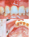
What is a erythroplakia?
How is it different from leukoplakia?
- Red, velvety eroded patch in the oral cavity that is at level or a little depressed with surrounding mucosa.
- Different from leukoplakia:
- less common
- epithelium is VERY atypical
- malignant transformation is HIGHER.
What are the histological changes of the epithelium seen in virtually all (90%) of erythroplakia?
- Severe dysplasia
- Carcinoma in situ
- Minimally invasive carcinoma
What are the histological changes of the epithelium in leukoplakias?
Hyperkeratosis over a thick, orderly acanthotic mucosal epithelium, but can undergo severe dysplasia and become CIS.
Around 95% of the cancers of the head and neck are of which type?
Remainder largely consists of which type?
- Squamous Cell Carcinoma (SCC) = majority (95%)
- Adenocarcinomas of salivary gland = remainder
The pathogenesis of squamous cell carcinoma is ____________.
Multifactorial
- Smoked tobacco & alcohol
- HPV (in oropharynx)
- Betel quid & paan (in India & Asia)
What are predisposing risk factors of squamous cell carcinoma of the lower lip?
- Actinic radiation (sunlight)
- Pipe smoking
The incidence of oral cavity SCC (particulary the tongue) is on the rise in whom?
Ppl < 40 YO with no known risk factors
In the oropharynx, as many as 70% of SCCs, particularly those involving the [tonsils, base of the tongue, and the pharynx] harbor what?
- Oncogenic variants of HPV
- Particularly HPV-16
Unlike the ________, HPV-associated SCC of the oral cavity is ________.
oropharynx
uncommon
Survival of SCC is dependent on a number of factors including the etiology of SCC.
What is the prognosis (5-year survival rate) of the “classic” (smoking and alcohol related) early-stage SCC?
Late stage?
- Good (80%)
- 20%
Patients with HPV (+) SCC have _______ long-term survival than those with HPV (-) tumors?
BETTER prognosis if p16+
- HPV (-) = worse prognosis
What is the basis of “Field Cancerization”?
- After repeated carcinogenic exposure, the entire superficial epithelium of the upper aerodigestive tract has a ↑ risk of developing pre-malignant lesions as a result of multiple genetic abnormalities
- Development of tumors decreases survival.
The 5-year survival rate of the first primary tumor is _______, but in the same people, second primary tumors are ___________.
- better than 50%
- most common cause of death
Which genetic mutations are commonly associated with the “classic - tobacco/alcohol” SCC subset?
- p53 pathway
- Proteins that regulate squamous cell differentiation (p63 and NOTCH1)
What is typically overexpressed in HPV-associated SCC’s?
Other common genetic alterations?
Have fewer and different genetic alterations.
- p16 (cyclin dependent kinase inhibitor) overexpression
- p53 inactivation –> E6 expression
- RB inactivation –> E7 expression
What is the molecular progression of oral SqCC?
Does it always progress in this manner?
NL => hyperplasia/hyperkeratosis => mild/moderate dysplasia => severe dysplasia/CIS => SCC
No, not always linear over a uniform period or time.
What is the most common genetic alteration associated with
- NL => hyperplasia/hyperkeratosis and mild/mooderate dysplasia?
- Mild/moderate dysplasia => severe dysplasia/CIS?
- Severe dysplasia/CIS => SCC?
- 9p21 (p16), 3p
- TP53
- Cyclin D (11q13); 4q, 6p, 8p, 13q, 14q
What is the typical age of onset and sex most affected by leukoplakia and erythroplakia?
- Age 40-70
- M 2x more than F
What are the 5 favored locations in the oral cavity for the development of classic SCC?
- Ventral surface tongue
- Floor of mouth
- Lower lip (associated w/ sun exposure and pipe smoking)
- Soft palate
- Gum
What is the morphology of oral SCC in the early stage?
- 1. Raised, firm, pearly plaques
- 2. Irregular, roughened or verrucous areas of mucosal thickening _(_may look like leukoplacia)
that are superimposed on obvi leukoplakia or erythroplakia
As early stages of SCC carcinoma enlarge, what do they look like?
Ulcerated, protruding mass that have irregular and indurated (rolled) borders.
What is the difference in progression of oral SCC and cervical cancer?
- Oral SCC: dysplastic lesions MAY OR MAY NOT progress into full-thickness dysplasia (CIS) before invading underlying CT.
- Cervical cancer: [full thickness dysplasia => invasion].
Is the degree of keratinization in oral SCC correlated with behavior?
No.
What are the favored sites for local metastasis of oral SCC?
Favored sites for distal metastasis?
- Local —> cervical LN’s
- Distal —> mediastinal LN’s, lungs, liver, and bones
- Often, when primary lesion is found distant metaseses has occured.
Overwhelming majority of odontogenic cysts are derived from what?
Remant of odontogenic epithelium within jaws.
Which type of cyst occurs when fluid accumulates between developing tooth and dental follicle, forming a cyst originating around the crown of an unerupted tooth?
Detingerous cyst
Radiographically, dentigerous cysts are seen as what type of lesions and most often associated with which teeth?
Treatment?
- - Unilocular lesion
- - Impacted third molar (wisdom) teeth
- Complete removal = curative
What is the significance of Keratocystic Odontogenic Tumors (OKC’s)?
Must be differentiated from other cysts due to its aggressive behavior
Keratocystic Odontogenic Tumors (OKC’s) are most often diagnosed between what ages and which sex is more commonly affected?
Form where in the oral cavity?
- Ages 10-40 yo; most often in males
- Posterior mandible
How do you ID OKC’s radiographically and histologically?
- Radiographically
- Well-defined unilocular or multilocular
- Histologically
- Cyst is lined by thin layer of keratinized stratified squamous epthilium with a prominant basal layer and corrugated epithelial surface

Multiple Keratocystic Odontogenic Tumors (OKC’s) occuring in a patient should prompt evaluation for what?
Nevoid basal cell carcinoma (Gorlin syndrome);
mutation in tumor suppressor PTCH on Chr 9q22
Tx of OKC
Surgical removal, because they are locally agressive and recurr
What is xerostomia?
↓ production of saliva, causing dry mouth and/or atrophy of papillae on the tongue with fissures and ulcers.
Xerostomia is a major feature of?
Sjorgren Syndrome
Complications of Xerstomia include increased rates of?
- Dental carries
- Candidiasis
- Difficulty swallowing and speaking
Xerostomia is most frequently a side-effect of what?
Meds
- Anti-cholinergic
- Anti-depressant
- Ant-psychotic
- Diuretic
- Anti-HTN
- Sedative, muscle rexant, analgesic
- Anti-histamine
__________ is an inflammation of the salivary glands, caused by what?
Sialadenitis, caused by trauma, infection or AI reaction
What is the most common type of inflammatory salivary gland lesion?
Mucoceles
- Mucoceles are caused by what?
- Most commonly found where in oral cavity and due to what?

- Trauma or blockage of a salivary gland duct, causing saliva to leak into the surrounding CT stroma.
- Most often LOWER LIP due to TRAUMA
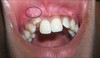
How do mucoceles present clinically (i.e., how do they look on examination)?
Patients often report what in regards to the size of the lesion?
- Fluctuant swellings of lower lip that have a blue translucent hue
- Often report hx of changes in size in assoc. w/ meals

What is the histological characteristics of Mucoceles?
Most common inflammatory cell present?
- Pseudocysts w/ cyst-like spaces, filled with mucin, lined by granulation tissue or fibrous CT
- MACROPHAGES

What is the Tx for Mucoceles?
Complete excision of the cyst and its accompanying minor salivary gland
What is a Ranula?
Epithelial-lined cysts that arise when duct of sublingual gland has been damaged
a 45 YO F presents with dry mouth followed by pain. Pain increases when eating. PMH dx Sjrogens sundrome. Which duct is asspocated with the involved gland?
Secretion of which of the following is most likely affected?
Stensen’s
- Parotid gland secretes what?
- Submucosa layer of the tongue secretes what?
- Salivary amylase
- Lingual lipase
The prescence of xerostomia in BMS (burning mouth syndrome) suggests what?
The disease may involve hypofunctioning of the parasympathetic NS, a target of OMT to normalize activity.
Bacterial sialadenitis, most often involves the __________ glands and is a common condition secondary to ___________
Bacterial sialadenitis, most often involves the submandibular glands and is a common condition secondary to sialolithiasis (ductal obstruction by stones)
What are the 2 most common organisms responsible for sialolithiasis leading to sialadentitis?
Unilateral or bilateral process?
1) S. aureus
2) S. viridans
- UNILATERAL -
A patient presenting with unilateral, sialadentitis with overt suppurative necrosis and abscess formation should raise suspicion of?
Sialolithiasis causing sialadentitis, likely due to S. aureus or S. viridans.
Overall, salivary gland neoplasms are _______________ and occur in whom?
What is the difference between when benign and malignant tumors appear?
- Relatively uncommon
- Adults (F)
- Benign (50-70s); malignant (later)
The likelihood of a salivary gland tumor being malignant is more or less ___________ proportional to the size of the gland
- Inversely
- *Smaller the gland = higher risk of malignancy
Majority of salivary gland tumors arise where?
Parotid gland
What is the most common tumor of the salivary gland and is it benign or malignant?
Pleomorphic adenoma – 50% of benign tumors, but 60% of tumors in parotid.
Pleomorphic adenomas contain a mixture of _________ and _________ cells
Pleomorphic adenomas contain a mixture of ductal (epithelial) and myoepithelial cells
- *MIXED tumor
Exposure to what increases risk for Pleomorphic Adenomas?
Associated with what genetic mutation?
- Radiation
- PLAG1 overexpression –> ↑ cell growth
How do pleiomorphic adenomas present clinically (i.e., mass where, any pain, and rate of growth)?
- Painless, slow-growing, mobile, discrete masses in the parotid/__submandibular gland or in the buccal cavity

What type of tumor is shown here and how can you tell?

-
Pleomorphic adenoma
- DOMINANT histological feature = GREAT heterogeneity
- Well-demarcated tumor w/ epithelial cells and myoepithelial cells arranged in ducts, strands or sheets within a chondroid matrix.
- DOMINANT histological feature = GREAT heterogeneity
In most cases of pleomorphic adenoma, is there epithelial dysplasia or mitotic activity?
No
What is the prognosis of a malignant pleomorphic adenoma (i.e., carcinoma ex pleomorphic adenoma)?
- Most aggressive of all salivary gland tumors
- Mortality rates of 30-50% at 5 years
Which benign tumor almost always occurs in the parotid gland?
Warthin Tumor (aka papillary cystadenoma lymphomatosum): a cystic tumor with alot of lymphocytes + germinal centers that almost ALWAYS occurs in parotid gland
When do Warthin tumors usually arise?
Which sex is most affected?
What is a major risk factor?
- 50-70s
- Males
- Smokers have 8x the risk
What are some of the distinct morphological characteristics of a Warthin tumor?
- Round to oval encapsulated mass (2-5 cm in diameter) in superficial parotid gland
- Lined by double layer of cells resting on dense lymphoid stroma, sometimes w/ GERMINAL centers
- Upper layer = palisading columnar cells w/ abundant, finely granular, eosinophilic cytoplasm.
- Granular bc many mT (oncocytic)
- Lower layer = cuboidal to polygonal cells

What is the reason for the granular appearance of the cytoplasm in the upper layer of cells seen in Warthin Tumors?
NUMEROUS mitochondria, feature referred to as “oncocytic”
What is the most common primary malignancy of the oral mucosal?
Mucoepidermoid carcinoma (makes up 15% of all salivary gland tumors)
Where do mucoepidermoid carcinoma occur most often?
60-70% occur in parotid gland, but also minor salivary glands
Which genetic mutation (i.e., translocation and gene products) is thought to play a key role in Mucoepidermoid carcinoma?
- Balanced (11;19) (q21;p13) translocation, creating MECT1-MAML2 fusion gene.
The prognosis of mucoepidermoid carcinoma depends on what?
GRADE
What are the histological characteristics of a mucoepidermoid carcinoma?
How large do they grow?
- Cords, sheets, or cystic configurations of squamous, mucous, or intermediate cells
- Grow to 8 cm in diameter and are circumscribed, but lack well-defined capsules and are infiltrate at margins

Which stain helps to visualize a mucoepidermoid carcinoma?
Mucin stains
What is the prognosis of both low-grade and high-grade Mucoepidermoid Carcinomas?
- Low grade —> 5-year survival of 90%
- High grade —> 5-year survival of 50%
Adenoid cystic carcinoma is most often seen where?
- Minor salivary glands (particularly the palatine glands); 50%
- May also be seen in major salivary glands (parotid and submandibular)
What is the morphology of Adenoid cystic carcinomas?
- Small, poorly encapsulated, infiltrative, gray-pink lesions that create a a cribiform pattern
- Small cells w/ dark, compact nuclei and scant cytoplasm
Describe the cancer seen here and its distinct morphology.

Adenoid cystic carcinoma of the salivary gland creating a Cribiform pattern
*Gaps between the cancer cells within the duct, with an appearance similar to the ‘holes in swiss cheese‘ or perhaps ‘ripples‘.
Adenoid cystic carcinomas cause what distinct symptom?
Pain, bc invade perineural spaces and metastize to bone, liver, and brain
