Brain Tumors I Flashcards
What is better: grading or staging? Why?
grading because brain tumors don’t often metastasize
Most common primary brain tumor?
astrocytomas
What is shown in this image?

Grade II astrocytoma
What is shown in this image?

Grade III astrocytoma
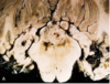
What is shown in this image?

Grade IV astrocytoma
What does this image show?

Grade II/IV astrocytoma
What is shown in this image?

Grade II astrocytoma
What does this image show?

H and E stain of grade II astrocytoma
What does this image show?

GFAP stain with positive tumor cells (stage II astrocytoma)
What is shown in this image?

Grade III astrocytoma/ anaplastic astrocytoma
What is shown in this image?

Grade III astrocytoma/ anaplastic astrocytoma
What is the most reliable indicator of a glioblastoma, grade IV?
necrosis
Genetic- Primary GBM
Occurs in older______
Amplification of ______ gene
MDM2 _______, p16 _____, PTEN mutations
Genetic- Primary GBM
Occurs in older adults
Amplification of EGFR gene
MDM2 overexpression, p16 deletion, PTEN mutations
Genetic- secondary GBM
____ patients
Arises from_____ tumor
Shares p53 ______ (and PDGF-A) amplifications with lower grade
Genetic- secondary GBM
Younger patients
Arises from lower-grade tumor
Shares p53 inactivation (and PDGF-A) amplifications with lower grade
Label the following image


Label the following image

GBM

What does this image show?

GBM with necrosis and vascular proliferation
Pilocytic Astrocytoma
►Occurs in children and young adults
►Often occurs in _____; can occur around ____ventricle, optic nerves, and cerebral hemispheres
Pilocytic Astrocytoma
►Occurs in children and young adults
►Often occurs in cerebellum; can occur around 3rd ventricle, optic nerves, and cerebral hemispheres
What is show in this image?

Pilocytic astrocytomas are often in posterior fossa, are cystic, and feature long, hair-like processes and Rosenthal fibers (protein globules or droplets).
What is shown in this image?

Pilocytic astrocytoma
Oligodendroglioma
►Arises from oligodendroglial cells
►Occurs most commonly in _____ matter
►Occurs in 4th or 5th decade; often with _____
Oligodendroglioma
►Arises from oligodendroglial cells
►Occurs most commonly in white matter
►Occurs in 4th or 5th decade; often with seizures
Oligodendroglioma morphology
Well-circumscribed
Cells
►Surrounded by _____(artifact) – “fried eggs”
►At border with grey matter, cells surround _____ – satellitosis
Delicate network of _____
Calcifications
____ mitotic rate
GFAP-positive cells (especially microgemistocytes)
Oligodendroglioma morphology
Well-circumscribed
Cells
►Surrounded by halo (artifact) – “fried eggs”
►At border with grey matter, cells surround neurons – satellitosis
Delicate network of capillaries
Calcifications
Low mitotic rate
GFAP-positive cells (especially microgemistocytes)
What does this image show?

Oligodendroglioma with perinuclear halos and satellitosis of neurons
Ependymoma
►Arise from ependymal cells
►______(in and around ventricles)
►Most common around _____ventricles in first 2 decades and in ____ _____ in adults
Ependymoma
►Arise from ependymal cells
►Central (in and around ventricles)
►Most common around 4th ventricles in first 2 decades and in spinal cord in adults
Morphology of ependymoma
- ____ pseudorosettes – result from cells sending processes (without nuclei) to vessels; this results in clear, perivascular zone
- _____rosettes – neoplastic cells appear to form primitive central canal-like structures
- Tends to _____ and not infiltrate
Morphology of ependymoma
- Perivascular pseudorosettes – result from cells sending processes (without nuclei) to vessels; this results in clear, perivascular zone
- Ependymal rosettes – neoplastic cells appear to form primitive central canal-like structures
- Tends to push and not infiltrate
What does this image show?
Ependymoma
Label the following image


Choroid Plexus Papilloma
- Papillary
- Hydrocephalus
- Noncommunicating
- Overproduction of _____
- Carcinoma can occur
- Cells are often S100, cytokeratin, and transthyretin (pre-albumin) ____
Choroid Plexus Papilloma
- Papillary
- Hydrocephalus
- Noncommunicating
- Overproduction of CSF
- Carcinoma can occur
- Cells are often S100, cytokeratin, and transthyretin (pre-albumin) positive
What is shown in this image?

Choroid plexus papilloma
Medulloblastoma
- Poorly differentiated/”undifferentiated”
- Arises in cerebellum
- Midline (vermis) – ____
- Lateral – _____
- “Small blue cells”: Homer-Wright rosettes can be seen
- _____ for synaptophysin
- Often shows ______ dissemination
- Genetic
- Commonly shows loss of 17p, sometimes with isochromosome 17q
- Genetic profile – prognostic value
- Survival – with radiation and chemotherapy – 75%
- Can metastasize to bone
Medulloblastoma
- Poorly differentiated/”undifferentiated”
- Arises in cerebellum
- Midline (vermis) – children
- Lateral – adults
- “Small blue cells”: Homer-Wright rosettes can be seen
- Positive for synaptophysin
- Often shows subarachnoid dissemination
- Genetic
- Commonly shows loss of 17p, sometimes with isochromosome 17q
- Genetic profile – prognostic value
- Survival – with radiation and chemotherapy – 75%
- Can metastasize to bone
What does this image show?

Medulloblastoma
What does the following image show?

primary CNS lymphoma
What does this image show?

CNS lymphoma invading blood vessels
Label the following images


What does this image show?

meningioma
What does this image show?

Metastases
What does the following image show?

Mestastases
Label the following image

What does this image show?

Neurofibroma
This is a plexiform neurofibroma involving peripheral nerve. The histology shows the wavy cytoplasm of tumor cells. The tumor often separates axons.
Neurocutaneous Syndromes/Phakomatoses
- Genetic diseases
- Often with skin, nervous system, and other organ system involvement (neurocutaneous syndromes)
- Mental retardation and/or seizures can occur
Neurocutaneous Syndromes/Phakomatoses
- Genetic diseases
- Often with skin, nervous system, and other organ system involvement (neurocutaneous syndromes)
- Mental retardation and/or seizures can occur
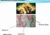
What do these images show?

Hemangioblastoma
Hemangioblastomas are often cystic and located in posterior fossa. The microscopic features include stroma cells that contain lipid (shown on oil red O stain). It is highly vascular. This tumor can secrete erythropoietin.
