Block 1 Flashcards


Sperm maturation arrest; do not see any 2’ spermatocytes or spermatids towards the center of the seminiferous tubule.


Cut surface of normal testes

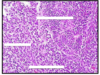
Testicular torsion: twisting of the spermatic cord, obstruction of thin-walled veins leads to hemorrhagic infarction; usually due to congenital failure of testes to attach to inner lining of scrotum (within processus vaginalis)

Hemorrhagic necrosis seen in testicular torsion

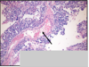
Cryptorchidism: undescended testes; fail to descend into scrotal sac;
complications: testicular atrophy, infertility, and increased risk for seminoma (CA).

Cryptorchidism: see testicular atrophy (yellow arrow)
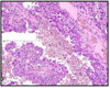
Bottom L: normal seminiferous tubule full of developing spermatogonia
Bottom R: cryptorchid seminiferous tubule; see no spermatogonia/spermatids –> infertility
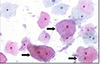
Seminoma:
Most common type of GCT
Large uniform “clear cell” tumor cells (red arrows)
Lymphocytic infiltration (green arrow)
Fibrous septa (yellow arrow)
Do NOT have hemorrhage or necrosis

Spermatocytic seminoma: rare, seen in older pts (54+), doesn’t arise from intratubular germ cell neoplasia
3 cell types:
1) small lymphocyte-like cells: yellow arrow
2) intermediate cells: red arrow
3) giant cells w/ 1+ nuclei: green arrow
Excellent prognosis; not related to cryptorchidism, serum tumor markers not elevated, usuall bilateral

Embryonal carcinoma:
gross path: hemorrhagic, necrotic, poorly circumscribed
histo: large highly pleomorphic cells; lots of pink cytoplasm; overlapping/indistince cell membranes
Poorest prognosis of all GCT’s; see elevated beta-HCG or AFP

Embryonal carcinoma w/ papillary growth; large pleomorphic cells, indistince cell membrane, lots of overlap; hemorrhagic
poorest prognosis

Yolk sac tumor; most common testicular tumor in kids/infants; see microcystic pattern on histo with multiple intercellular holes (“sieve-like” pattern)
tumors secrete AFP, so see elevated serum levels

Yolk sac tumor: relatively uniform cells with clearish pink/vacuolated cytoplasm;
see Shiller-Duval bodies: (yellow arrow) central BV surrounded by tumor cells; looks like primitive glomeruli
Hyaline-like globules: (black arrows) contains AFP and alpha1-antitrypsin

Mature teratoma: see cartilage (red “A”), ducts/glands (yellow arrow), and hair follicles (black arrows)
Made of 1+ tissues from different germinal layers
2 age peaks: <4 y.o and 20’s-40’s

Immature teratoma: undifferentiated spindle cells, primitive small round blue cells; poorly differentiate, poorer prognosis.
pre-pubertal teratoma in males is BENIGN, post-pubertal teratomas in males are MALIGNANT

Choriocarcinoma: tumor of syncitiotrophoblasts and cytotrophoblasts; grossly appears as hemorrhagic tumor; on histo see areas of hemorrhage
rarely pure tumor, usually seen in mixed GCT.
Marked elevation in beta hCG

Choriocarcinoma:
A) syncitiotrophoblasts: large multinucleated cells with pink cytoplasm
B) cytotrophoblasts: polygonal cells with clear cytoplasm, bland nucleus, well define border
C) beta-HCG + stain of choriocarcinoma

Mixed GCT: most common after seminoma; prognosis based on worst component (i.e. embryonal)

Left: hypospadias—urethral opening on ventral surface of penis; 1/300 live births
Right: Epispadias: abnormal urethral opening on dorsal aspect of shaft; even rarer

Peyronie’s dz: localized fibromatosis of penile shaft resulting in painful erections

Penile Infections:
Left: HSV
Right: Syphillis chancre

Condyloma accuminata (genital warts); due to HPV 6 & 11

Penile carcinoma = SCC; related to HPV infection (serotypes 16 and 18); circumcision is protective; uncommom in USA, more common in Africa & Asia
Tx: surgical removal with adjuvant RT to groin lymph nodes for more advanced lesions

SCC of the penis; well differentiated SCC’s make lots of keratin (PINK); poorly differentiated SCC’s do not; keratin pearls

Normal prostate; smooth, walnut-sized, 20-25 mL

Prostate normal histology:
Glands: basal cells (red arrow), luminal/secretory cells (yellow)
Stroma: smooth muscle (blue “x”)

BPH: see hyperplasia of transition zone and periurethral zone; can lead to bladder outlet obstruction (BOO)

BPH nodule: see increase in glands and stroma, but all normal appearing

BPH: see hyperplasia of prostate tissue, but otherwise normal appearing

High Grade Prostatic Intraepithelial Neoplasia (HGPIN): several architectural forms: flat (top), tufting (bottom L), regular (bottom R)
histology: luminal cell crowding, hyperchromasia, clumping, and prominent nucleoli
HGPIN on a bx means 20-25% risk of carcinoma on subsequent bx’s (should re-check in 6 month)

high grade basal cells, seen in patchy distribution

1 CA in men, #2 killer of men w/ CA
Prostate Adenocarcinoma (CaP)
Heterogenous and multifocal appearance

Prostate adenocarcinoma: multifocal and heterogenous

Prostate Adenocarcinoma: benign and tumor cells share same compartment


Prostate Cancer:
haphazard architecture w/ small invasive glands
Loss of basal cells
hyperchromatic, enlarged nuclei
prominent nucleoli
“blue intraluminal mucin”
intraluminal crystalloids
perineural invasion

Primordial Follicle; oocyte surrounded by single layer of granulosa cells; arrested in 1st prophase of meiosis for up to 50 yrs, and recruited to develop after puberty

Primary unilaminar follicle; oocyte in prophase I and secretes glycoproteins to made zona pellucida; follicular cells a monolayer of cuboidal cells with FSH receptors
Are gonadotropin-independent, and are stimulated to develop from primordial follicle by paracrine factors

Primary multilaminar follicle; oocyte still in prophase I; stratified layer of granulosa cells surround and have FSH receptors; oocyte and granulosa cells connected by gap jxns; stroma cells form theca layer

Antral Secondary Follicle; atrum = fluid collection amidst granulosa cells; LH stimulates androgen production by theca cells; FSH stimulates granulosa cells to growth, and synthesize E, Inhibin, IGF-1, and activin.
Cohort of antral follicles will grow (in response to gonadotropins) and 1 will be selected for ovulation as dominant follicle.

Mature Graafian Follicle; dominant follicle that continues to grow; oocyte surrounded by GC’s and suspended in fluid = cumulus oophorus
Oocyte still in prophase I but primed to continue meiosis
Big increase in E due to FSH and follicular factors
Vascularization of theca layer

Corpus Luteum; remnant of dominant follicle s/p ovulation; LH creates and maintains CL.
GC’s luteinize (fill with fat) and produce Progesterone, E, and Inhibin A
Decrease in FSH halts further follicular development

Ovary with fully developed CL; note yellow color of CL due to lipid

Corpus albicans; the remnant of a regressing CL (luteolysis)
See drop in P (endometrium shed) and drop in Inhibin (and secondary increase in FSH and follicular development).

Menstrual Phase of uterine cycle, D1-D5; E & P fall –> spiral arterioles constrict –> fxn’l layer of endometrium becomes ischemic –> hemorrhage and necrosis forming menses

Proliferative Phase of Uterine Cycle; D6-D14; E levels increase stimulating growth/activity of myometrium; see regeneration glands, stroma, and spiral arterioles in basal endometrium
cells possess ER, and estradiol stimulates synthesis of PR

Secretory Phase of Uterine Cycle; D15-D28; very vascular and lots of nutrient rich fluid secreted;
A) endometrial glands: coiled with squiggly sides; lined by pale epithelial cells secreting fluid rich in glycoproteins, sugars, and aa’s.
B) myometrium: activity suppressed by high P
C) spiral arterioles: increase in #
D) endometrial stroma: thick and edematous

Normal breast: terminal duct/lobular unit (TDLU)

Fibroadenoma: well-defined palpable lump w/ smooth borders in young women (<35 y.o); most common benign breast tumor
path: circumscribed stromal/epithelial tumor (fibroepithelial), with bluish-colored stroma and slit-like ductal areas
NO increased risk of CA

Fibrocystic changes: hormone-mediated; seel dilated cysts lined with metaplastic apocrine cells; grossly breasts are lumpy-bumpy;
No increased risk of CA in non-proliferative fibrocystic changes: apocrine metaplasia, fibrotic stroma, cysts
Slight increased risk of CA if proliferative: epithelial hyperplasia, sclerosing adenosis, radial scar

Lactational change/adenoma: see foamy bubbly cytoplasm secreting lipid/protein. Adenomas are well circumscribed, palpable, mobile masses with benign epithelial elements. No increased risk of CA

Gynecomastia: proliferation of stroma and ducts (also hyperplasia), but no lobules/acini

Breast Implant Capsule: see silicon/saline implant in center, surrounded by a fibrous pseudocapsule; also see macrophages/histiocytes with ingested silicon

Fat necrosis: seen with trauma, s/p surgery; presence of anuclear fat cells, with inflammatory infiltrates; can calcify and scar and be mistaken for CA, so have to look at closely.

Proliferative Fibrocystic Change: Usual Ductal Hyperplasia (UDH); benign, low risk of CA;
see ducts full of proliferating cells that are hyperplastic but otherwise normal appearing (not malignant)

Papilloma: fibrovascular stalk; can see bloody discharge from infarction; slight cancer risk 1.5-2x; grossly see bloody nipple discharge, usually non-palpable

Radial Scar: see radiating architecture that can mimic CA (radiographically and pathologically); really hard to dx on bx and usually require special stains

Ductal Carcinoma In Situ (DCIS):
green arrow: normal cells
yellow arrow: tumor cells
SEe expanded ducts filled with cells with larger nuclei/nucleoli, mitotic figures, but contained still within the basement membrane

DCIS: yellow arrow pointing to a comedonecrosis (area of high grade necrosis, usually calcifies); all DCIS tumors are bound by cell membrane

Infiltrating Ductal Carcinoma:
yellow arrows: DCIS w/ calcification
green arrows: invading ductal carcinoma; see jagged/stringy appearance of stroma due to malignancy
bottom R photo: FNAB stain (doesn’t differentiate in-situ from invasive)

Infiltrating ductal carcinoma; see malignant epithelial infiltrates amongst fat cells and stroma;
desmoplastic stroma (yellow arrow)

LCIS ( green arrow); contained within basement membrane
Infiltrating Lobular Carcinoma (red arrows): see single-file lines of cells; can see loss of Ecad on IHC, which prevents cells from forming round globules.

Left: Mucinous Carcinoma
Right: Tubular Carcinoma
both have “Good” prognosis
breast

Medullary carcinoma: high grade/invasive, but good prognosis
well circumscribed on imaging
breast

Micropapillary carcinoma: invasive, worse prognosis due to propensity for lymph nodes
breast

Invasive Ductal Carcinoma
lymph node

Lymph Nodes:
Left: metastatic tumor (yellow arrow) and lymphocytes (white arrow)
Right: Keratin IHC helps detect really focal tumors
breast

Paget’s Dz: epidermal adenocarcinoma (skin involvement), most often associated with in-situ or invasive ductal carcinoma in underlying breast (seen in 2% of mammary CA)
Clinically presents as a rash or erosion of nipple
Histology: glandular tumor cells with pale fluffy cytoplasm, amongst epithelial cells; Her-2/neu + staining in Upper R image.
breast

Inflammatory Carcinoma: tumor invades/obstructs dermal lymphatics leading to redness/warmth/edema of breast, peau d’orange appearance; very poor prognosis
breast

Angiosarcoma: rare complication of radiation (+/-mastectomy); malignant tumor of vascular tissue;
Stewart-Treves = angiosarcoma in skin of lymphedematous area poast-mastectomy/axillary dissection
May present as bruising on the breast

Cervical transformation zone: transition from stratified squamous epithelium (of vagina) to transitional epithelium (uterus)
What you evaluate on a pap smear

Pap smear:
squamous cells and endocervical glandular cells
Red cell are mature
Blue cells are immature
Dark, fluffy cells (arrow) are from the T-zone
Compare nucleus:cytoplasm ratio

Clue cells; squamous cells covered by gardnerella organism, have dusty appearance; sign of BV

Fungal forms seen on pap smear; often seen in pregnant women

Trichomonas seen on pap smear; tiny blue dots with “halos” can see swimming around on slide.

HSV seen on pap smear; most genital HSV is HSV 2; on path see multinucleation and marginating chromatin

Cervical Bx/Pap smear
1) Normal/negative
2) ASCUS
3) LSIL
4) HSIL

Dysplasia of cervix at squamocolumnar jxn
cervical bx

Moderate cervical dysplasia: CIN2

Adenocarcinoma In Situ (glandular lesion) of endocervical glands; not invasive; related to HPV; bottom slides are stained for p16 (tumor marker).
vulva

VIN 2+ (vulvar intraepithelial neoplasia); leads to surgical excision of the carcinoma in situ;
Left= H&E slide, can’t tell if just atrophy vs. HGSIL
Right= stained for p16 a tumor marker–>VIN
clinical sxs may be white, itchy vulva
vulva

Lichen Sclerois; autoimmune condition targeting the vulva; by this H&E image unable to distinguish LS vs. SCC.
vulva

Extra-mammary Paget’s Dz; see adenocarcinoma in-situ from glandular cells beneath the epithelium. NOT melanoma.
Grossly see pigmented spots on vulva.
vulva

Melanoma of vulva; looks similar to Paget’s dz but has melanin.
vulva

Vulvar cancer; depth of invasion is really important; only 1 mm invasion neede to metastasize to broad ligament, pelvic lymph nodes and peri-aortic nodes.
endometrium

Secretory endometrium: normal part of endometrium cycle; see pink, bubbly glandular cells; may mimic a polyp on US
endometrial mass

Endometrial polyp: benign growths that enlarge with estrogen stimulation; histologically are mixed dilated and small, with fibrous stroma, thick-walled BV’s, and simply hyperplasia
Section of endometrium

Chronic endometritis; see characteristic plasma cells on histopath; associated with retained placenta, IUD, or gonorrhea infection
Endocervical mass

Benign polyp: hypocellular stroma
Uterine mass

Leiomyoma (fibroid):
A) normal uterine tissue
B) fibroid: well circumbscribed, well defined border, tissue is disorganized, but no angiolymph invasion—>BENIGN

Ovarian tumors:
Left= borderline tumor; little trees growing in the cyst
Right= malignant; solid, variably colored tumor
Ovarian tumor

papillary serous cystadenoma; finger-like projections lined by single layer of uniform ciliated epithelial cells
mostly benign
ovarian tumor

Ovarian mucinous tumor; intestinal type; goblet cells, most common, risk of jelly belly
ovarian tumor

Ovarian mullerian mucinous tumor with endocervical-like mucosa; usually no goblet cells, no risk of mets or jelly belly
Ovarian tumor

Endometrioid adenocarcinoma; invasive
Ovarian tumor

Clear cell carcinoma; looks a lot like yolk sac tumor as well as CCC of kidney
Most often malignant
Ovarian tumor

Brenner tumor; resembles renal pelvic tumor (bladder urothelium); almost always benign
ovarian tumor

Ovarian teratoma
Ovarian tumor

Top L: matura teratoma w/ struma ovarri (thyroid tissue)
Bottom R: immature teratoma; see rosettes on path, can be low grade or high, but has metastatic potential
Ovarian tumor

Yolk Sac (Endodermal sinus) tumor; a germ cell tumor characterized by elevated serum AFP (staining + for AFP in magnified view), as well as Schiller Duval bodies; excellent prognosis w/ chemo
ovarian tumor

Yolk sac tumor; with Schiller Duval body (papillary structure w/ fibrovascular core, lined by tumor cells with clear cytoplasm and dark malignant-appearing nuclei); looks a lot like a clear cell carcinoma;
See elevated AFP (serum) and in younger pts (how to differeniate from CCC)
Ovarian tumor
Dysgerminoma; the female equiv. of a seminoma; on histo see “fried eggs and lymphocytes”
ovarian tumor

Granulosa Cell Tumor (adult); see sheets of small tumor cells wiht grooved/coffee-bean nuclei; also see Call-Exner bodies (glandular-like structures with hyalin in middle); usually seen in postmenopausal women with PMB (estrogen producing tumor)
ovarian tumor

Granulosa cell tumor; see sheets of uniform tumor celsl with coffee-bean/grooved nuclei; all GCT’s stain positive for Inhibin; usually seen in older women with PMB.
Ovarian tumor

Sertoli-Leydig cell tumor; usually occurs in younger women (20-40) with assoc. androgen effects (i.e. virilization)
histo: tubule-like glands lined by sertoli cells that stain for inhibin
Ovarian tumor

Stromal tumors: fibroma, leiomya, thecoma==all are benign
ovarian tumor

Fibroma, Leiomya=solid white tumor of ovary; bening
Meig’s syndrome is a solid white ovarian mass with pleural effusion, maybe ascites as well as elevated CA-125; looks malignant at first, but once you get histo/cytopath see it’s benign.