Anti-arrhythmics - Elam Flashcards
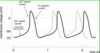
Which drugs cause torsade? What is it?

- Torsade: polymorphic vtach initiated by early after-depolarization (EAD) in setting of prolonged QT (i.e., prolonged phase 2 and 3 repolarization of AP)
- Seen with (drugs with class III activity):
1. K+ channel blocking antiarrhythmics (class III); rarely with Amiodarone
2. Tricyclic anti-depressants (Imiprimine)
3. Na+ channel blockers (Procaine, Quin)
4. Non-sedating anti-histamines (Terfenidine withdrawn from market) - Exacerbated by hypokalemia, low HR
- Initiated by a triggered VPC from an EAD and leading to re-entry
Describe the normal automaticity of the pacemaker cells.
- SA, atrial, AV, ventricular conducting fibers spontaneously depolarize (pacemaker activity)
- Pacemaker currents mediated by activation of inward Na+/K+ pacemaker (hyperpolarization or funny) channel
- SA, AV node: depolarization results in opening of inward Ca channels (L-type)
- Atrial, ventricular fibers depolarization mediated by inward Na channel
Flecainide (Class IC)
- Action: long-acting Na+ blocker (>10s); suppresses automaticity, INC ERP in atria, ventricles
-
Use: SV arrhythmias (afib, aflutter, SVT) and NO structural heart disease
1. Contraindicated in pts w/structural heart disease (pro-arrhythmic in CHD, LV dysfunction)
2. May be used in young people to treat congenital causes of afib or flutter (after trying everything else)
Dronedarone
- Non-iodinated congener of Amiodarone approved for tx of paroxysmal atrial fibrillation or maintenance of sinus rhythm following cardioversion
- Contraindicated in patients with decompensated CHF
How do class I antiarrhythmics suppress arrhythmias?

- Suppress ectopic pacemakers and interrupt re-entry by blocking Na+ channels
- Phase 0 depolarization slowed -> slowed conduction and suppressed ectopic pacemakers
- Reduced availability of Na+ channels INC mem voltage required for new depolarization -> INC refractory period
- Depolarization sodium-dependent in atrial/ventricular myocytes
Vtach long-term tx. EF 30% 4 months after tx, so high-risk for sudden cardiac death due to vtach or vfib.
Treatment?

- Implantable defibrillator (no dosing concerns)
- Could give Amiodarone or Sotalol if defibrillator was not an option for some reason
Dofetilide/Ibutilide
- Pure class III (no activity from other classes) anti-arrhythmics primarily used to tx atrial arrhythmias
-
Dofetilide: used to interrupt re-entry in the atrium
1. Major AE: polymorphic vtach (torsade)
2. Contraindication: pts with long QT interval -
Ibutilide: IV only for acute termination of atrial flutter or fibrillation
1. Torsade de pointes in 3-8% of pts
Digoxin
- Antiarrhythmic effects mediated via INC vagal N activity:
1. Reduces SA node automaticity, AV conduction
2. Controls ventricular rate in SV arrhythmias
3. Interrupts re-entry in AV node - Direct effect (arrhythmogenic): INC normal automaticity, delayed afterdepolarizations (DAD; APC, VPC), paroxysmal atrial tachycardia with block, vtach (Na/K ATPase inhibition with Ca overload)
Beta-blockers (Class II)
- Block/slow AV node, slowing HR in SV arrhythmias
1. Metoprolol, Esmolol - Suppress ventricular premature beats (VPB’s) related to SYM activity
1. Propranolol, Acebutolol - Rate control in atrial flutter/fibrillation; prevents recurrence of PSVT
1. Atenolol - Remember: beta-adrenergics INC Ca in cytosol and SR (via upregulation of cAMP)
- NOTE: can tx SYM overactivity in mitral prolapse
Amiodarone (Class III)
- Action: K+ channel blockade; secondary Na+, Ca+ channel blockade and B-adrenergic blocking
- Use: atrial, ventricular arrhythmias (esp. life-threatening, i.e., vtac, vfib, cardiac resuscitation)
- Side effects: pulm fibrosis (worst), photosensitivity derm, corneal microdeposits, hypo/hyperthyroidism, prox muscle weakness, hepatitis (monitor LFT’s)
- Metabolism/prep: IV/oral; slow elim b/c accumulates in fat tissue (CYP3A4)
What is Wolff Parkinson White syndrome?

- Congenital direct connection b/t atrium and ventricle, called accessory pathways or bypass tracts
1. Allow impulses from the atria to be conducted directly to ventricle as well as through AVN - Asymetric activation of ventricle via this bypass tract reflected on ECG by short PR interval and delta wave
- Bypass tract allows for rapid conduction of impulses to ventricle during atrial arrhythmias, which can induce vfib; also allows reentry b/t bypass tract and AVN, leading to rapid SV tachys
- Definitive tx for pts w/arrhythmias in WPW is to interrupt circuit by radiofrequency catheter ablation of bypass tract
What are the mechanisms of arrhythmias?
- Abnormal impulse generation (automaticity) or abnormal impulse propagation (re-entry)
- Specific mechanisms: (1) enhanced normal automaticity, (2) enhanced abnormal automaticity, (3) triggered automaticity, (4) re-entry (organized or disorganized)
- Aim of therapy of arrhythmias is to reduce ectopic pacemaker activity and modify conduction or refractoriness in re-entry circuits to prevent re-entry
Vtach in pt with coronary disease. 57-y/o WM with chest pain and palpitations. Cardiac enzymes elevated and ECG confirms acute non-ST MI. Admitted to CCU, where telemetry shows runs of monomorphic vtach.
Mechanism and treatment?

- Organized reentry via premature ventricular beat (monomorphic vtach, unlike torsade)
- Amiodarone, Lidocaine, Procaineamide
- NOT Fleconide
How is automaticity influenced by the ANS?

- SA/AV nodes innervated by ANS (no vagal innervation below AV node)
- Rate of phase IV depolarization:
1. INC by SYM stimulation (NE)
2. DEC by vagal stimulation (Ach);also hyperpolarizes SA/AV node cells
3. DEC by action of adenosine receptors - Primary effect of these influences on AV node is to modify conduction of impulses
- Ca channel blockers reduce rate of firing of SA/AV node conduction by reducing depolarization due to Ca influx
Paroxysmal supraventricular tachycardia. Narrow QRS, regular activation, no identifiable atrial activity (p wave) preceding QRS.
Mechanism and treatment?

- P wave buried, but would be inverted if seen; AV node bc narrow QRS
- Adenosine, Verapamil, Esmolol, Digoxin
Class IV antiarrhythmics
- Non-dihydropyridine Ca2+ channel blockers: Verapamil, Diltiazem
- Reduce SA node automaticity and AV conduction
- AE’s: SA, AV block, impaired myocardial contractility, hypotension
- Contraindicated in pts with CHF, sinus brady, or AV block (prolonged PR interval)
- Little effect on electrophysiology of fast conduction tissues (atria, ventricles) under normal circumstances
- Dihydropyridine Ca2+ blockers have little antiarrhythmic effect at clinically used doses
What are arrhythmias due to abnormal “triggered” automaticity?

- Abnormal impulse generation after normal action potential
- Early afterdepolarization (EAD) interrupts phase 3 repolarization -> trigger long QT-related arrhythmias, i.e., torsade
- Delayed afterdepolarizations (DAD) interrupt phase 4 repolarization, and occur as a result of calcium overload in digitalis toxicity
What are the characteristics of re-entrant arrhythmias? How do antiarrhythmic drugs interrupt this?
- Characteristically rapid (>140bpm) and sustained
- Can occur at any location in the heart:
1. Atrium: flutter, fibrillation
2. AV node: PSVT
3. Ventricle: vtach, vfib - Antiarrhythmic drugs interrupt re-entry by changing the properties of the re-entry circuit: 1) conduction velocity, 2) refractory period, 3) covert unidirectional block to complete block
What is vtach? How is it treated?

- Arrhythmia that results from organized re-entry of impulses, within the ventricle
- For sustained vtach, tx of choice is cardioversion, then drugs to prevent recurrence of re-entry
- Class I (procainamide or lidocaine) would interrupt re-entry by slowing conduction through the circuit and increasing refractory period
- Class III (sotolol or amiodarone) would do so by prolonging repolarization and increasing ERP
Class II anti-arrhythmics
- Beta-adrenoreceptor blockers (Proprano, Metopro)
- Electrophysio effects: reduce enhanced automaticity related to catecholamines and ischemia
- Reduce atrial, ventricular arrhythmias in pts with coronary heart disease, and improve survival
- Used to tx symptomatic premature ventricular contractions in pts w/ and w/o structural heart disease
- Slow AV node conduction by blocking + influence of catecholamines, esp. exercise-induced increase in ventricular rate in atrial fibrillation
- Mitral valve prolapse tx
How do Na+ channel blockers interrupt re-entry?

- Bind Na+ channel during open and inactivated states
- Decreased rate of rise in phase 0 depolarization results in slower conduction of impulses through re-entrant circuits
- Delayed voltage-dependent recovery of Na+ channels increases ERP
- Both effects result in interruption of re-entry (circus movement)
21-y/o med student with palpitations and tachy. Denies syncope or family hx of sudden cardiac death. Frequent premature beats noted on cardiac auscultation, but exam is otherwise normal. ECG shows frequent premature atrial beats (APB’s).
What is mechanism, and best treatment?

- Enhanced automaticity
- Caffeine a common cause (b/c it is a PDE inhibitor)
- Inverted p wave, so not coming from SA/AV: Sotalol (inc risk of torsade) or Flecanide would fix this, but consider beverage choices first
1. If symptoms persisted, could treat with BB
Describe the electrophysiology of the normal cardiac rhythm.

- ECG is the body surface manifestation of depolarization and repolarization waves of the heart
- P wave generated by atiral depolarization
- AV node depolarization electrically silent; manifested as delay in conduction through AV node (PR interval)
- QRS generated by ventricular muscle depolarization
- T wave reflects ventricular repolarization
Know this.

Voltage-gated Na+ channels mediate Phase 0 depolarization in atrial/ventricular myocytes.


