Anatomy of the Thorax Flashcards

1
Q

How do you call the inferior and superior ends of the thorax?

A

Superior thoracic aperture (open, allows continuity with neck)

Inferior thoracic aperture (closed by diaphragm)

2
Q

Which three structures are involved in breathing movement?

A

Diaphragm

Thoracic wall (muscles)

Ribs (Bucket handle, pump mechanism)

3
Q

How many times does a rib articulate with vertebral collum?

A

Most (II-IX) 3 times: (three facets)

Own vertebra

Vertebra above

transverse process of own vertebra

4
Q

How many ribs are there and how are they classified?

A

Ture ribs 1-7

False Ribs (8-10)

Free /Floating Ribs (11+12)

–> Total: 12

5
Q

What are the borders of the superior thoracic aperture?

Which major structures pass through it?

A

Posterior: T1 vertebra

Superior Rib 1

Anteror: Superior border of manubrium

6
Q
A
7
Q

How do you call the joint between the transverse process of a vertebra and its related rib? How is the other joint (between the vertebral body) and rib called?

A
  1. Costotransverse joint
  2. Costovertebra joint
8
Q

How is a costotransverse joint stabilised?

A

By three ligaments:

Costotransverse ligament (medial to joint)

lateral costotransverse ligament (lateral to joint)

Superior costotransverse ligament (superior surface of the rib to the transverse process above)

9
Q

How are the connections between Sternum and ribs I-VII called?

What is different between joint Rib I and others?

A

Sternocostal joints

I: Not synovial but fibrocartilaginous connection

II - VII: Synovial

10
Q

How are the connections between costal cartilages of adjacent ribs called?

A

Interchondral joints

11
Q

Synovial joint

A

Glenk mit gelenkflüssigkeit und Kapsel

12
Q

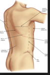
What is the endothoracic fascia?

A

Connective tissue posterior to ribs+ intercostal spaces

Sepparates structures (muscles, ribs, spaces etc.) from pleura

13
Q

Where and in what order are intercostal vein, artery and nerve located?

A

Are located in the costal groove (comma-shaped ribs)

Colateral bonds often also superior to inferior rib below

Superior to inferior:

Vein

Artery

Nerve (often not protected by groove anymore)

14
Q

What are the three layers of intercostal muscles?

How are they innervated?

A
  1. External intercostal muscles
  2. Internal intercostal muscles
  3. Inntermost intercostal muscles

Innervated by Intercostal nerves T1-T11

15
Q

External intercostal muscles (orientation, function)

A

Inferior margin of the rib above to superior margin of rib below

Orientation: Inferior medially

Inspiration, superior movement of ribs

16
Q

Internal intercostal muscles (orientation, function)

A

Lateral edge of costal grove rib above to sperior margin of tib below

Orientation: Inferior laterally

Function: Expiration, inferior movement of ribs

17
Q

Innermost intercostal muscles

A

Medial edge of superior costal grove to internal superior margin of rib below

Orientation (like internal) Inferiorlaterally

Function: Supports internal

18
Q

What are the borders of the safe tiangle?

A

Superior border of rib

4th or 5th intercostal space, between anterior axillary and mid-axillary line (pec and latissimus dorsi)

19
Q

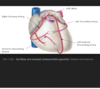
Which structures pass the diaphragm at which levels?

A

VIII: Inferior vena cava

X: Oesophagus

XII: Aorta

20
Q

Which nerves innervate the diaphragm?

A

Innervated by phrenic nerve (C3,4,5)

21
Q

What are the medial, superior and inferior borders of the pleural cavities?

A

Superior: above rib 1 into the root of neck

inferior: just above the costal margin
medial: mediastinum

22
Q

How are the two sides of the pleura called?

A

Visceral pleura: attached to the lung surface

Parietal pleura: wall of pleural cavity

23
Q

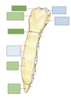
What shows the purple and blue colour?

A

Purple: marking of the lungs (visceral pleura)

Blue: Pleural cavity( parietal pleura)

–> a lot of space to extend

24
Q

What happens to the pleura at the hilum of the lung?

A

It deflects and visceral + parietal pleura are continous

25
Blue= Structrues Green= Borders Orange= Surface
26
What is a bronchiopulmonary segment?
A segment of the lung which is supplied by one tertiary bronchus (10 per lung)
27
How is the hilum of the lung organised? What does it contain?
28
How do the vagus and phrenic nerves pass the hilum?
Phrenic= anteriorly Vagus= posteriorly
29
How are the main structures (vein, artery, bronchus) in the hilum generally organised?
Artery= superior Vein= inferior Broncus= posterior
30
Right Lung
31
What does the oblique fissure separates and what are its surface markings?
Spinous process of vertebra TIV, the lateral crossing of 5th intercostal space laterally, follow rib VI anteriorly Separates inferior from superior lobe (in both lungs)
32
What does the horizontal fissure separate and what are it's surface marks?
Separates superior from middle lobe in the right lung Fourth intercostal space from sternum until it meets oblique fissure as crossing rib V
33
How do you call the projection of the left lung over the heart?
Lingula of left lung
34
How is the lowest tracheal ring called (when trachea bifurcates)? Why is it important?
Carina (hook shape) --\> can be disturbed by swelling of lymph nodes nearby
35
Trachea borders + characteristics
CVI in the lower neck to TIV/TV flexible tubes with c-shaped cartilage rings
36
How are the bronchi after the first 3 divisions called?
1st division: Main bronchi (right (steeper)+ left 2nd division: lobular bronchi, each supplies one lobe 3rd division: segmental bronchi: each supplies one bronchopulmonary segment
37
What are the contensts of the superior mediastinumg? (from anterior to posterior)
From anterior to posterior: * •Thymus * •Phrenic Nerves * •Great veins * •Main lymphatic trunks * •Vagusnerves * •Great arteries * •Trachea and main bronchi * •Upper oesophagus
38
In which segments can you divide the mediastinum?
Superior Inferior: * Anterior * Middle * Posterior
39
Where do you divide the mediastinum into the superior and inferior mediastinum?
At the level of the sternal angle
40
Explain the route of the superior Vena Cava into the heart
**Formed by** (asymmetric) **Brachiocephalic** veins + recieves blood from **azygous vein** + **pericardial/mediastinal veins** It begins **posterior to the lower edge of the right first costal cartilage** (junction of two barchiocephalic veins)
41
Wich vessels joint to give rise to the right and left brachiocephalic veins?
It is formed by **Subclavian and internal jugular veins**
42
Explain the routes and fusion of the right and left brachiocephalic veins
Right: Begins posterior to the medial end of the right clavicle, passes vertically downward Left: Begins posterior to medial end of the left clavicle, passes over behind manubrium and joins RBCV at lower border of 1st costarl cartilage
43
What vessel is the posterior wall of the thorax and abdomen drained by? Explain, how an which bigger vessel it joins
The Azygous vein: * it arches over right lung root to drain directly into the SVC
44
Which the vessel branch of the aortic arch? In which order? Which surface structure relate to them?
1. Brachiocephalic trunk (devides into right common carotid and right subclavian, largest of the three), behind manubrium of sternum 2. Left common carotid artery 3. Left subclavian
45
What is the ligamentum arteriosum? What is its relevance?
It is the ductus arteriosus in fetal circulation --\> connects pulmonary trunk and arch of the aorta to allow bypass of pulmonary circulation (closed in adults)
46
Explain the relations of the pumonary trunk, the aorta, the bronchi and the Superior vena Cava in the superior mediastinum
From Anterior to posterior: SVC ascending aorta right pulmonary artery Bronchus Aortic arch
47
Explain the relationship of the aortic arch to the trachea
Trachea lies behind & between brachiocephalicand left common carotid arteries Aorta branches over left main bronchus
48
Where does the phrenic nerve originate from?
In cervical plexus: C3,4,5 --\> (also innervates diaphragm)
49
Which nerve supplies sensation of the ## Footnote –central tendon of the diaphragm –mediastinalpleura –pericardium –peritoneum of central diaphragm
The phrenic nerve
50
Explain the route of the right phrenic nerve in relation to the brachiocephalic vein, SVC and the heart
•Right phrenic nerve reaches diaphragm lying on surface of: –rightbrachiocephalicvein –superior vena cava –right side of heart and pericardium - in front of lung root
51
Explain the route of the left phrenic nerve in relation to the aorta, hilum of lung and pulmonary trunk
1. Cross arch of the aorta 2. Passes anteriorly to hilum of lung 3. Passes over pulmonary trunk and over the heart
52
Explain the route of the right and left vagus nerve in relation to the great vessels and the heart
Passes lateral to common carotid arteries --\> both break up into branches of the oesophagus **Left vagus:** * passes anterior of the aortic arch, * crosses behind root lung gives off left recurrent laryngeal nerve – recurs (=turns back) around ligamentum arteriosumn and aortic arch **Right vagus:** * lies on trachea * crosses behing the root of lung * •Recurrent laryngeal branch – recurs (turns back) around right subclavian artery
53
What is the transverse pericardial sinus? What does it surround?
It is a reflection of the pericardial sack (at posterior wall of pericardial sack), surroundingt the arteries: aorta and pulmonary trunk (superiorly)
54
What is the oblique pericardial sinus? What does it contain?
It is a reflection of the pericardial sac surrounding the veins (Superior, Inferior Vena cava, pulmonary veins), posterior to the left atrium
55
What is the coronary sulcus?
It circles the heart and separates the atria from the ventricle It contains the right coronary artery, coronary sinus and circumflex branch of left coronary artery
56
What are the anterior and posterior interventricular sulci?
They separate the two ventricles (one on anterior side, one on posterior side)
57
Where does the coronary sinus returns blood to the heart?
Via the lower, posterior part in the right atrium
58
What is the right atrioventricular orifice
It is the opening of the heart between the right atrium and ventricle --\> closed by the tricuspid valve
59
What is the crista terminals?
It is a smooth muscular ridge beginning on the roof of the right atrium (anterior to opening of SVC), extends down the lateral wall to anterior part of inferior vena cava
60
What is the right/left auricle?
Part of the atrium extending over chamers of heart
61
How are the ridges called that cover the inner surface of the right atrium?
They are called Pectinate muscles, originating from the Crista Terminalis
62
Explain the structure and function fo the papillary muscles in the right atrium
These are small muscles, attached to **choradae** **tendinaea,** (tendons) which are attached to the free edge of the tricuspid valve
63
Explain the structure of the tricuspid valve : What is the commissures, how are the individual cusps called
Anterior, septal and posterior cusp --\> Based on their position Are continuous as their base (commissures)
64
What necrosis of a papillary muscle lead to? Why?
It can might lead to prolapse of the related valve --\> can't be hold in place anymore
65
Explain the structure of the pulmonary valve How does its sturcture prevent it from opeining after ventricular contraction
Three semilunar cusps --\> Left, right and anterior semilunar cusp When closed, the pulmonary sinused fill with blood --\> prevent it from opening again
66
How do the chordae tendineae and the tubercular carneae in the left ventricle differ from the ones in the right ventricle?
There are only two pairs of chordae tendinaea and papillary muscles --\> Mitral valve = two cusps Tubercular carneae are a little less prominent
67
In which cusps of the aortic valves are openings for which coronary vessels?
Right Cusp: Right coronary artery Left= Left coronary artery Noone in the posterior aortic sinus
68
What are the three most important coronary arteries?
The right coronary artery Left coronary atery * Anterior interventricular branch (Left anterior decending) * Left circumfles
69
What does right dominance in the context of the heart mean?
Posterior intraventricular branched arise from the right coronary artery --\> Right coronary artery supplied more of the apex of the heart (more common)
70
How many intercostal nerves are there? What are teir branches?
11 Intercostal and 1 Subcostal nerves **Anterior primary rami:** * supplies the intercostal space **Lateral cutaneous branch** * Devides into posterior and anterior branch **Anterior Cutaneous branch** * Devides into lateral and medial branch
71
Which nerve/ nerves supply the Peritoneum and pericardium ?
The phrenic nerves
72
Where do the sympathetic nerves supplying the heart and Lungs emerge from? What is their route to get to the viscera?
T2 – T4, passing through cervical and upper thoracic ganglia of sympathetic trunk Many of their synapses are in micro-ganglia in the pulmonary and cardiac plexuses rather than in trunk ganglia
73
What does the pulmnary plexus consists of?
It consists of both, the sympathetic and parasympathetic nerves supplying the lungs and bronchi
74
Where do the right and left vagus nerves come, which route do they take in regards to the esophagus?
They left vagus nerves tends to coil to the front of the esophagus (anterior vagal trunk) , the right vagus nerves tends to coil to the back of the oesophagus (posterior vagal trunk)
75
Where does lymph from the thoracic wall drain into?
Lymph drains into nodes associated with: –internal thoracic arteries (parasternal) –ribs(intercostal) –diaphragm(diaphragmatic)
76
What is the thoracic duct? Where does it begin? Which route does it take? Where does it joint the systemic circulation?
Main channel of lymph draining most of the body Begins at **cisterna chyli**, begins at L2 Ascends on right of midline, crosses over at T5 to left of midline It empties into the junction of left internal jugular and left subclavian
77
Summarise the lymphatic drainage of the heart
•Follow the coronary arteries and drain into: * –Brachiocephalic * Tracheobronchial
78
Summarise the lymphatic drainage of the Lungs
•Tracheobronchial * –Around bronchi and trachea * –From within lung through hilum * –Unite with vessels from parasternal and brachiocephalic nodes anterior to brachiocephalic veins to form: **Bronchomediastinal(**left and right)
79
What kind of tissue is a mamillary gland actually?
It is a modified apocrine sweat gland
80
How is the breas supplied with blood?
Branches of the **lateral thoracic artery,** **internal thoracic artery, thoracoacromial artery, thoracodorsal artery** **and** **intercostal arteries** Nipple normally supplied by **internal thoracic**
81
Summarise the nerve supply of the breast
* Mainly - anterolateral and anteromedial branches of thoracic intercostal nerves T3-T6. There is also innervation from the supraclavicular nerves to the upper and lateral parts of the breast. * The nipple - dominant supply from the lateral cutaneous branch of T4.
82
Summarise the lymphatic drainage of the breast
* 75% is drained into axillary lnodes * Most other into thoracic artery