Abnormal placentation/bleeding in pregnancy Flashcards
Placenta accreda
(general definition)
- “Abnormal placental attachment due to absence of decidua basalis and incomplete development of fibrinoid (Nitabuch) layer”
- Fibrinoid tissue deposits (nitabuch) incomplete → abnormal/increased invasion of trophoblasts into decidua basalis → bound to uterus (doesn’t separate after birth)
- One of the most serious complications of pregnancy → risk of maternal mortality
- U/S picks up 80-90% of time with experienced sonographers
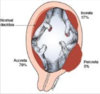
Name/describe 3 variations of placenta accreda
- Accreda vera - adherence to myometrium
- Increda - growth INTO the myometrium
- Percreta - growth through uterine wall and adheres to other structures and maybe bladder/bowel (depending on where it attaches)
- Bonus:
- Partial accreda – placenta can being to separate (aka abrupt) → see bleeding
Placenta accreda
Incidence + risk factors
- Incidence 1 in 533 birth,
- Risk factors:
- Placenta previa
- Prior c/s or myomectomy (scar on uterus)
- Also: increased parity, maternal age, submucosal fibroids, female fetus
Management/Complications of placenta accreda
- Cesarean at 34-0 to 35-6
- Risk of massive post partum hemorrhage in 2/3 patients, requiring cesarean and hysterectomy
Vasa previa
- Fetal vessels run through the membranes over the cervix unprotected by the umbilical cord or placental tissue
- Associated with velementous insertion but more rare.
- 0.04-0.02% pregnancies
- Associated low lying pregnancies, multiple gestation, resolved previa
-
Must plan cesarean before membranes rupture
- sometime between 34-6 to 36-0
Placenta previa
Definition, types
- Placenta implanted very close to or over internal Os of uterus
- Types: Complete, partial, marginal, low lying (not really a previa)

Risk factors for placenta previa
- Advanced maternal age > 35
- Multiparity
- Prior cesarean section - 0.3-0.7%, higher with 1st section and goes up with each subsequent one
- Infertility treatments
- Smoking
- Unexplained elevated alpha-fetoprotein (AFP)
- Multiple gestation
- Short interpregnancy interval
- Prior uterine curettage
Main symptom of placenta previa
- Painless vaginal bleeding
- Usually self limiting and presents in late 2nd or early 3rd
- 1st episode often stops after a few hours
- Subsequent bleeds tend to become worse (deattachment from LUS)
- Some may experience pain/cramping due to uterine irritibility
- Don’t do a digital exam on someone that has painless vag bleeding who hasn’t had an ultrasound showing no previa (ie if you don’t know if they have a previa). Speculum exam is ok
- Hard to tell if/when they might bleed again or be delivered
Placenta previa monitoring/management
- Serial US to assess placental location and fetal growth
- Avoidance of cervical examinations and intercourse
- Activity restrictions
- Counseling regarding labor symptoms and vaginal bleeding
- Dietary and nutrient supplementation to avoid maternal anemia
- Early medical attention if any vaginal bleeding occurs.
- Asymptomatic (no bleeding) → expectant mgmt as long as compliant and live close to hospital
- Bleeding previa → hospitalization/monitoring/stabilization
- < 34 wks → steroids
- Cesarean
- H/H, Ferritin if chronic
Placental abruption
- Antepartal decidual hemorrhage leading to premature separation of the placenta.
- Often caused by rupture of maternal vessels in the decidua basalis, where it comes into contact with the anchoring villi of the placenta.
- Bleeding is almost always maternal in origin.
- 1% of all pregnancies
Placental abruption risk factors
- Usually defect in maternal vessels
- HTN (chronic, gestational, PEC), interpregnancy interval < 1 year doubles risk
- PPROM
- Smoking - dose dependent relationship with # cigs smoked
- Trauma
- Cocaine
- Older maternal age
- Polyhydramnios
- Multiple gestation
- Fibroids
- Thrombophelias
Peak occurance of placental abruption
- 24-26 weeks
- Recurrence 5-17% higher with more episodes
Placental abruption presentation and differentials
- Vary markedly → can make dx difficult
- Acute vs chronic
- Overt vs concealed
- Severity
- Hard abdomen
- Colicky cramping pain that is on/off that makes her irritates, anxious, uncomfortable,
- Vaginal bleeding
- Maternal tachycardia or non-reassuring FHR
- Grades:
- 1, 2 (more vag bleeding),
- 3 (severe bleeding and maternal VS changes, risk of DIC)
Differentials: previa, cervical bleeding/infection/cancer, vag trauma
Placental abruption potential complications
- Maternal
- Significant blood loss →
- Shock
- Consumptive coagulopathy
- Renal failure
- Death
- Couvelaire uterus - blood seeping into the uterine musculature
- High recurrence rate in subsequent pregnancies.
- Significant blood loss →
- Fetal
- Decreased oxygenation →
- Cerebral compromise
- Stillbirth.
Amniotic bands
- Amniotic sacs folds and constricts areas of the fetus → amputation of fingers, limb, palate issues (if comes across face)
- Can be assiciatied with miscarriage
- Not likely to reoccur
Placental abruption management
- Ultrasound evalutation - retroplacental clotting indications abruption
- Normal U/S does not rule out abruption
- CBC, Type and screen, Coags, FIbrinogen
- IV access
- Contonuous FHR monitoring
- Rhogam if Rh neg (Kleihauer-Betke test to determine dose)
- Management depends on GA and maternal and fetal statuses
- Small abruption < 34 wks → expectant mgmt
- At/near term → delivery (induction/augmantation ok as long as closely monitored)
- Obstetrical emergency → REFER
Bleeding in the 1st half of pregnancy
Etiologies
- Non-pregnancy causes: cervical polyp, sex, sti’s, hemorrhoids, hematuria
- Subchorionic bleed/hematoma - bleeding due to seperation of chorion to uterine lining. Usually self-resolve and good outcome if occurs early in 1st tri and is small
- Fibroids - bleeding if intramural fibroid twists on its stalk or placenta implants over one. Increase risk of pregnancy loss
- Spontaneous pregnancy loss - may manage expectantly, use misoprostol, or aspiration. Rhogam for Rh negative
- Ectopic
- Gestational trophoblastic disease (hydatidiform mole) - chorionic villi don’t develop properly → not viable pregnancy → normal pregnancy process turns into benign tumor, can also be malignant
Rhogam
- CHECK BLOOD TYPE the minute of a report of 1st or 2nd trimester bleeding → need to prevent isoimmunization if mother is Rh negative
- If FOB is Rh + (D antigen) fetus can be Rh+ → any blood mixing → maternal isoimmunization → can cause hemolytic anemia in fetus/hydrops in future pregnancy → may never be able to carry Rh+ fetus again
- Rhogam – antibodies that compete with other antibodies to bind to the D antigen → prevents mom from having a reaction to foreign antigen
- Rhogram dosing: any dose lasts 12 weeks
- 50 mcg = microdose given at < 12 weeks for 1st trimester.
- Lasts for 12 weeks (needs to be redosed after 12 weeks if there is another bleed)
- 300 mcg dose at 28 weeks
- KB test (fetal screen) – looks at certain volume of blood – determines percentage of fetal cells – if increased give a bigger dose (ie if placenta abruption)
- 50 mcg = microdose given at < 12 weeks for 1st trimester.
- Give post partum Rhogam dose within 72 hours if baby is Rh+
- Blood product – risk of contaminants and theoretical risk of mad-cow, More common: injection site reactions, fever, allergic reaction to IgA in rhogam
Bleeding during second half of pregnancy
Etiologies
- Placenta previa
- Placental abruption
Bleeding during second half of pregnancy
General managment
- Review Rh status
- Rhogam if Rh negative
- KB test
- Review placental location on US reports or perform US
- Avoid digital exam unless US has r/o previa
- Transvaginal US best
- Assess uterine tone
- Vital signs
- Potential hospitalization
Identify scope of practice for CNM/WHNP when caring for the woman with placental/fetal growth abnormalities in the third trimester
- 2nd + 3rd trimester bleeding almost always abnormal → MD collaboration or referral
