9: Pulm 2 Flashcards
define: diffuse interstitial lung disease
- Heterogeneous group of disorders
- Interstitial inflammation (alveolar septae)
- _**Fibrosis_ – stiff or restricted lung morphology
- Reduced compliance with more effort needed to expand the “stiff” lungs
diffuse interstitial lung disease:
pathogenesis
- Inhaled or blood-born toxins –> Epithelial/endothelial injury
- Recruitment/activation of inflammatory cells including macrophages, neutrophils and
- Fibroblasts (alveolar walls/spaces)
- Release of injurious (oxidants/ cytokines) and fibrogenic (FGF,IL-1, TGF-β1) mediators
- Interstitial fibrosis and chronic inflammation

What are the FIBROSING DISEASES w/in the Diffuse Interstitial Lung Disease
- Idiopathic pulmonary fibrosis, older males.
- Collagen vascular diseases (SLE, RA, systemic sclerosis)
- Pneumoconioses: non-neoplastic lung reactions to inhaled mineral dusts (coal dust, silica, asbestos), organic particulates, chemical fumes/vapors.
- Therapeutic drugs, therapeutic radiation
What are the GRANULOMATOUS DISEASES w/in the Diffuse Interstitial Lung Disease
- sarcoidosis
- hypersensitivity pneumonitis
4 categories of Diffuse Interstitial Lung Disease
- FIBROSING disease
- GRANULOMATOUS disease
- PULMONARY EOSINOPHILIA
- PULMONARY ALVEOLAR PROTEINOSIS (more rare)
which histologic pattern is associated with Idiopathic pulmonary fibrosis?
Usual interstitial pneumonitis (UIP): is a chronic fibrosing interstitial lung disease
- Pathologic correlate of clinical syndrome of idiopathic pulmonary fibrosis.
- UIP can be pathologic diagnosis, whereas IPF is clinical term (idiopathic pulmonary fibrosis)
- End result is FIBROSIS OF THE LUNG (irreversible)
define: idiopathic pulmonary fibrosis (IPF)
Progressive interstitial fibrosis with hypoxemia
IDIOPATHIC PULMONARY FIBROSIS:
etiology and epidemiology
- Etiology: UNKNOWN
- theory is that it’s caused by repeated cycles of epithelial injury and activation by unknown antigen –>
- chronic inflammation and fibroblastic/myofibroblastic proliferation
- Epi:
- OLDER adults of either gender (mostly >50 years)
USUAL INTERSTITIAL PNEUMONIA (UIP)
histopathology
- histopath:
- interstitial fibrosis in PATCHWORK PATTERN
- FIBROBLASTIC FOCI
- parenchymal scarring

USUAL INTERSTITIAL PNEUMONIA (UIP):
end stage
end stage: honeycomb lung

(characteristic appearance of variably sized cysts in a background of densely scarred lung tissue)
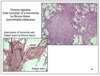
which histo pattern is associated w/ the following images?

Usual intersitial pneumonia
Since Idiopathic Pulmonary Fibrosis is a diagnosis of exclusion (50% of interstitial fibrosis), what are the other disease in which Usual Interstitial Pneumonia (UIP) pattern is seen?
Similar pattern can be seen in later/end stage of diseases :
- Pneumoconioses
- Hypersensitivity pneumonitis
- Collagen vascular diseases
Non-specific interstitial pneumonia (NSIP):
how are subtypes classified?
on basis of histology – divided into:
- cellular pattern
- fibrosing pattern
define: Non-specific interstitial pneumonia (NSIP)
Uniform and diffuse alveolar septal expansion by lymphocytes or collagen or both
which condition was previously known as BOOP?
(bronchiolitis obliterans-organizing pneumonia)
organizing pneumona (OP): common pathologic finding in lung
- characterized by the presence of polyp-like collections of fibroblasts within airspaces,
- referred to as fibroblastic plugs or Masson bodies

what are the major causes of ORGANIZING PNEUMONIA
- Infections (localized finding at the edge of another histologically obvious process such as fungal granuloma)
- Minor part of the pathologic findings of another process
- Connective tissue disease
- Drug-related
- Bronchial obstruction
- Idiopathic (Cryptogenic organizing pneumonia)
what are the causes of PNEUMOCONIOSES?
- Mineral dusts (e.g: coal, silica, asbestos, Beryllium)
- Organic dusts (e.g: mold [aspergillus], bird droppings, cotton)
- Fumes and Vapors (e.g: nitrous oxide, sulfur dioxide, ammonia, benzene)
what factors affect toxicity and fibrogenic activity in PNEUMOCONIOSES?
- Physical properties of the particles (size/shape)
- Physiochemical reactivity, solubility
- Concentration
- Duration of exposure
- Effectiveness of clearance mechanisms
how do the physical properties of particles affect toxicity and fibrogenic activity in Pneumoconiosis?
- Particles 1-5 µm are greatest danger as get lodged at bifurcation points in distal airways
- Elicit macrophage (MO) phagocytosis → release of mediators/cytokines → inflammation and fibrosis
- RECALL: MO should be in lung for protection
define, and epidemiology:
anthracosis
- DEF: Accumulation of coal-dust in lungs, pleura, and lymph nodes
- No significant reaction
- Asymptomatic
- Epi: Smokers & urban dwellers
define: Simple Coal Workers’ Pneumoconiosis (CWP)
and symptoms
- Coal macules (coal-dust laden macrophages 1-2 mm, peribronchial) and coal nodules (> 2 mm) in a background of collagen deposition
- Sxs:
- Cough
- Black sputum
- No dysfunction
define: COMPLICATED Coal Workers’ Pneumoconiosis (CWP)
and effect on respiration
- Complicated CWP (progressive massive fibrosis - PMF)
- Progressive severe fibrosis in a background of simple CWP
- RESPIRATORY INSUFFICIENCY results
COMPLICATED Coal Workers’ Pneumoconiosis (CWP):
histo features
Multiple nodules and black scars > 2cm (dense collagen and black pigment) with central necrosis

define: Caplan’s Syndrome
- a combination of rheumatoid arthritis (RA) and pneumoconiosis
CAPLAN’S SYNDROME:
diagnosis, course, associated w/ which minerals/conditions?
- dx: Nodular lesions with central necrosis and peripheral fibrohistiocytic inflammation
- nodules are seen on xray
- course: Faster progression/more severe
- assoc: carbon, silicosis and asbestosis
sources of exposure of Silicosis:
what is the most damaging/fibrogenic of these causes?
- **Most damaging/fibrogenic to the lung: crystalline forms such as QUARTZ
- Other causes (occupational):
- Mining (gold, tin, copper, coal)
- Quarrying
- Sandblasting, stone cutting
- Metal grinding
- Manufacture of ceramics
what specific disease does SILICOSIS increase the susceptiblity of?
silicosis –> increased susceptibility to TUBERCULOSIS
(bc it interferes w/ immune function –> macrophage clearance is decreased)
SILICOSIS:
histopathology
- Nodular fibrosis
- Hyalinized whorls of collagen
- Scant inflammation
- Birefringent silica particles
- Coalescence of nodules/large areas of dense scars (progression even after cessation of exposure)
- Concomitant anthracosis

SILICOSIS:
radiology
- Eggshell calcification on chest x-ray
ASBESTOSIS:
histology
- Mineral fibers containing crystalline hydrated silicates
- curled flexible serpentines
- straight stiff amphiboles (more pathogenic)
- ferruginous bodies (iron coating)** important clue on histology
- Diffuse interstitial fibrosis
- Pleural effusions/fibrous adhesions
- Hyalinized fibrocalcific pleural-plaques
what is the KEY histological finding to confirm ASBESTOSIS?
FERRUGINOUS BODIES (iron coating)
ASBESTOSIS:
pathogenesis
- Fibers ingested by macrophages leads to:
- activation of macrophages and neutrophils
- release of enzymes and fibrogenic cytokines
- deposition in interstitium and lymphatics
- direct stimulation of fibroblast collagen
- Progressive fibrosis even after cessation of exposure
what are occupational exposure causes of asbestosis?
- Mining
- Pipefitters and plumbers
- Textile industry
- Shipbuilding
- Constructions
- Insulations
asbestosis acts synergistically with what to increase risk of carcinoma (but not mesothelioma)
cigarette smoking
(act synergistically to inc risk of carcinoma)
asbestosis has strong association with which tumors?
- Strong association with malignant mesothelioma and bronchogenic carcinoma
- (Generates free radicals and adsorbs toxic chemicals, affecting nearby mesothelium or bronchial epithelium)

what condition is this gross anatomical image of?
PLEURAL PLAQUES;
common manifestation of asbestos exposure

key word associations of:
- anthracosis
- silicosis
- asbestosis
- Anthracosis: complicated coal workers pneumoconiosis
- Silicosis: occupations exposed to silica; large collagenized nodules in lungs; seen under microscope
- Asbestos: can cause pleural plaques, fibrosis (asbestosis), mesothelioma, lung cancer
SARCOIDOSIS:
define
- Non-caseating granulomas (many tissues and organs)
-
Disordered immune regulation in genetically predisposed individuals when exposed to certain environmental agents
- CD4+ TH1 cells play a big role in formation of granulomas
SARCOIDOSIS:
epidemiology
- Common disease, with unknown etiology
- Young adults (females)
- Geographic/ethnic variations (African-americans)
SARCOIDOSIS:
clinical presentation
- bilateral, symmetrical, enlargement of lymph nodes (aka “potato lymph nodes”)
- Peripheral lymphadenopathy/hepatosplenomegaly
- may be asymptomatic (incidental on chest xray)
- Usually lung involvement with hilar adenopathy (characteristic chest x-ray)
- Insidious onset with respiratory and constitutional symptoms (fever, night sweats, weight loss)
-
Acute onset with fever, erythema nodosum, polyarthritis
- isolated cutaneous or ocular lesions
- Usually lung involvement with hilar adenopathy (characteristic chest x-ray)
SARCOIDOSIS:
key histopathological findings
- non-caseating granulomas
- Pulmonary granulomas increase and eventually replaced by diffuse interstitial fibrosis of the lung
Sarcoidosis is a DIAGNOSIS OF EXCLUSION;
how do you get a definitive diagnosis?
you need a transbronchial biopsy for a definitive diagnosis
(granuloma is circled)
SARCOIDOSIS:
clinical course
- 70% - recover w/ minimal or no residual disease
- 20% - permanent lung or ocular dysfunction
- 10% progress w/ severe interstitial pulmonary fibrosis and cor-pulmonale and death
HYPERSENSITIVITY PNEUMONITIS:
define
- Intense, prolonged exposure to inhaled organic antigen
- Type III/IV hypersensitivity reactions
- Inflammation w/ gradual fibrosis
HYPERSENSITIVITY PNEUMONITIS:
causes
- Farmer’s lung: thermophnilic actinomycetes in hay
-
Pigeon breeder’s lung: proteins from bird feathers and excreta
- mold at home, or bird droppings
-
Humidifier/air-conditioner lung: thermo-philic bacteria
- hot tub lung
HYPERSENSITIVITY PNEUMONITIS:
histopathology;
what is key feature?
- Non-caseating poorly formed interstitial granulomas - (key feature)
- Interstitial pneumonitis
- Fibrosis
- Organizing pneumonia reaction
- May progress to severe interstitial fibrosis

PULMONARY ALVEOLAR PROTEINOSIS (PAP):
very rare disease; what is the clinical manifestation?
- Sputum (chunks of gelatinous material)
- Cough
- Progressive respiratory difficulty
PULMONARY ALVEOLAR PROTEINOSIS (PAP):
histopathology
- Alveolar spaces filled with dense, amorphous, PAS-positive, lipid containing, surfactant-like material
- autoimmune disease (whole lung undergoes lavalge in the patient)

pulmonary alveolar proteinosis:
two common forms of the disease?
- autoimmune
- secondary (associated with hematopoietic disorders, malignancies and immunodeficiency
states)
lung disease can result from several causes:
what are the drug-induced lung diseases?
- Bronchospasm (Aspirin, Beta-antagonists)
- Pulmonary edema
- Chronic pneumonitis/fibrosis (amiodarone/bleomycin)
- Hypersensitivity pneumonitis (methotrexate)
lung disease can result from several causes:
what are the radiation-induced lung diseases?
(acute and chronic)
- Acute radiation pneumonitis (1-6 months.)
- fever, dyspnea, radiologic infiltrates
- Lymphocytic alveolitis, hypersensitivity pneumonitis
- Respond to steroids
- Chronic radiation pneumonitis
- DAD and atypical type II pneumocytes
- Progression to interstitial fibrosis
what does CHRONIC radiation-induced lung diseases look like on histology?
can look like fibrotic lung disease
lung disease can result from several causes:
what are complications of therapy following LUNG TRANSPLANT?
- Infections (immunocompromised host)
- Acute rejection - we want to recognize and tx this as early as possible
- Chronic rejection
- Lymphoproliferative diseases
ACUTE rejection of lung transplant:
timing, sxs, tx
- Timing: early weeks to months post transplant
- Sxs: fever, dyspnea, cough, infiltrates on CXR
- perivascular and peribronchiolar mononuclear cell infiltrates
- Tx: increased immunosuppression
CHRONIC rejection of lung transplant:
timing, sxs, tx
- Timing: 6 - 12 months post transplant
- Sxs: dyspnea, cough, –> bronchiolitis obliterans (BO, aka popcorn lung)
- Chronic rejection –> total occlusion of bronchiole by fibrous tissue (BO)
- BO is key histological finding
- Tx: of BO disappointing