8: Pulm 1 Flashcards
Normal Lung anatomy:
conducting zone versus respiratory zone?
CONDUCTIVE
- Trachea
- Main stem bronchi
- Bronchi
- Bronchioles
- Terminal Bronchioles
RESPIRATORY - these 3 layers form pulmonary acinus; structure is simpler for gas exchange
- Respiratory Bronchioles
- Alveolar ducts
- Alveoli

Histology of LARGE AIRWAYS (trachea/bronchi)
- pseudostratified, tall columnar, ciliated epithelium - cilia to move things out
- goblet cells - to make mucus to capture pathogens
- basal cells
- neuroendocrine cells - receive neuronal input –> put hormones into blood
- submucosal mucous glands
- **cartilage - key feature of conductive zone

histology of SMALL AIRWAYS (bronchioles)
- lack of cartilage
- lack of submucosal glands
- gradually thinner epithelium
- gradually less mucous cells
- non ciliated columnar clara cells (terminal bronchioles)

Type I versus Type II pneumocytes?
- Type I pneumocyte: forms part of the barrier across which gas exchange occurs
- Type II pneumocyte secretes surfactant;/ acts to repair larger, cuboidal cells and occur more diffusely
Alveoli:
composition of alveolar SURFACE,
composition of alveolar LINING
- Alveolar SURFACE:
- 95% type I pneumocytes; 5% type II pneumocytes
- Alveolar LINING:
- 40% type I pneumocytes, *60% type II pneumocytes

Alveoli:
structure
- capillary network
- fusion of BM and endothelium and epithelium (gas-exchange areas)
- pores of Kohn (b/w alveoli)
- macrophages
- surfactant layer
- interstitium
define: atelectasis
Incomplete expansion of the lung, or collapse of previously inflated lung leading to loss of lung volume
effect of Atelectasis on function?
- Reduces oxygenation (ventilation-perfusion imbalance)
- Predisposes to infection
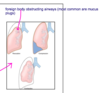
Types of Atelectasis?
-
Resorption (obstruction) - most common are mucus plugs
- airways are obstructed there is no further ventilation to the lungs and beyond
- early stages, BFcontinues and gradually the oxygen and nitrogen get absorbed
-
Compression (relaxation)
- The loss of negative pressure in pleura permits the lung to relax, due to elastic recoil.
-
Contraction atelectasis
- compression of parts of the lung by fibrotic changes in the pleura
-
Patchy (micro-atelectasis)
- occurs in the absence of surfactant, such as can occur in newborns

Hemodynamic Pumonary Edema is the accumulation of fluid in the lungs caused by the disruption of Starling’s forces;
What are the causes?
- Increase hydrostatic pressure (left-sided HF, volume overload, PV obstruction, etc.)
- Decrease oncotic pressure (hypoalbuminemia)
- Lymphatic obstruction
- Accumulation of fluid in dependent basal regions of lower lobes
What are the causes of Edema due to microvascular injury?
- capillary hydrostatic pressure not elevated
- primary injury to vascular endothelium and/or alveolar epithelium
- leakage of fluids into interstitial space and then alveolar space
- Non cardiogenic pulmonary edema
causes of microvascular injury?
- infections (viruses, mycoplasma, etc.)
- inhaled gases (oxygen, cyanides, smoke, etc)
- liquid aspiration (gastric acid and contents)
- drugs and chemicals
- shock, trauma, sepsis, radiation
- pancreatitis, uremia, TTP, DIC, etc.
Histologic findings of Pulmonary Congestion and Edema?
- engorged capillaries
- granular pink precipitate in alveolar spaces
- microhemorrhages
- hemosiderin-laden macrophages and fibrin
- fibroblastic plugs (repair) and interstitial fibrosis (chronic)
acute phase versus subacute phase
histo findings of pulmonary congestion and edema
- ACUTE:
- congestion of capillaries
- edema fluid (granular precipitate) in alveolar spaces
- SUBACUTE
- hemosiderin laden macrophages – stains golden brown in lung
- fibrin in alveolar spaces

histo features of ORGANIZATION/REPAIR phase in pulmonary congestion/edema?
- immature fibrous tissue (plugs) in alveolar spaces

Adult Respiratory Distress Syndrome (ARDS):
causes
-
Acute respiratory failure/ acute lung injury - MOST COMMON CAUSE OF ARDS
- *Specifically in pt w/ SEPTIC SHOCK
- Decreased lung compliance
- Hypoxemia refractory to oxygen therapy
Adult Respiratory Distress Syndrome (ARDS):
diagnosis, course, mortality
- Dx: Bilateral radiologic opacities
- Frequent superimposed infections
- Course: Progression to multi-organ failure
- Mortality over 50% (v high mortality)
what is this histologic pattern and what pulmonary disease does it correlate with?

DIFFUSE ALVEOLAR DAMAGE (DAD): assoc w/ ARDS (adult resp distress syndrome)
- EARLY (injury phase) of DAD
- edema, +/- hemorrhage
- fibrinous exudate
- hyaline membranes (fibrin-rich layer with necrotic cells)
- mild interstitial inflammation
- fibrin microthrombi

which phase of diffuse alveolar damage is pictured below?

TYPE II PNEUMOCYTE HYPERPLASIA;
part of the repair (organizing) phase
which phase of diffuse alveolar damage is pictured below?

INTERSTITIAL/ AIRSPACE fibroblastic proliferation (fibrous plugs);
with marked thickening of alvolar septae
(part of Repair (organizing) phase)

presenting clinical symptoms of PULMONARY EMBOLISM?
- Chest pain
- Dyspnea (difficult or labored breathing)
- Tachypnea (abnormally rapid breathing)
- Hemoptysis (coughing of blood or blood-stained mucus)
sources of pulmonary embolism?
what is MOST COMMON source?
- MOST COMMON SOURCE OF PE: DEEP VENOUS THROMBOSIS (DVT)
- Other sources
- pelvic vein thrombi
- foreign body emboli
- bone marrow emboli
- amniotic fluid emboli
- air emboli
pulmonary embolism:
PREDISPOSING FACTORS
venous stasis
hypercoagulable state
endothelial injury
pulmonary embolism:
RISK FACTORS
- immobilization
- obesity
- pregnancy
- estrogenic oral contraceptives
- hereditary clotting disorders
how does pulmonary embolism compromise respiration and hemodynamics?
- Respiration
- Clot in lungs –> ventilated segment is NOT perfused –> compromised resp
- Hemodynamics
- Clot –> increased resistance to pulmonary blood flow (vasoconstriction) –> hemodynamic compromise
Consequences of PE depends on size of embolus and adequacy of bronchial circulation;
Compare Large and Small emboli consequences
- LARGE emboli (5%)
- saddle embolus - sitting at bifurcation of pulmonary trunk
- –> sudden death OR CV collapse
- SMALL emboli (60-80%)
- asymptomatic, OR
- transient chest pain, hemoptysis
- repeated small emboli –> may have same effect as large emboli
Consequences of PE depends on size of embolus and adequacy of bronchial circulation;
Consequences of MIDDLE-SIZED EMBOLI
Middle sized emboli (20-35%) – PULMONARY INFARCTION
- Hemorrhagic
- pleural-based
- wedged-shaped
- fibrinous exudate on pleural surface
- form contracted scar w/ resolution
- predominantly lower lobes

Chronic Obstructive Pulmonary Disease (COPD):
hallmark sign, and pathogenesis
- Hallmark of COPD: decreased expiratory flow rate (hard to get air out of lungs)
- (Total lung capacity is normal or increased)
- Path: Chronic/recurrent airflow obstruction
- narrowed/small airways
- loss of elastic recoil
what is most common trigger for COPD and Emphysema?
Chronic injury such as **CIGARETTE SMOKING
define: emphysema
- abnormal permanent enlargement of airspaces distal to terminal bronchioles
- w/ destruction of alveolar walls
(no structure left to lung bc structure is destroyed –> filled w/ spaces of air)
emphysema:
gross and histo morphology
- gross: voluminous lung, as if inflated balloon
- histo:
- enlarged airspaces
- thin alveolar walls
- compressed septal capillaries
- rupture of walls

compare the types of emphysema:
centriacinar (centrilobular) and panacinar (panlobular)
-
Centriacinar (centrilobular)
- Central/proximal acinus (Distal alveoli spared )
- Apical segments/upper lobes
- Smokers
-
Panacinar (panlobular)
- All portions of acinus
- Lower basal zones
- Alpha-1-antitrypsin deficiency
- Worse in smokers
- Paraseptal (distal acinar)
- Irregular

which type of emphysema is caused by Alpha-1-antitrypsin deficiency ?
panacinar (panlobular) emphysema

there are 2 theories of pathogenesis of emphysema:
descirbe the protease-antiprotease hypothesis
- Irritation (smoke particles)
- Activation of macrophages
- Recruitment of neutrophils
- Release/enhancement of neutrophilic elastase
- Inactivation of a1-AT
- Destruction of alveolar wall

there are 2 theories of pathogenesis of emphysema:
descirbe the oxidant-antioxidant imbalance hypothesis
- Lung has a substantial amount of antioxidants (glutathione, superoxide dismutase)
- Agents that contain abundant oxidants can deplete these stores
- Net effect is oxidative damage to antiproteases, giving a functional deficiency

how does emphysema affect lung function?
- decreased gas-exchange area
- reduction of diffusion capacity
- small airway collapse (loss of support structures) –>
- airflow obstruction
- clinically apparent after reduction of 25% of lung volume
clinical presentation of pure emphysema:
- exertional dyspnea –> (walking capacity decreases –> requires oxygen)
- minimal cough or sputum
- use accessory muscles –> barrel chest (huge, expanded lungs)
- hyperresonance (occurs in the chest as a result of overinflation of the lung)
- depressed diaphragm
chronic bronchitis is defined by duration and trigger factor:
what are these definitions?
- Persistent cough w/ sputum for 3 months in at least 2 consecutive years
- Trigger: Chronic irritation (tobacco smoke)
effects of chronic bronchitis on mucus glands and bronchi/bronchioles
- Mucous glands hypertrophy/ hypersecretion
- Bronchitis and bronchiolitis (inflammation of the bronchi and bronchioles)
- Secondary infections
histopathology of CHRONIC BRONCHITIS
- Hyperemia and edema of mucous membranes
- Mucinous secretions/casts filling airways
- Mucous glands hyperplasia in trachea and bronchi
- Bronchial/bronchiolar mucous plugging
- Bronchial and bronchiolar epithelium with squamous and goblet-cell metaplasia
- Inflammation
- Fibrosis
Chronic bronchitis:
clinical presentation
- Copious mucoid sputum
- Hypercapnia (elevated CO2)
- Hypoxemia (low O2)
- Cyanosis (bluish color to skin)
Associated conditions of CHRONIC BRONCHITIS
- Cor pulmonale (RHF)
- **Secondary bacterial infections - predisposed to secondary bacterial infections
- Pulmonary hypertension
define: asthma
- Chronic OBSTRUCTIVE, reversible inflammatory disorder of airways
- causes recurrent episodes of wheezing, breathlessness, cough
mechisms of Asthma, and types
- Paroxysmal contraction of bronchial smooth muscle in response to various stimuli
(hyperresponsiveness)- Increased mucous secretion
- Reversible airway narrowing
- Types of Asthma
- ATOPIC
- NON-ATOPIC
ATOPIC asthma = allergen-triggered asthma;
what are key cells found in this type of asthma?
MAST CELLS and eosinophils; usually in response to allergen
immediate phase (minutes), and late phase (hours)

ATOPIC ASTHMA:
cause, epidemiology
- IgE mediated hypersensitivity reaction <– triggered by environmental pathogens
- Epi: most common type of asthma
- begins in childhood
- often w/ family hx
Recall: other atopic diseases incl
ATOPIC ASTHMA:
pathogenesis
-
T-helper 2 lymphocytes play a critical role in initial sensitization
- Excessive reaction against environmental antigens
- Cytokines produced by T Helper-2 cells account for most of the asthma features
- Subsequent IgE-mediated reaction to inhaled allergens elicits an immediate response and a late-phase reaction
which cytokines are produced by T helper 2 cells in atopic asthma; and what are their functions?
- IL-4 stimulates IgE production which binds mast cells in the mucosal surface
- IL-5 activates eosinophils
- IL-13 stimulates mucous production
NON-ATOPIC ASTHMA:
triggers, etiology
- Triggers: respiratory tract infections, chemicals, and drugs
- Etiology: NO FHx
- unknown primary etiology
- theory is virus-induced inflammation –> hypersensitive airways
ASTHMA:
gross and micro morphology
- GROSS: overinflated lungs, patchy atelectasis; airways w/ mucous plugs
- Micro: AIRWAY REMODELING
- Edema
- Sub-basement membrane thickening
- Inflammation with eosinophils (bronchitis-olitis)
- Hypertrophy of smooth muscle and mucous glands
- Curschmann’s spirals (whorled mucous plugs mixed with epithelial shed)
- Charcot-Leyden crystals (debris of eosin. Membranes)
ASTHMA:
clinical presentation
- Acute dyspnea
- Wheezing
- Reversible (spontaneous or with therapy)
-
Status asthmaticus - emergency
- hypoxia
- hypercapnia
- acidosis
- fatal
define: BRONCHIECTASIS
Chronic necrotizing obstructive lung infection –> affecting bronchi and bronchioles
effect of Bronchiectasis on lungs?
- Leads to abnormal dilatation of airways with destruction of the muscle and elastic supporting tissue
- Basal segments (worse drainage)
what are the conditions associated w/ bronchiectasis?
(anything that affects function of cilia)
- Congenital or hereditary conditions including cystic fibrosis, intralobar sequestration, immunodefeciency states and primary ciliary dyskinesia.
- Postinfectious conditions including necrotizing pneumonia (mycobacterium and staphylococcus aureus), viruses (adenovirus and infleunza), and fungi (aspergillus).
- Bronchial obstruction (tumor, foreign body and mucous impaction)
- Others, such as collagen vascular disease, inflammatory bowel disease, chronic lung rejection post-transplantation and GVHD
BRONCHIECTASIS:
histopathology
- Dilatation of airways
- Severe necrotizing acute and chronic inflammation (bronchitis/bronchiolitis)
- Squamous metaplasia
- Fibrosis
- Abscesses
BRONCHIECTASIS:
clinical presentation
- cough
- fever
- abdundant purulent sputum
- obstructive respiratory insufficiency (dyspnea/cyanosis)
- cor-pulmonale (Right heart failure)
- Metastatic brain abscesses
- Amyloidosis
BRONCHIECTASIS:
complications
- Cor pulmonale (right heart failure)
- Brain abcscesses
- Amyloidosis
