12_Peds Path 1 Flashcards
why do we separate pediatric diseases from adult?
- Genetic origin
- Not genetic:
- Unique to children
- Take distinctive forms in children
epidemiology of childhood disease mortality?
if patient survives beyond one year, likely to survive

annual incidence of congenital anomalies (structural defects present at birth)?
120,000 babies born w/ birth defect each year in US (1:33)
some (cardiac or renal) defects may not be clinically apparent until later
5 types of morphogenesis errors?
- malformations
- deformations
- disruptions
- sequence
- malformation syndrome
define: malformations
primary errors of morphogenesis; intrinsically abnormal developmental process;
usually multifactorial
what are the 2 possible manifestations of malformations?
- single body system (e.g. congenital heart disease)
- multiple coexisting malformations involving many organs and tissues
polydactyly vs syndactyly
- polydactyly: one or more extra digits
- syndactyly: fusion of digits’
little functional consequence if occurs in isolation
if cleft lip and cleft palate are isolated anomaly, likely compatible w/ life.
When would cleft lip likely be INCOMPATIBLE with life?
If it’s a sign of an underlying malformation syndrome, e.g. trisomy 13
(related to other health effects such as cardiac defects)
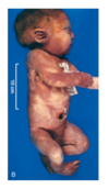
what does a severe degree of external dysmorphogenesis of the head indicate?
associated w/ severe internal anomalies (e.g. maldevelopment of brain and cardiac defects);
in this case, the mid-face structures are fused or illformed;
often a lethal malformation
define: disruption
results from secondary destruction of an organ or body region that was previously normal in development;
- potential environmental causes;
- NOT heritable
how do malformations AND disruptions differ?
disruptions arise from extrinsic disturbance in morphogenesis;
whereas malformations are primary errors/intrinsically abnormal development
what type of congenital anomaly is amniotic bands?
type of disruption;
-
rupture of amnion –> “bands”
- encircle, compress, and attach to parts of developing fetus
- occurs when the inner membrane (amnion) ruptures, or tears, without injury to the outer membrane (chorion).
- The developing fetus is still floating in fluid but is then exposed to the floating tissue (bands) from the ruptured amnion. This floating tissue can become entangled around the fetus.
pathogenesis of deformations
(type of extrinsic disturbance of development)
localized or generalized compression of the growing fetus by abnormal biomechanical forces –> structural abnormalities
*COMMON: 2% of newborn infants
most common cause of deformations?
pathology?
uterine constraint;
during weeks 35-58 of gestation –> rapid increase in fetus size outpacing growth of uterus –> relative decrease in amniotic fluid
what maternal conditions can cause uterine constraint, and subsequently deformation?
- first preganncy
- small uterus
- malformed (bicornuate) uterus
- leiomyomas
what fetal conditions can cause uterine constraint, and subsequently deformation?
- multiple fetuses
- oligohydramnios (less cushioning ofr baby)
- abnormal fetal presentation
define: sequence;
what are the 3 initiating events?
- def: multiple congenital anomalies that result from secondary effects of a single localized aberration in organogenesis; initiating events incl:
- Malformation
- Deformation
- Disruption
oligohydramnios is decreased amniotic fluid;
what are the potential maternal, placental, and fetal causes?
- maternal: chronic leakage of amniotic fluid due to rupture of amnion
- placental: uteroplacental insufficiency from maternal hypertension or severe toxemia
- fetal: renal agenesis

oligohydramnios (potter) sequence:
pathology, and resulting phenotype
Oligohydramnios –> fetal compression –> classic phenotype:
- flattened facies (facies is distinctive facial expression or appearance )
- positional abnormalities of hands /feet
- hips may dislocate
- compromised growth of chest wall and lungs

malformation syndrome:
define / pathology
presence of several defects that can’t be explained on basis of single localizing initiating error in morphogenesis;
path: single causative agent –> simultaneously affecting several tissues (e.g. viral infection or specific chromosomal abnormality)
define: agenesis
complete absence of an organ or its anlage (rudimentary basis of a particular organ or other part)
define: hypoplasia
underdevelopment of an organ
difference between aplasia and atresia
- aplasia: incomplete development of an organ
- atresia: absence of an opening, usually of hollow visceral organ or duct (e.g. of intestines and bile ducts)
causes of congenital malformations in humans?
genetic
environmental
multifactorial
unknown

pathogenesis of congenital anomalies is complex and poorly understood;
what are the 2 general principles?
- timing of the prenatal teratogenic insult
- interplay b/w environmental teratogens and intrinsic genetic defects
which period of development do internal/external factors most often affect fetus?
**embryonic period, which is the critical phase of growth
(3-8 weeks of development)

two types of perinatal infections?
transcervical (ascending) infections
transplacental infections
define: transcervical (ascending infections)
spread from cervicovaginal canal;
acquired in utero or during birth
define: transplacental** infections**
gains access to fetal bloodstream by crossing the placenta via the chorionic villi (hematogenous transmission)
how does fetus acquire infection in transcervical infections?
- by “inhaling” infected amniotic fluid into lungs
- passing through infected birth canal during delivery
Fetal infection –> inflammation of placental membranes (chorioamnioitis) umbilical cord (funisitis)
examples of conditions w/ transcervical as typical mode of spread ?
- bacterial infections (e.g., α-hemolytic streptococcal infection)
- viral infections (e.g., herpes simplex)
- pneumonia
- sepsis
- meningitis
when do transplacental infections occur?
may occur at any time during gestation or at the time of delivery via maternal-to-fetal transfusion (e.g. HBV & HIV);
mnemonic for transplacental infections?
TORCH :
- Toxoplasma ( T)
- Other (O) microbes (e.g., Treponema pallidum)
- Rubella virus (R)
- Cytomegalovirus (C)
- Herpesvirus (H)

effects of early TORCH infections?
CHRONIC SEQUELAE
- growth restriction
- mental retardation
- cataracts
- congenital cardiac anomalies
effects of later TORCH infections?
tissue injury & inflammation
- encephalitis
- choriorenitis
- hepatosplenomegaly
- pneumonia
- myocarditis
prematurity:
definition, epidemiology
- def: gestational age less than 37 weeks
- epi: 2nd most common cause of neonatal mortality (2nd only to congenital anomalies)
difference b/w prematurity and fetal growth restrictions?
- premature infants weigh less than normal (<2500), but appropriate for gestational age
- fetal growth restrictions (1/3 of infants weighing <2500 gm BORN AT TERM; undergrown, rather than immature)
what are risk factors for prematurity?
- prematurity rupture of membranes
- intrauterine infxn leading to chorioamnionitis
- structural abnormalities of the uterus, cervix, and placenta (e.g. bicorunate nucleus)
- multiple gestation
what complications may result from prematurity?
- respiratory distress syndrome (RDS) aka “hyaline membrane disease”
- necrotizing enterocolitis (NEC)
- sepsis
- intraventricular and germinal matrix hemorrhage
- long-term sequelae, incl developmental delay
small-for-gestational-age (SGA) infants suffer fetal growth restriction.
what are the causes of fetal growth restrictions?
- fetal
- maternal
- placental
- unknown
fetal factors causing fetal growth restrictions?
conditions that intrinsically reduce growth potential of fetus despite an adequate supply of nutrients from the mother
-
chromosomal disorders
- cause 17% of fetuses evaluated for growth restriction
- 66% of fetuses w/ documented Ultrasonographic malformations
- congenital anomalies
- congenital infections
what should be considered in all growth-restricted neonates?
transplancental infections (TORCH);
cause is intrinsic to fetus –> growth retardation is symmetric (affecting all organs equally)

what are the placental factors that cause fetal growth restrictions?
any factor that compromises the uteroplacental supply line;
- placenta previa (low implantation of placenta)
- placental abruption (separation of placenta from decidua by retroplacental clot)
- placental infarction
which factors of fetal growth restriction cause symmetric retardation?
which cause asymmetric?
- SYMMETRIC growth retardation if cause is INTRINSIC to fetus (fetal factors)
- affects all organ systems equallty
- ASYMMETRIC (e.g. brain spared relative to visceral organs) if cause is placental or maternal causes of growth restriction
what is most common causes of growth deficit in small-for-gestational-age (SGA) infants?
maternal factors
-
avoidable influences
- narcotic abuse
- alcohol intake
- heavy cigarette smoking
- maternal malnutrition (prolonged hypoglycemia)
-
drugs causing fetal growth restriction
- teratogens (phenytoin, dilantin)
- non-teratogenic agents
-
others
- vascular disease (preeclampsia, “toxemia of pregnancy”
- chronic HTN
Respiratory distress syndrome (RDS) in infants is MOST COMMON CAUSE of respiratory insufficiency bc “membranes” form in peripheral air spaces;
what are the causes of RDS in newborn?
- excessive sedation of the mother
- fetal head injury during delivery
- aspiration of blood or amniotic fluid
- intrauterine hypoxia secondary to compression from coiling of umbilical cord around the neck
incidence and mortality of Respiratory Distress Syndrome in infants in US?
- Incidence: 24,000 cases reported annually in US
- Mortality: now 900/yr, down from 5000/year due to improvements in management
pathophysiology of Respiratory Distress Syndrome?
- premature infant
- reduced surfactant synthresis, storage, release (due to underdevelopment)
- decreased alveolar surfactant –>
- inc alveolar surface tension –>
- ATELECTASIS–> uneven perfusion & hypoventilation
- hypoxemia & CO2 retention
- acidosis
- pulmonary vasoconstriction
- pulmonary hypoperfusion –> endothelial and epithelial damage
- plasma leak into alveoli –>
- fibrin + necrotic cells –> HYALINE MEMBRANE
-
decreased diffusion gradient –> HYPOXEMIA & CO2 RETENTION
13.

microscopic morphology of hyaline membrane disease?
- alternating atelectasis & dilation of alveoli
- eosinophilic thick hyaline membranes lining dilated alveoli

clinical features of respiratory distress syndrome includes maturity, birth weight, & promptness of institution of therapy.
how is RDS prevented?
- delaying labor until fetal lungs are mature
- induce maturation of fetal lungs at risk
- analyze amniotic fluid phospholipids (est level of surfactant)
- prophylactic admin of exogenous surfactant at birth to extremely premature infants
respiratory distress syndrome:
complications
- uncomplicated –> recover in 3-4 days
- affected infants require oxygen
- complicated –> long-term use of high conc of O2 of ventilators can cause:
- retrolental fibroplasia (retinopathy of prematurity)
- bronchopulmonary dysplasia (BPD)
necrotizing enterocolitis (the wall of the intestine is invaded by bacteria, which cause local infection and inflammation that can ultimately destroy the wall of the bowel);
epidemiology?
most commonly in premature infants;
occurs in 1 out of 10 very low birth weight infants (<1500 grams)
what is thought to be the cause of necrotizing enterocolitis (NEC)?
associated w/ enteral feeding;
some postnatal insult sets cascade –> tissue destruction
(likely infectious agents involved, but no single pathogen is linked to disease)
what are the effects of inflammatory mediators in necrotizing enterocolitis?
platelet activating factor (PAF) –>
- increases mucosal permeability
- promotes enterocyte apoptosis
- compromises intercellular tight junctions
which organs/segments are involved in necrotizing enterocolitis?
what is the gross anatomical morphology of affected segments?
*see image
- at postmortem exam, you may see entire small bowel markedly distended w/ thin wall –> implies IMPENDING PERFORATION

what does the congested portion of ileum correspond to?
what are the arrows pointing to?

- congested portion –> areas of hemorrhagic infarction and transmural necrosis seen on microscopy
- submucosal gas bubbles (pneumatosis intestinalis) – indicated by arrows
microscopic findings of necrotizing enterocolitis?

clinical course and dx of necrotizing enterocolitis?
- course: bloody stools –> abdominal distention –> circulatory collapse
- dx; abdominal radiographs showing gas w/in intestinal wall (pneumoatosis intestinalis)
outcomes of necrotizing enterocolitis?
- Early detection –> can be managed conservatively
- 20-60% require operative intervention (incl resection of necrotic segments of bowel)
- Assoc w/ high perinatal mortality
- Infants who survive often develop post-NEC strictures from healing fibrosis


