8 - Lung Cancer, Pleural Disease and ILD Flashcards

1
Q

What are some diseases of the pleura?

A
  • Pneumothorax
  • Pleural effusion
  • Empyema
  • Pleural tumours
  • Pleural plaques
  • Pleural thickening
2
Q

What are some of the causes of a pneumothorax?

A

1. Spontaneous (primary with no lung disease or secondary with lung disease)

2. Traumatic

3. Tension

4. Iatrogenic (post central line or pacemaker insertion)

3
Q

What are some primary and secondary causes of a spontaneous pneumothorax?

A

Primary: young thin men with ruptured subpleural bulla

Secondary: asthma, COPD, TB, lung fibrosis, CF, Marfan’s, EDS

4
Q

What are some iatrogenic causes of a pneumothorax?

A
  • Subclavian CVP line insertion
  • Pleural aspiration
  • Transbronchial biopsy
  • Liver biopsy
  • Positive pressure ventilation
5
Q

What are some risk factors for developing a pneumothorax?

A
  • Pre-existing lung disease
  • Height
  • Smoking
  • Diving
  • Trauma/Chest procedure
  • Conditions e.g Marfan’s
6
Q

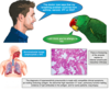
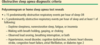
What are the signs and symptoms of a pneumothorax?

A

Symptoms: if small asymptomatic, sudden onset dyspnea, pleuritic chest pain

Signs: reduced expansion, hyperresonance to percussion, diminished breath sounds, if tension pneumothorax trachea is deviated to opposite side

7
Q

How is a pneumothorax managed? (not tension)

A

NEW GUIDELINES

8
Q

How is a tension pneumothorax managed?

A

Large bore cannula into 2nd ICS MCL then insert a chest drain

Do this BEFORE a CXR

9
Q

What are the two different types of pleural effusion?

A

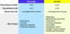
Transudate: protein concentration <25g/L

Exudate: protein concentration >35g/L

10
Q

What are some of the causes of pleural effusions?

A

Transudates: heart failure, cirrhosis, constrictive pericarditis, hypoalbuminaemia (peritoneal dialysis or nephrotic syndrome), hypothyroidism, mitral stenosis, PE, Meig’s syndrome

Exudates: infection, inflammation (RA, pancreatitis) malignancy, Yellow nail syndrome, drugs

11
Q

What is Meig’s syndrome?

A

Right pleural effusion and ovarian fibroma

12
Q

How you do you tell the difference between a transudative and exudative pleural effusion?

A

Light’s Criteria

if borderline protein use this, only needs one or more of the following to be an exudate

13
Q

What are the signs and symptoms of a pleural effusion?

A

Symptoms: asymptomatic, pleuritic chest pain, dyspnea

Signs: decreased expansion, stony dull percussion, diminished breath sounds, decreased vocal resonance, if large may have tracheal deviation away from effusion

Look for stigmata of other disease to try and determine cause

14
Q

What investigations should you do for a pleural effusion?

A
  • History and exam

- CXR

- Diagnostic apsiration

- Pleural biopsy (CT guided or thoracoscopic)

  • ECG
  • Bloods: FBC, U+Es, LFTs, CRP, Bone profile, LDH, clotting
  • ECHO is supect heart failure
  • Staging CT if suspect exudative
15
Q

What does a pleural effusion look like on CXR?

A
  • Blunting of costophrenic angles
  • Shadows with menisci
16
Q

What should you send a pleural aspiration off for?

A

Chemistry: protein, glucose, pH, LDH, amylase

Bacteriology: microscopy and culture, TB stain

Cytology

Immunology: rheumatoid factor, ANA, complement

17
Q

Why should you not put in a chest drain for a pleural effusion before a well established diagnosis?

When would an urgent chest drain only be indicated?

A

Only put in if diagnosis is well established. Draining all the fluid off can hinder the opportunity to get pleural biopsies

Urgent chest drain only if underlying empyema (pH<7.2 or visible pus on aspirate)

18
Q

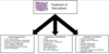
How do you manage a pleural effusion?

A
  • Therapeutic aspiration/drainage via tap or intercostal drain. Repeat as many times as needed. Use chest drain if empyema.

- Pleurodesis

  • Surgery if persistent collections and increasing pleural thickness
19
Q

How do you treat a transudate pleural effusion?

A
20
Q

What is interstital lung disease?

A

Umbrella term for a number of conditions that primarly affect the lung parenchyma in a diffuse manner

Usually have chronic inflammation or progressive interstitial fibrosis

21
Q

What are some of the underlying diseases causing interstitial lung disease?

A
  • Usual interstitial pneumonia (UIP)
  • Non-specific interstitial pneumonia (NSIP)
  • Sarcoidosis
  • Drug induced
  • SLE
22
Q

What are some clinical features of interstitial lung disease?

A
  • Dyspnea on exertion
  • Paroxysmal non-productive cough
  • Abnormal CXR
  • Restrictive pattern on spirometry
23
Q

What are some blood tests that should be ordered when a patient is diagnosed with ILD from a lung biopsy?

A
  • ANA
  • ENA
  • Rheumatoid factor
  • Anti-GBM
24
Q

What is the most common cause of idiopathic pulmonary fibrosis and what are the signs and symptoms of this?

A

UIP (usual interstitial pneumonia)

Symptoms: dry cough, exertional dyspnea, malaise, arthralgia, weight loss

Signs: cyanosis, finger clubing, reduced chest expansion, fine inspiratory crepitations, may have pulmonary hypertension

25
What will idiopathic pulmonary fibrosis show on blood tests and CXR?
**Bloods:** lowered PaO2 on ABG, increased CRP, may have ANA or RhF+ **CXR:** decreased lung volume, bilateral lower zone shadows, honeycomb lung
26
What is the management for idiopathic pulmonary fibrosis (UIP)?
- Oxygen - Pulmonary rehabilitation - Opiates - Consider lung transplant - Consider palliative care
27
What is extrinsic allergic alveolitis? (a.k.a hypersensitivity pneumonitis)
Repetitive **inhalation of allergens** (e.g fungal spores or avian proteins) that a patient has **already been sensitised** to so this provokes a **hypersensitivity reaction** In acute phase alveoli are filled with acute inflammatory cells. If diagnosed early and remove allergen can reverse process. If chronic can lead to granuloma formation and obstructive bronchiolitis
28
What are some causes of extrinsic allergic alveolitis?
Exposure to mould and spores - Farmers and Mushroom worker's lung - Bird/Piegeon Fancier lung - Malt worker lung - Sugar worker lung
29
What are the clinical features of extrinsic allergic alveolitis?
**Acute (4-6h post exposure):** Fever, rigors, myalgia, dry cough, dyspnea. Reversible and resolves spontaneously after 1-3 days **Chronic (months-years):** Finger clubbing, dyspnea, weight loss, Type 1 Respiratory failure, Cor Pulmonale. Less reversible
30
How do we manage extrinsic allergic alveolitis?
**Acute:** remove allergen and give O2, PO prednisolone **Chronic:** allergen avoidance or wear facemask, long term steroids, seek compensation
31
What are some causes of fibrotic shadowing on x-ray?
32
What is sarcoidosis and what is the aetiology of this?
**Multisystem inflammatory granulamotous disorder.** (Non-caseating granulomas). Usually affects respiratory system but can affect other areas Associated with HLA-DRB1 and DQB1 alleles **_Affects_:** age 20-40 years, Afro-Caribbeans
33
What does sarcoidosis do to the respiratory system?
- Pulmonary infilitrates of fibrosis - Dry cough - Progressive dyspnea - Chest pain - Decreased exercise tolerance
34
What are some extra-thoracic manifestations of sarcoidosis?
- Lymphadenopathy - Hepatosplenomegaly - Uveitis - Conjunctivitis - Erythema nodosum - Arrhythmias - Renal stones - Spinal syndromes - Bone cysts - Hypercalcaemia
35
What investigations should you do for sarcoidosis?
**- Lung function tests** (obstructive until fibrosis then restrictive) **- CXR** (4 stages) **- Bloods** (increased ACE, increased Ca, increased ESR) **- Urinary calcium** (increased) **- ECG/ECHO/Cardiac MI** (may show arrhythmias) **- CT/MRI head** (neurosarcoidosis) ***- Tissue biopsy*** (diagnostic showing NCGs)
36
What would blood tests show for sarcoidosis?
- Raised ESR - Lymphopenia - Raised LFTs **- Raised Ca, raised ACE** - Raised urinary Ca
37
What are the stages of pulmonary sarcoidosis?
Based on CXR 0 - Normal 1 - BHL 2 - BHL and Pulmonary Infiltrates 3 - Peripheral pulmonary infiltrates alone 4 - Pulmonary fibrosis
38
What is the diagnostic test for sarcoidosis?
**Tissue biopsy** of lung (thoracoscopic), liver or lymph nodes showing **non-caseating granulomas** Bronchoalveolar lavage can help diagnosis
39
How is sarcoidosis managed?
**- No treatment:** if just BHL often resolves spontaneously **- NSAIDs and bed rest:** if acute sarcoidosis **- Immunosuppressants and Anti-TNFa:** severe disease **- Corticosteroids**: see future flashcard **- Lung Transplant:** severe disease
40
What are some indications for steroid use in sarcoidosis?
- Parenchymal lung diseasse - Uveitis - Hypercalcaemia - Neurological or cardiac involvement Give prednisolone PO for 4-6 weeks
41
What are the treatment principles for ILD in general?
42
What are some causes of bilateral hilar lymphadenopathy?
43
What is the prognosis of lung cancer?
Biggest cause of cancer death worldwide 5 year survival is 13%
44
What are some risk factors for developing lung cancer?
- Smoking - Airflow obstruction - Increasing age - Family histroy - Asbestos - Radiation
45
What are some signs and symptoms of lung cancer?
**Symptoms:** cough, haemoptysis, dyspnea, chest pain, weight loss, anorexia, hoarse voice **Signs:** cachexia, anaemia, clubbing, supraclavicular lymphadenopathy, pleural effusion, metastatic signs (bone tenderness, hepatomegaly,)
46
What are some complications of lung cancer?
- Laryngeal nerve palsy - Phrenic nerve palsy - SVC obstruction (plethoric face and tortorous veins) - Horner's Syndrome (Pancoast tumour) - SIADH - Hypercalcaemia - Lambert Eaton syndrome - Cushing's - Addison's (metastases)
47
What is the WHO performance status?
Indication of patients level of fitness
48
What are some tests done once lung cancer is suspected to aid diagnosis?
**- Bloods:** FBC, U+Es, LFTs, INR **- CXR** **- Biopsy** **- Staging CT or PET scan** to detect small metastases that may not be seen on CT - **Bone scan** **- Lung function tests:** to assess whether lobectomy
49
What are some things you may see on a CXR with lung cancer?
- Hilar enlargement - Peripheral nodule - Pleural effusion - Consolidation - Lung collapse - Bony secondaries
50
What lung cancer patients are PET scans used in?
If patient is **surgical candidate** and CT was suggestive of **low stage** Helps detect small metastases not seen on staging CT
51
What are some ways of obtaining a biopsy/histological samples from a suspected lung cancer?
**- Bronchoscopy:** CT guided **- CT biopsy** **- US guided neck node FNA:** cytology if lymphadenopathy **- Thoracoscopy:** for cytology of pleural effusion
52
What are the different histologies for lung cancer?
**- Small cell lung cancer (Oat cell)** **- Non small cell lung cancer:** SCC, adenocarcinoma, large cell carcinoma, bronchoalveolar cancer **- Other:** carcionoid tumour
53
How does small cell lung cancer usually present?
Arises from endocrine cells that secrete polypeptide hormones so leads to **para-neoplastic syndromes** - **Cushing's from ACTH secretion** **- SIADH** **- Gynaecomastia** **- Labert Eaton Syndrome**
54
What paraneoplastic syndromes do non small cell lung cancer present with?
**- Hypercalcaemia of malignancy** (PTHrP) - Occurs with squamous cell carcinoma which is the most common NSCLC
55
What is the prognosis with SCLC and NSCLC?
**NSCLC:** 15% 5 year survival overall. Stage 1 after resection 60-75%, Stage 2 after resection 30-55%, Stage 3 3%, Stage 4 1% **SCLC:** Rapid growth so often too extensive for surgery at diagnosis. 4-12 weeks survival untreated, combination chemotherapy 6-15 month survival
56
What are the principles of treatment in SCLC and NSCLC?
**_SCLC: (often palliative)_** - Consider surgery if early stage - Chemotherapy +/- radiotherapy if well enough - Palliative radiotherapy for bronchial obstruction, bone pain, SVC obstruction - Can do pleural drainage/pleurodesis for pleural effusions **_NSCLC:_** - Curative lobectomy if low stage - Radical radiotherapy - Chemo-radio for more advanced
57
What are the clinical features of malignant mesothelioma?
Tumour of mesothelial cells that often occurs in the pleura. Related to previous asbestos exposure, often 45 years before. **Signs/Symptoms:** chest pain, dyspnea, weight loss, finger clubbing, recurrent pleural effusions, lymphadenopathy
58
How is mesothelioma diagnosed and managed?
**_Dx:_** - Biopsy via thoracoscopy - CXR/CT may show pleural thickening and effusion - Bloody pleural fluid **_Mx:_** - Chemotherapy - Pleurodesis or indwelling pleural drain - Can get compensation
59
What is obstructive sleep apnoea?
**Upper airway obstruction during sleep** due to intermittent closure of pharyngeal airway Causes sufficient **sleep fragmentation** so **daytime somnolence** Often obese middle aged men
60
What is the pathophysiology of obstructive sleep apnea?
Upper airway patency depends on **pharyngeal dilator muscle** activity. Muscles relax during sleep. Can have a small pharyngeal size whilst awake so when relaxing during sleep can cause critical narrowing OR Excessive narrowing with relaxation during sleep
61
What are some causes of small pharyngeal size and excessive narrowing of the airway during sleep?
**_Small pharyngeal size:_** - Fatty infiltration of pharyngeal tissues due to increased neck fat - Large tonsils - Craniofacial abnormalities - Extra submucosal tissue e.g myxoedema **_Excessive Narrowing:_** - Obesity can enhance muscle dilator relaxation - Neuromuscular diease e.g stroke, MND - Muscle relaxants e.g alcohol, sedatives - Increasing age
62
What are some of the symptoms of obstructive sleep apnea?
- Daytime somnolence (Epworth \>9) - Loud snoring - Nocturia - Decreased libido
63
Why can OSA cause CVS damage?
- Every arousal there is a rise in BP of about 50mmHg - Daytime hypertension too
64
What is the Epworth sleepiness scale?
Points for 0 (never doze), 1 (slight chance), 2 (moderate chance), 3 (high chance) \>9 in OSA
65
How is OSA diagnosed?
**- Overnight oximetry** or video recordings **- Limited sleep study (most common):** oximetry, snoring, body movement, heart rate, oralnasal flow, chest movements, leg movements **- Full polysomnography:** limited study plus EEG, EMG
66
What is classified as significant sleep apnea?
15 or more episodes of apnea during 1h of sleep
67
What are some complications of OSA?
- Pulmonary hypertension - Type II respiratory failure - Hypertension
68
How is OSA managed?
**- Weight loss** - Avoid smoking and alcohol - Mandibular advancement device **- CPAP** via nasal mask - Surgery to relieve obstruction e.g tonsils CONSIDER OCCUPATION AND WHETHER TO INFORM DVLA
69
What driving advice should you give to a patient with OSA?
- Do not drive whilst tired, stop for nap - One diagnosed patient needs to notify DVLA
70
How does CPAP help OSA?
- Usually nasal mask - Upper airways held open by 10cm H2O pressure to prevent airway collapse, stops sleep fragmentation so less daytime somnolence - Also opens collapsed alveoli
71
What is the difference between CPAP and BIPAP?
**CPAP:** - Supplies constant positive pressure through inspiration and expiration so not ventilatory support **BIPAP:** - Form of NIV and ventilatory support as two levels of positive pressure
72
How do SABA and LABAs work and what are some of the side effects of these?
- Tremors - Headache - GI upset - Palpitations - Tachycardia - HypoK
73
How do SAMAs and LAMAs work, what are some examples of these and what are the side effects?
- Dry mouth - Constipation - Headache - Be careful in BPH and acute closure glaucoma
74
How do xanthines work for Asthma, what are some examples and what are the side effects of these?
75
How do ICS work for Asthma and COPD, what are some examples of these and what are the side effects?
76
How do oral corticosteroids work for Asthma, what are the side effects and what are some examples of these?
77
What are some comorbid conditions associated with lung cancer?
- COPD - Hypertension - Type 2 DM - Cardiovascular disease
78
Where does lung cancer typically metastasise to?
- Liver - Bones - Brain
79
Why is lung cancer prognosis so poor?
Often advanced and metastasised at presentation
80
How do you diagnose a pleural effusion?
- CXR/CT/US - Thoracocentesis
81
What are some symptoms of ILD?
- Exertional dyspnea - Dry cough
82
What are the common types of ILD?
- Idiopathic - Drug related - Autoimmune
83
What are some drugs that cause ILD?
- Methotrexate - Amiodarone - Ciprofloxacin - Nitrofurantoin - Bleomycin
84
What stage of ILD is this?
Stage 4: pulmonary fibrosis
85
How is a pleural effusion aspirated safely?
Under US guidance to check not puncturing other important organs
86
When a patient is sent on a 2 week wait referral for suspected lung cancer, what test are they given?
- Pre-staging CT scan - At appointment break bad news and explain the need for biopsy/histology - Work out performance status - Discuss at MDT
87
What does this CXR show?
- Poor cardiac borders due to lung parenchymal disease - Diffuse alveolar shadowing ILD!!!!
88
What is the diagnostic imaging for ILD?
**High resolution CT**
89
What are the high risk characteristics for a pneumothorax?
1. Haemodynamic compromise (tension pneumothorax) 2. Significant hypoxia 3. Bilateral pneumothorax 4. Underlying lung disease 5. 50 or over with significant smoking history 6. Haemopneumothorax