3 - ECGs and Bradyarrhythmias Flashcards

1
Q

What is the protocol for reading an ECG?

A
  1. Confirm patients name and ECG date

2. Rate

3. Rhythm

4. Axis

5. P waves

6. Intervals: PR interval, QRS complex, QT interval, ST segment, T waves

  1. R wave progression
2
Q

What time interval does each of the following on an ECG represent:

  • 1 small box
  • 1 large box
A

Small box: 0.04 seconds

Large box: 0.2 seconds

1 second is represented by 5 large boxes

3
Q

How do you calculate the rate on an ECG? (assuming speed is 25mm/s)

A

Regular: 300 ÷ Number of Big squares between R-R

Irregular: Number of QRS complexes on rhythm strip (10 seconds) x 6

Normal is 60-100bpm

4
Q

How do you work out the rhythm of an ECG?

A

Use card method to mark position of 3 successive R waves and see if all intervals equal

Can be irregularly irregular or regularly irregular or sinus arrhythmia (p waves but irregular)

5
Q

What is sinus rhythm?

A
  • All QRS complexes preceded with a P-wave
  • Regular rhythm
  • Between 60-100bpm
6
Q

What is sinus arrhythmia?

A

Slight but regular lengthening and then shortening of RR intervals. All QRS have P waves so sinus node still working

Common in young people, lengthening and shortening corresponds to breathing

7
Q

What is the difference between AF and atrial flutter?

A

AF: has no p-waves and is irregularly irregular

Atrial flutter: sawtooth baseline with no discernible p-waves but it is regular

8
Q

What is the axis on an ECG and what is a normal axis?

A

Describes the direction of depolarisation across the heart, should spread from 11 to 5 o clock (-30 and +90)

Need to look at JUST LIMB LEADS/ leads I, II and III. There should be positive deflections for a normal axis.

Most positive deflection should be in II and most negative should be aVR

9
Q

How do you work out axis on ECG easily?

A

Normal: Lead II or I most positive

Left deviation: aVL/Lead I most positive

Right deviation: Lead III most positive

10
Q

What are the causes of right and left axis deviation?

A

Right: right ventricular hypertrophy, PE, anterolateral MI, WPW, left posterior fasicle block

(normal in very tall individuals, associated with pulmonary oedema as RVH)

Left: conduction abnormalities, left anterior hemiblock, inferior MI, WPW, LVH

11
Q

How can you distinguish right and left axis deviation on an ECG?

A

Right: lead I becomes negative and lead II, III/aVF become more positive (Lovers Returning)

Left: lead III and II become negative and lead I more positive (Lovers Leaving)

12
Q

What are the normal time values for the following and where are these intervals on ECG:

  • PR interval
  • QRS complex
  • QT interval
  • ST interval
A

PR: start of P to start of QRS. 0.12-0.2s (3-5 small squares)

QRS: <0.12S

QT: start of QRS to end of T. Should be 0.38-0.42s

ST interval: end of S to start of T

13
Q

After looking at rate and rhythm on ECG you look at P waves. What are you looking for?

A
  • Are they present?
  • Are they followed by a QRS
  • Should be upright in II, III, aVF but upside down in aVR
  • Flat, flutter or chaotic baseline?
14
Q

What is the normal PR interval and what can shorten and lengthen this?

A

3-5 small squares (0.12-0.2 seconds)

Prolonged: Delayed AV conduction e.g heart block

Shortened: fast AV conduction via accessory pathway e.g WPW or SA node in different place

15
Q

What is a normal QRS complex like and what can cause changes to the QRS complex?

  • Width
  • Height
  • Morphology
A

Should be <0.12s with Q waves being <0.04s wide and <2mm deep

Prolonged QRS: bundle branch block, metabolic disturbance, ventricular origin

Tall QRS (>5mm in limb leads, >10mm in chest leads): LVH

Pathological Q-Waves: following MI

16
Q

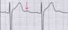
What are the QRS complexes on this ECG showing?

A

Delta wave which is common in Wolf Parkinson White Syndrome

Sign that ventricles are being activated earlier than normal from a point distant to the AV node. Early activation spreads slowly across myocardium causing slurred upstroke of QRS

17
Q

WPW cannot be diagnosed with the delta wave alone. What other ECG abnormality has to be present?

A

Tachyarrhythmia + Delta Wave

18
Q

When is a Q wave pathological?

A

> 25% the depth of the QRS that follows it or > 2mm in height and >1mm (40ms) in width.

Single Q wave is ok, need to look for Q in whole territory for evidence of previous MI e.g look at all inferior leads

19
Q

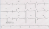
What is wrong with the QRS complexes in this ECG and why might this have occured?

A

Poor R wave progression

R wave should go from small to big from V1 to V6. Transition of S>R to R>S should be around V3/V4

Poor lead position or previous MI

20
Q

What is the J point?

A

Where the S wave joins the ST segment

21
Q

What is the Osborn wave (J wave)?

A

Positive deflection of the J point due to hypothermia, SAH or SEVERE hypercalcaemia

22
Q

What is a normal ST segment and what is ST elevation/depression?

A

Should be isoelectric

ST elevation: greater than 1 mm (1 small square) in 2 or more contiguous limb leads or >2mm in 2 or more chest leads.

ST depression: >0.5mm in >2 contiguous leads

23
Q

What is the cause of ST elevation and ST depression?

A

Elevation: full thickness myocardial infarction

Depression: myocardial ischaemia

24
Q

What do T waves represent and what leads are they normally inverted on?

A

Ventricular repolarisation

Usually inverted in aVR, V1 and V2 and sometimes V3

Abnormal if inverted in I, II, V4-V6

25
What are the causes of the following: - Tall T waves (\>5mm in limb leads and \>10mm in chest leads) - Inverted T waves - Biphasic T waves - Flattened
**Tall:** hyperkalaemia or STEMI **Inverted:** ischaemia, general illness, bundle branch block, PE **Biphasic:** Ischaemia and hypokaelaemia **Flattened:** hypokalaemia
26
What is a U wave and what causes these?
**\> 0.5mm deflection after the T wave** usually in V2 or V3 Seen in **electrolyte imbalances, hypothermia** and **secondary to antiarrhythmic therapy** (such as digoxin, procainamide or amiodarone) Seen larger in slower bradycardias
27
What leads are the most positive in normal cardiac axis, left axis deviation and right axis deviation?
**Normal:** II **Left:** aVL **Right:** III
28
What does ST elevation across all leads represent?
**Pericarditis** Saddle shaped
29
What are some causes of sinus bradycardia?
- IHD - Thyrotoxicosis - Hypothermia - Increased ICP - Cholestasis
30
What are some causes of the following: - ST elevation - ST depression - T wave inversion
31
What are some ECG changes in a PE?
- Sinus tachycardia - RBBB - Right axis deviation - S1Q3T3
32
What is the Digoxin effect on ECG?
**- Down-sloping ST depression** **- Short QT interval** **- Inverted T wave** in **V5-V6** - Any arrhythmia e.g ventricular ectopics and nodal bradycardia
33
What heart territory do all of the 12 leads of the ECG cover and what vessel supplies them?
34
How do hyper and hypokalaemia present on ECG?
**_Hyper_** - Tall tenting T waves - Widened QRS - Absent P waves - Sine **_Hypo_** - Small T waves - U waves - Prominent P waves
35
How do hypo and hypercalcaemia present on ECG?
**_HypoCa_** - Long QT - Small T waves **_HyperCa_** - Short QT
36
What are some continuous ECG monitoring methods that can be used to diagnose paroxysmal arrhythmias that may be missed on a single ECG?
**Telemetry:** Inpatient, signals watched by staff on screens so if dangerous arrhytmia can help immediately. Used for those at high risk of arrhythmias e.g post STEMI **Exercise ECG:** BP also taken whilst doing standardised exercise **Holter Monitor:** Worn for 24h for a week whilst going about normal life **Loop recorder:** Placed under the skin (e.g Reveal) and record short period before the event, e.g patient has syncope, can press the button when regains consciousness. Good if events happen months apart **Pacemakers and ICDs**
37
How can we split arrhytmias into different categories?
- Bradycardia - Narrow complex tachycardia - Wide complex tachycardia - AF and Atrial flutter MEMORISE ALGORITHM ON PHOTO
38
What are some causes of arrhythmias in general?
Cardiac or Non-cardiac
39
How do patients with arrhythmias present and what questions do you need to ask in the history?
**Presentation:** palpitations, chest pain, presyncope/syncope, hypotension, pulmonary oedema, asymptomatic **History:** take history of palpitations including onset/offset/nature, associated symptoms (e.g chest pain, breathlessness), drug history, family history of cardiac disease or sudden death
40
What are the different management options in arrhythmias?
**Conservative** e.g cut down alcohol **Medical** e.g betablockers **Interventional** e.g pacemakers, ablation of accessory pathways, ICDs
41
Why is syncope during exercise a worrying presentation?
Patient may have syndrome predisposing them to sudden cardiac death e.g long QT syndrome
42
What are some tests you can do when a patient presents with palpitations that is likely to be due to an arrhythmia?
**- Bloods:** FBC, U+Es, Glucose, Ca, Mg, TFTs - **ECG**: look for short PR (WPW) or long QT **- 24h ECG monitoring** or continuous ECG monitoring **- ECHO** for structural heart disease - Exercise ECG
43
How do you classify bradycardic arrhythmias based on the aetiology? (\<60bpm) IMPORTANT FLASHCARD TO UNDERSTAND BRADYCARDIAS
Based on the pacemaker at fault (**sinus node or AV node)** **Sinus Node**: sinus bradycardia, sick sinus syndrome (tachy-brachy), sinus arrest, vasovagal syncope **AV Node:** First degree AV block, Second degree AV block (Mobitz I or II), Complete AV block
44
What are some causes of sinus bradycardia?
**- Drugs** (b-blockers, amiodarone, digoxin) **- Hypothyroidism** **- Hypothermia** **- Sleep Apnoea** **- Increased intracranial pressure** **- Athletic** **- Rare:** rheumatic fever, viral myocarditis, amyloidosis, haemochromatosis, pericarditis
45
Most bradycardias cause by sinus node disease are asymptomatic. If they are symptomatic what is needed?
Pacemaker
46
What are some symptoms of bradycardia?
- Asympyomatic - Fatigue - Nausea - Dizziness - Syncope - Chest pain - Breathless
47
What is relative vs absolute bradycardia?
**Absolute:** any heart rate less than 60bpm, can be normal for sleeping athletes **Relative:** a heart rate too slow for the haemodynamic state of the patient, doesn't have to be below 60bpm. (systolic BP\<90, HR\<40, poor perfusion, poor urine output)
48
How is symptomatic bradycardia managed acutely? (especially after MI)
1. Do ECG and check electrolytes (K+,Mg2+, Ca2+) 2. Connect to cardiac monitor/telemetry **3. Address cause** e.g correct electrolytes, give glucagon if cause by B-blocker 4. If patient has adverse signs give **atropine IV 500mcg** every 3-5 minutes up to 3mg 5. If atropine not working do **transcutaneous pacing**
49
What is sick sinus syndrome caused by and what are the symptoms?
Usually due to **sinus node fibrosis**, often in the elderly. This can cause sinus bradycardias or tachyarrhythmias like AF Can get **tachy brachy syndrome** where they alternate between tachy and bradycardic rhythms **_Symptoms:_** syncope, presyncope, light-headedness, palpitations, breathlessness
50
How is sick sinus syndrome diagnosed and managed?
##Footnote **- VTE prophylaxis** is episodes of AF detected **- Permanent pacemaker** if symptomatic bradycardia or sinus pauses - Hard to treat tachy-brachy syndrome as treating one increases the risk of the other
51
What are some causes of 1st, 2nd and 3rd degree heart block?
**1st and 2nd:** normal variant, athletes, sick sinus syndrome, IHD especially inferior MI, acute myocarditis, digoxin, beta-blockers **3rd:** IHD especially ***inferior MI,*** ***digoxin toxicity,*** ***hyperkalaemia,*** idiopathic fibrosis, congenital, aortic valve calcification, cardiac surgery infiltration by tumours/granulomas/parasites
52
How does first degree AV block present on ECG and how do you manage it?
**PR interval \>0.2 seconds** with no missed beats - Check for toxicity if on digoxin - No treatment needed unless symptoms of dizziness or syncope then cardiac monitoring should be done to identify higher degrees of heart block
53
How does second degree AV block Mobitz Type I present on ECG and what type of patients may this present in?
**Progressive lengthening of the PR interval followed by drop in QRS then pattern resets** Can occur in ***young fit patients*** with high vagal tone during the night. Also often after ***inferior MI***
54
What is another name for Mobitz Type 1 Second degree AV block?
Wenckeback phenomenom
55
How do we managed Mobitz Type I Second Degree Heart block?
Rarely proceeds to complete heart block so **no therapy needed** If syncope or diziness do cardiac monitoring for higher degrees of heart block
56
How does second degree AV block Mobitz Type II present on ECG and how is it managed?
**Constant PR interval but then sudden drop of QRS** Can be defined as ratio, e.g 2:1 would be 2 normal for every 1 dropped **Management:** risk of progression to complete heart block so permanent pacing needed if drugs excluded
57
How does 3rd degree complete AV block present on ECG?
**No relationship between P and QRS waves** as no conduction between atria and ventricles Patient is very **bradycardic** due to ventricular rhythm taking over and can develop haemodynamic compromise Can be **broad complex escape rhythm** (block below the AV node and not well tolerated) or **narrow complex escape rhythm** (block above AV node, often congenital, well tolerated)
58
How is 3rd degree AV block managed?
**Urgent permanent pacing** within 24 hours, unless they are likely to recover normal conduction such as they have had a recent coronary event
59
What are the main causes of 3rd degree heart block and what medicaions can you administer for this?
**- Digoxin toxicity** **- Inferior STEMI** (often resolves in hours or days) **- Severe hyperkalaemia** - Give ***calcium chloride*** for hyperK to stabilise membrane - If haemodynamically unstable can give ***atropine*** 0.5mg up to 3mg - Can try ***isoprenaline*** if atropine not working
60
What is a junctional rhythm and how does it present on ECG?
Has **regular narrow QRS waves but no P waves** or P waves hidden in QRS Due to electrical activation occuring near to or within the AV node rather than the SA node
61
What does this ECG show?
Intermittent sinus bradycardia on the rhythm strip Has a tachycardia on other leads so **tachy-brady syndrome** Issue at SA node as when AF it is conducted to ventricles
62
What does this ECG show?
2:1 heart block (Mobitz Type II)
63
What does this ECG show?
Complete heart block as no relationship with QRS. Narrow escape rhythm
64
What does this ECG show?
Complete AV block with broad escape rhythm More dangerous than narrow escape rhythm as whole AV node not working!!!! Shows as a bundle branch block
65
How do pacemakers show on an ECG?
**One pacing wire in ventricle:** one spike followed by broad QRS **Two pacing wires:** will have spike then p wave and spike followed with broad QRS
66
Where do you place the ECG leads on a patient?
V7 placed at posterior axillary line Make sure you clean the skin with wipe and shave if hairy before placing on leads
67
What does a widened QRS complex mean?
\>0.12 seconds mean it is originating from the ventricles Slower pace than if from atria
68
How does RBBB on ECG appear and what are the causes of this?
Due to right ventricle being activated by the left ventricle. They cannot be activated at the same time as there is an obstacle in the RBBB. **- Prolonged QRS** **- M Pattern in V1** **and V1 positive** **- Bunny ears** **- Wide slurred S wave in V6, hence the W pattern** **- T wave inversion V1-V3** **- Normal axis** **Causes:** normal variant, RVH, cor pulmonale, PE
69
How does LBBB appear on ECG and what are the causes of this?
**Causes**: if new onset STEMI, HTN, cardiomyopathy, idiopathic fibrosis, aortic stenosis **- Prolong QRS** **- V1 QRS negative (look at chest leads to see when positive)** **- M pattern in V6** **- Dominant S (W) in V1** **- Left axis deviation** Cannot comment on ST segment or T waves if LBBB
70
What are some ways of remembering the ECG findings in LBBB and RBBB?
LBBB: **W**I**LL**IA**M** RBBB: **M**A**RR**O**W**
71
What is bifasicular block and how does it show on ECG?
Conduction block between AV node and two of the three fasicles **RBBB + left or right axis deviation**
72
What is trifasicular block and how does it present on ECG?
Usually bifasicular block plus 1st degree heart block - RBBB - Left or right axis deviation - Prolonged PR interval
73
Complete heart block may be intermittent so may not be seen on ECG. What can be seen on ECG that could indicate a poor conducting system and could mean complete heart block?
**Bi or trifasicular block**
74
What are the causes of low voltage QRS (\<5mm in all leads)
- COPD - Hypothyroidism - PE - BBB - Pericarditis - Pericardial effusion
75
What is the issue with bundle branch block when wanted to read an ECG for a patient with acute chest pain?
Can mask ST changes and T wave issues
76
What is normal axis, left deviation, right deviation?
**Normal:** lead II most positive **Left deviation:** lead aVL most positive **Right deviation:** lead III most positive
77
What does R wave progression mean and what can late R wave transition indicate?
Transition of more positive R than S should happen in V3 **_If late:_** - Prior anterosepctal MI so infarct - Inaccurate lead placement - LVH - Dilated cardiomyopathy
78
What is this ECG abnormality?
Looks like ST elevation but it is just high take off
79
What is the QTc interval?
QT interval corrected for heart rate
80
If there is a prolonged QRS, what is the first thing you should do?
Look in V1 to see if BBB!! V1 negative in LBBB V1 positive in RBBB
81
What do very tall R waves indicate?
LVH
82
What are some causes of LVH?
- Hypertension - Aortic stenosis - Hypertrophic cardiomyopathy
83
What do small alternating R waves on an ECG indicate?
**Pericardial effusion** as the heart is swinging in the sac
84
What is the normal QT interval and what are some causes of long QT and short QT?
\<0.44s or 2 large squares **Long:** MI, LVH, HypoK, HypoMg, HypoCa, DKA, Drugs, RBBB/LBBB **Short:** HyperCa
85
When is T wave inversion normal?
In leads avR and III if in isolation
86
A cardiologist has asked you to start oral amiodarone for a patient who has previously been admitted with ventricular tachycardia. What tests is it important to ensure the patient has had prior to starting treatment
**- TFT** **- LFT** **- U+Es:** risk of hypoK **- CXR:** risk of pulmonary fibrosis
87
What is the treatment algorithm for tachycardias?