7. the biology of cancer Flashcards
what kind of disease is cancer?
sporadic (but different to sporadic genetic) and familial (genetic predisposition)
only disease of somatic mutation
Metaplasia
- normal in appearance but in the wrong place, usual from an adjacent tissue layer.
Adenomas/polyps/warts
larger growths of dysplastic cells. (adenoma more advanced than polyps)
The features of cancer
- Proliferation: grow independently of signals
- Immortality: avoid senescence/telomere shortening
- Avoiding cell death: apoptosis, they don’t do it (as well as necrosis)
- Angiogenesis: they must be fed
- Metastasis: many activities needed
multistage evolution of cancer
- what gives clones a growth advantage
- is cancer clonal?
- is cancer homogenous or heterogenous?
Sequential mutations give clones of cells a growth advantage
Cancer is clonal: all cells share some mutations of their common ancestors
BUT, they also develop subclones
Heterogenous
Eg. A mole can have abnormal mutations but needs 6 or 7 more mutations in specific genes for it to divide
Eventually get the strongest clone that survives and divides
All starts from a single cell
‘driver’ mutations
mutations that DO affect function of genes that regulate proliferation, apoptosis, immortality
- ‘passenger’ mutations
All other mutations that are no relevant to the promotion of cancer
(PROTO)-ONCOGENES:
promote cell proliferation, gain of function mutations in cancer
Oncogenes promote proliferation (via restriction point)
TUMOUR SUPRESSOR GENE:
inhibit events leading to cancer, loss of function mutations in cancer
4 checkpoints of cell cell
the restriction point in cell cycle
- G1 phase- signals from outside cell effect this
- DNA damage checkpoints in late G1 and G2
- Metaphase checkpoint (spindle attachment checkpoint) in M- alignment
Two processes play a role in the intrinsic limit in the number of times a cell lineage can divide
scenesence: cells in G0, don’t proliferate (normally cells cant go past 50 divisions, then stops dividing)
apoptosis
Senescence
- Metabolically active, irreversibly lost ability to re-enter cell cycle
- Normal cells have finite proliferative capacity (Hayflick limit), stop dividing and go into replicative senescence
- Therefore, cancers must avoid senescence if they are to keep growing
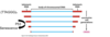
Telomere shortening
telomeres are repetitive sequences
Normally shortening results in senescence.
Cells only bypass senescence and reach ‘crisis’ if key tumour suppressors such as P53 are inactivated
what does telomere loss lead to
chromosome instability
Especially fusion between ends of sister chromatids which are then torn apart at anaphase
Hybrid chromosomes generated as the cells repair itself it finds another part of chromosome to bind to
Normally these will die due to gene disruption
Excess telomere shortening: leads to crisis and apoptosis
crisis: damage will make it unviable, cells will undergo apoptosis if they can

what is TERT and what does cancer do to it
what else is switched on
- In cancer a rare cell reactivates TERT to become cancerous- mutation to TERT gene
- Adds telomeres back on
- switches on telomerase
- Only germ and stem cells have telomerase switched on but almost all cancer cells have telomerase switched on


apoptosis
triggers
how does it kill cell
Triggered by damage to cell/DNA, stress, oncogene activation.
Uses energy to kill cell without releasing contents and so avoid inflammation – uses caspases.
Don’t want to release cell contents due to enzymes
Usually macrophage comes and digests it
P53 also regulator of apoptosis

Two key genes that control proliferation, senescence and apoptosis:
p 53
Rb- senses whether signal tells cell to proliferate
Sustained Angiogenesis
- Newly arisen tumours must promote angiogenesis to survive
- Hypoxia Induced Factor 1alpha
- If hypoxic- pathway triggered to encourage blood vessel growth (this is a normal pathway)
what do many cancer produce that effects vasculature
VEGF (vascular endothelial growth factor):
induces new vessel growth and production of Endothelial Precursor cells in bone marrow which travel to the tumour.
Antibodies to block VEGF Blocking these vessels have not been very successful but sometimes used as a form of therapy
what is the vasculature like in cancer cells
Vascularization is disorganized- imbalance of secreted factors stimulating growth over differentiation.
Tumour sprout new branches- tissue nearby isn’t very good and can turn leaky (spread is easy)
Leaky due to imperfect cell/cell junctions
Tissue Invasion and Metastasis
- Loss of adhesion: E-cadherin inactivation
- Activation of endogenous metalloproteinases
- Invasion of leaky blood vessels (or lymph system)
- Blood flow: millions travel, few survive (immune surveillance)- different tumours spread to different places
Secondary Tumours
- Capillary beds
- Tumours are big so get stuck and get trapped in capillary beds in the next tissue along
- ‘Seed and Soil’ theory:
- Not all tumours can survive in all environments so spread to specific tissues
- If it can spread to next tissue along it stays there and survives otherwise it goes onto the next to see if theres a good environment
- Cells have capacity to live elsewhere prior to migration
- Cells acquire capacity to live elsewhere after migration:
- Sequential passage in Xenograft, can select for secondary site specificity
