7 - Less Common Neurological Disorders Flashcards
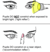
What is the presentation of Horner’s syndrome?
Triad of miosis, partial ptosis and anhydrosis
Due to interruption of the face’s sympathetic supply (oculosympathetic pathway)
Miosis leads to anisocoria

What is the aetiology of Horner’s syndrome?
Lesion anywhere from first to third order neurone
1st Order (Brainstem)
- Stroke (Medullary), MS, SOL, Syringomyelia (Bilateral), Cervical Cord trauma
2nd Order (Thoracic Outlet)
- Pancoast tumour, Thoracic outlet lesion, Brachial plexus injury, Thoracic aneurysm
3rd Order (Carotid)
- Carotid artery dissection, Cavernous sinus pathology, neck mass

How can you tell if a Horner’s syndrome is due to a carotid artery dissection?
Will be acute and painful!!!

What is the degree of anhidrosis in Horner’s syndrome?
Depends on the neurone affected

What investigations should you do if a patient has Horner’s syndrome?
Prompt evaluation for underlying cause
- CT angiography: look for carotid dissection as risk of stroke
- CT chest: look for Pancoast tumour
- MRI spine: spinal cord lesions
- MRI head: look for brainstem or cavernous sinus pathology

If a Horner’s disease is very subtle, what test can you do to confirm diagnosis?
- Cocaine Drops: no dilation if sympathetic chain involved
- Apraclonidine: dilation due to dennervation sensitivity
- Hydroxyamphetamine: Localises lesion, if doesn’t dilate it is 3rd order lesion. Stimulates release of noradrenlaine from post ganglionic neurones

How is Horner’s syndrome managed?
Treat underlying cause
- If acute and painful this is a neurological emergency as could be carotid artery dissection need to be sent to stroke unit (HASU)
What is the best way to do a neurological examination of a patient in a coma?

What pathology can arise at the cerebellopontine angle?

- Acoustic Neuroma/Vestibular Schwanomma (80%)
- Lipoma
- AV malformations
- Haemangioma

What is an acoustic neuroma? (vestibular schawnomma)
Benign cerebellopontine angle tumour of the schwann cells of the vestibular nerve
Usually unilateral and grow very slowly (1-2mm/year)
Bilateral in Neurofibromatosis Type 2

How do acoustic neuromas typically present?
- Unilateral sensorineural hearing loss
- Vertigo
- Tinnitus
- Facial nerve palsy
- As they grow larger they can cause ipsilateral V, VI, IX and X enrve palsies and ipsilateral cerebellar signs. Can also have signs of increased ICP

How are acoustic neuromas investigated and managed?
Ix
- Serial gadolinium-enhanced MRI
- Audiogram
Mx
- Surveillance with serial MRIs every 6/12 for 2 years then 2 years then every 5 years
- Microsurgery
- Radiotherapy

What are some of the risks with microsurgery for an acoustic neuroma?
- CSF leak and meningitis.
- Cerebellar injury.
- Stroke.
- Epilepsy.
- Facial paralysis (V and VII)
- Hearing loss (IX)
- Balance impairment (IX)
- Persistent headache
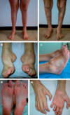
What is subacute combined degeneration of the spinal cord?
Vit B12 deficiency causes degeneration of the dorsal columns and the corticospinal tracts
Peripheral neuropathy (loss of fine touch, 2-point discrimination, proprioception, and vibration sensations) but pain and temperature fine
Combination of LMN and UMN signs

How does subacute combined degeneration of the cord typically present?
Signs and symptoms
- Symmetrical pins and needles, numbness progressing to weakness
- Visual impairment
- Change in mental state
- Bilateral spastic paralysis
- Sensations diminished (pressure, vibration, proprioception)

What signs may you elicit on examination of a patient with subacute combined degeneration of the cord?
- +ve Romberg sign (e.g falls in low light and shower)
- + Babinski sign (UMN)
- Absent ankle jerk reflex (LMN)
- Brisk knee jerk reflex (UMN)
- Unsteady ataxic gait
- Spastic paralysis
- Loss of vibration sensation

What other symptoms can B12 deficiency cause apart from subacute combined degeneration of the cord?
- Optic atrophy
- Anaemia
- Cognitive impairment

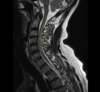
How is subacute combined degeneration of the cord investigated and managed?

Ix
- Serum B12 and Folate levels
- FBC
- MRI spine T2
Mx
- B12 replacement either IM, SC or sublingual with hydroxycobalamin
- Folate replacement
- Can take 3-6 months to improve, sometimes deficit is irreversible

What is a cavernous sinus syndrome?
How can it present?
Any pathology of the cavernous sinus leads to certain symptoms to paralysis of CN’s in the sinus
Causes: Cavernous Sinus Thrombosis, Cavernous Sinus Tumours, Carotid Cavernous Aneurysms
Presentation: Ophthalmoplegia, proptosis, ocular and conjunctival congestion, trigeminal sensory loss and Horner’s syndrome.

What are some causes of a cavernous sinus thrombosis and how does it present?
Causes: spread of infection from sinuses or boil/folliculitis on face
Presentation: headache, chemosis, oedema of eyelids, proptosis, painful opthalmoplegia, fever

How is a cavernous sinus thrombosis investigated?
- Bloods: thrombophillia screen
- Non-contrast CT: hyperdensity in the affected sinus.
- CT venogram: look for filling defect (‘ empty delta sign’)
- MRI-T2: look for thrombus

How is a cavernous sinus thrombosis treated?
- IV antibiotics
- LMWH
- Corticosteroids for swelling
- Surgical drainage of any pus
How do intracranial venous thrombi present in general?
- Presentation is variable but headache, confusion/drowsiness, impaired vision, and nausea/vomiting.
- Seizures, reduced consciousness, focal neurological deficitis, cranial nerve palsies, and papilloedema

What are some causes and some differential diagnoses for an intracranial venous thrombosis?
Causes of hypercoagulable state: Pregnancy, COCP, Trauma, Local infection, Tumours, Malignancy, dehydration
Differentials:
- SAH
- Meningitis
- Encephalitis
- Stroke
- Intracranial abscess
How are intracranial venous thrombi investigation and managed?
Ix
- MRI/CT (will show infarction not compatible with arterial infarct)
- CT venogram
- Bloods for thrombophillia screen
- LP if no CI
Mx
- Anticoagulate with LMWH
- If raised ICP decompresive hemicraniectomy
- Address risk factors
- Elevate head 30-40 degrees

What is the most common type of pituitary tumour and what are some conditions that increase the chance of developing a pituitary tumour?
- Usually a benign tumour of the anterior lobe of pituitary (microadenoma)
Conditions:
- MEN1
- Carney Complex
- McCune-Albright Syndrome

How are pituitary tumours classified?
Microadenoma = <1cm diameter
Macroadenoma = >1cm diameter
Giant adenomas = prolactinoma with >4cm diameter
Can further be classified by the hormone that they secrete e.g prolactin, GH, no hormone secreted, ACTH, gonadotropin and TSH
What are the clinical features of a pituitary tumour?

Mass Effect
- Non-specific headaches
- Bitemporal hemianopia (optic chiasm compression)
- CN V1, V2, III, IV, and VI palsies (cavernous sinus compression)
- CSF rhinorrhea (rare)
Hormonal Effect
- See image

What is pituitary apoplexy, how does it present and how is it managed?

NEUROSURGICAL EMERGENCY
- Spontaneous haemorrhage into a pituitary tumour or ischaemia of pituitary gland
- Sudden and severe headache, loss of consciousness, neck stiffness, vomiting, photophobia
Mx: IV hydrocortisone and emergency surgical resection to preserve vision

What are the differential diagnoses for a pituitary tumour?
- Craniopharyngioma (derived from pituitary gland embryonic tissue, supracellar, in children, cystic on imaging)
- Meningioma
- Cerebral metastasis
- Arachnoid cysts
How are pituitary tumours investigated?
- Gold standard: MRI
- High resolution CT
- PRL levels: over 2000 suggests prolactinoma
- IGF1: test GH levels
- ACTH, FSH, LH and TFTs

How are pituitary tumours managed?
Conservative
- Hormone replace if hyposecretion
- Dopamine agonsits (Cabergoline) for prolactinoma
- Somatostatin analogue (Octreotide) for GH
Surgical (Mainstay)
- Transphenoidal surgery to remove
- Stereotactic radiotherapy for recurrence

What are the complications of transphenoidal surgery?
- CSF leak
- Meningitis
- Epistaxis
- Diabetes Insipidus
Evaluate hormones after surgery after surgery to see if any need replacing
What are some causes of falsely elevated PRL?
- Stress
- Pregnancy
- Certain antipsychotics and SSRIs
- Hypothyroidism
- CKD
- Status epilepticus
What is Charcot-Marie Tooth disease and what causes it?
Group of inherited peripheral neuropathies affecting sensory and motor neurones
Autosomal dominant
Type 1 (more common): demyelinating, PMP22
Type 2: axonal

How does Charcot Marie Tooth disease present?
Usually presents in puberty, length dependent so affects feet first then hands when knees involved
- High foot arches (pes cavus)
- Distal muscle wasting (“inverted champagne bottle legs”)
- Weakness in lower legs, particularly loss of ankle dorsiflexion
- Weakness in hands
- Reduced tendon reflexes
- Reduced muscle tone
- Peripheral sensory loss

What are the 3 main signs to look out for on OSCE examination of a patient with Charcot Marie Tooth syndrome?

1. Pes Cavus (high-arched feet)
2. Symmetrical distal muscle atrophy (inverted champagne legs due to peroneal atrophy, claw hands)
3. Thickening and Enlargement of nerves

What is a pneumonic to remenber the causes of peripheral neuropathy?
A – Alcohol
B – B12 deficiency
C – Cancer and Chronic Kidney Disease
D – Diabetes and Drugs (e.g. isoniazid, vincristine, amiodarone and cisplatin)
E – Every vasculitis

How is Charcot Marie Tooth syndrome investigated in order to make a diagnosis and how is it managed?
Ix
- NCS (reduced conduction velocity in type 1)
- Genetic testing
Mx
- Incurable so give supportive care. Most will need walking aids but does not alter life span or mean will end in wheelchair
- Physiotherapy
- Podiatrist for foot symptoms and insoles
- Orthopaedic surgeons

What is the cause of Creutzfeldt Jakob Disease?
Neuodegenerative disease caused by prions (misfolded protein of PrPc) that can turn other proteins into prions
Spongiform changes in brain (tiny cavities and tubulovesicular structures)
Causes:
- Sporadic
- Inherited
- Variant (BSE) from contaminated CNS tissue
- Iatrogenic from contaminated instruments, corneal transplants, blood transplant

How does CJD present?
- Rapidly progressive dementia
- Psychiatric impairment
- Myoclonus
- Eye signs (diplopia, hallucinations, cortical blindness)

How is CJD investigated and managed?
Ix
- Tonsil/olfactory mucosal biopsy
- CSF gel electrophoresis showing 14-3-3 protein
- MRI can distinguish between variant and sporadic
Mx
- No cure, death in around 6-12 months with sporadic, slightly longer with variant and genetic so palliation
- Regulations to reduce spread of BSE and iatrogenic transmission

What is Wernicke’s encephalopathy?
Thiamine (B1 deficiency) with a triad of

- Confusion
- Ataxia
- Opthalmoplegia (nystagmus, lateral rectus or conjugate gaze)
What are some causes of Wernicke’s encephalopathy?

- Chronic alcoholism
- Eating disorders
- Malnutrition
- Prolonged vomiting e.g hyperemesis gravidarum, chemo

How is Wernicke’s encephalopathy diagnosed and managed?
- Clinical diagnosis
Mx (Emergency)
- Urgent IV Pabrinex (Thiamine) to prevent change to Korsakoff’s
- PO Thiamine until no longer at risk
- Abstain from alcohol
- If hypoglycaemic give thiamine before glucose as glucose can precipitate Wernicke’s

What is the prognosis with Wernicke’s encephalopathy?
- Untreated will lead to death
- Can progress to Korsakoff’s psychosis and need long term institutional care

What is Argyll-Robertson pupil?
Neurosyphillis (prostitute pupil)
A pupil that is constricted, unreactive to light but reacts to accomodation

Apart from neurosyphillis, what are some other causes of Argyll-Robertson pupil?

- Lyme disease
- HIV
- Diabetes
- MS
- Sarcoidosis

HIV and AIDS can have lots of neurological presentations. What are some examples of these?
Acute HIV infection:
- Transient aseptic meningoencephalitis
- Myelopathy
- Neuropathy
Opportunistic infections:
- Toxoplasma Gondii: cerebral abscess, ring enhancing lesion
- Cryptococcus neoformans: chronic meningitis and seizures
- CMV: encephalopathy
- Syphillis
- TB
Tumours
- Cerebral lymphoma: EBV
- B-Cell Lymphoma
Neuropathies
- Due to HIV or ARV e.g proximal myopathy, painful sensory peripheral neuropathy

What prophylaxis is given to people with AIDs to prevent opportunistic infection of Toxoplasma Gondii?
AIDS when CD4<200
At CD4<100 give trimethoprim prophylaxis
What is chronic HIV associated neurocognitive disorder?
- HIV associated dementia
- CD4<200
- Progressive behavioural changes, memory loss, poor attention, bradykinesia
What is a syrinx and what are some causes of this?
Tubular cavity of the central canal of the cervical cord (syringomyelia) or brain stem (syringobulbia). Essentially, there is a collection of CSF within the spinal cord.
Causes: (blockage of CSF from 4th ventricle)
- Arnold-Chiari malformation
- Basal arachnoiditis
- Massess e.g cysts. tumours
- Myelitis
- Cord trauma

How may syringomyelia present?

Onset often sudden after being static for years at around age 30 e.g on coughing or sneezing as increase pressure causes extension of syrinx
- Bilateral sensory loss: of pain and temperature but preserved others, usually over trunk and shoulders
- Wasting/weakness of hands: can lead to claw hand
- Horners syndrome: can be bilateral so difficult to see
- UMN leg signs
- Charcot’s joints: as lost proprioception

What is syringobulbia?
Brainstem involvement of syrinx
- Nystagmus
- Tongue atrophy
- Dysphagia
- Pharyngeal weakness
- CNV sensory loss

How is syringomyelia investigated and managed?
Ix
- MRI: look for Chiari malformation, how big is syrinx
Mx
- Chiari Malformation: Decompression at Foramen Magnum to promote flow of CSF and prevent syrinx dilation
- Drain syrinx surgically

What is hypoxic ischaemic encephalopathy and what is the cause of this?
Brain damage from ante or perinatal hypoxia
Ischaemia results in irreversible brain damage, both from primary neuronal death (immediate) and secondary reperfusion injury (delayed).
Causes: placental abruption, cord compression during delivery, failure to breathe on delivery

How may Hypoxic ischaemic encephalopathy present in a neonate?

Depends on severity of hypoxia
- Mild: irritability
- Severe: hypotonia, prolonged seizures, poor responses

How is HIE diagnosed and managed?
Ix
- EEG monitoring
Mx
- Respiratory support
- Anticonvulsant therapy
- Fluid balance ad electrolyte monitoring and inotropes.
- Induce mild hypothermia to prevent further damage by secondary reperfusion injury (therapeutic hypothermia)
What is the prognosis with HIE?
- Cerebral palsy
- Death
- Cognitive impairment
Assess degree of damage with MRI
What is neurofibromatosis?
Genetic condition that causes nerve cell tumours (neuromas) to form throughout the nervous system
Tumours are benign but cause neurological and structural issues
What is type 1 neurofibromatosis and how does it present?
Autosomal dominant NF1 gene on Chromosome 17 that codes for neurofibromin which is a tumour suppressor
At least 2 of 7 of CRABBING:
C – Café-au-lait spots (6 or more) measuring ≥ 5mm in children or ≥ 15mm in adults
R – Relative with NF1
A – Axillary or inguinal freckles
BB – Bony dysplasia such as Bowing of a long bone or sphenoid wing dysplasia
I – Iris hamartomas (Lisch nodules) (2 or more) are yellow brown spots on the iris
N – Neurofibromas (2 or more)
G – Glioma of the optic nerve

How is type 1 neurofibromatosis (Von Recklinghausen’s disease) investigated and managed?
Ix
- PURELY CLINICAL
- Genetic testing if doubt
- CT MRI if concerned about lesions in cord or brain
Mx
- Yearly cutaneous survery and measurement of BP
- Dermal neurofibromas can be excised if troublesome
- Geneti counselling
- No cure

What are some complications of type 1 neurofibromatosis?
- Learning disability
- GI bleeds
- Epilepsy
- Sarcoma

What is type 2 neurofibromatosis and how does it present?
Autosomal dominant NF2 gene on chromosome 22. Merlin protein that supresses schwann cell growth so NF2 causes schwanommas
- Cafe au lait spots
- Bilateral acoustic neuromas (tinnitus, vertigo, sensorineural hearing loss)
SENSORINEURAL HEARING LOSS OFTEN FIRST SIGN

How is type 2 neurofibromatosis investigated and managed?
Ix
- MRI
Mx
- Hearing tests yearly from puberty in affected families. If abnormality do MRI
- Neurosurgical removal of vestibular schwannoma but risk of hearing loss os and facial palsy
What is the pathophysiology of Lambert-Eaton syndrome?

Similar features to myasthenia gravis
Autoimmune or Paraneoplastic (Small Cell Lung Cancer)
Antibodies to Voltage-Gated Ca channels on pre-synaptic membrane so impaired influx of calcium so Ach not released

How may Lambert Eaton syndrome present?
- Symptoms develop slowly (gait first then eyes)
- Proximal muscle weakness (usually legs)
- Intraocular muscles affected causing diplopia, the levator muscles in the eyelid causing ptosis
- Oropharyngeal muscles causing slurred speech and dysphagia
- Autonomic dysfunction (dry mouth, blurred vision, impotence, dizziness)
- Hyporeflexia which improves after exercise (post-tetanic potentiation)

How is Lambert Eaton syndrome investigated?
- EMG: similar to MG but amplitude increases post-exercise
- Bloods for anti-voltage-gated calcium channels
- CXR and CT chest: rule of SCLC
How is Lambert Eaton syndrome managed?
Manage underlying cause
- Amifampridine: blocks voltage-gated potassium channels in the presynaptic cells, which in turn prolongs the depolarisation of the cell membrane allowing Ach to be released
- IV Immunoglobulin or Plasmapheresis: if severe respiratory impairment or bulbar weakness
- Do regular CXR/High Resolution CT as symptoms may precede cancer by 4 years
What is idiopathic intracranial hypertension?
Disorder characterised by features of raised ICP (LP opening pressure >25) but no mass causing this raised ICP
Clinical Features: headache, papilloedema and visual loss
What are some risk factors and associated conditions for IIH?
Risk Factors
- Female
- Obese
- Reproductive age
Associations
- Drugs: COCP, steroids, Vitamin A, Lithium, Tetracyclines
- Sleep apnea
- PCOS
- SLE
- Cushing’s
- Hypoparathyroidism

The cause of IIH is unknown, what are some of the theories about how this disease occurs?
- Intracranial venous hypertension: due to venous sinus narrowing/stenosis
- Raised intra-abdominal pressure from central obesity: felt to increase central venous pressure and subsequently leads to an increase in ICP
- Altered sodium and water retention
- Impaired CSF reabsorption: occurring secondary to excess vitamin A

What are the signs and symptoms of idiopathic intracranial hypertension?
Symptoms
- Raised ICP headache (e.g worse on bending down)
- Transient visual loss
- Diplopia and blurred vision
- Pain behind eyes
Signs
- CN6 Abducens palsy
- Papilloedema
- Peripheral visual field loss and large blind spot
What investigations are done to get to a diagnosis of idiopathic intracranial hypertension?
- BP: due to headache
- Pregnancy test
- Opthalmoscopy: papilloedema
- LP: with raised opening pressure, check no CI
- Neuroimaging: rule out other causes preferabbly MRV
Differentials: SOL, venous sinus thrombosis, obstructive hydrocephalus, decreased CSF production, increased CSF production

How is IIH managed?
Conservative
- Weight loss
Medical
- Acetazolamide (poor SE not often tolerated e.g peripheral paraesthesia, anorexia and metallic dysgeusia)
- Furosemide or Topiramate
Surgical
- Repeated Therapeutic LPs: if medical therapy not tolerated
- Optic nerve sheath fenestration: if prominent visual symptoms
- Shunting: ventriculoperitoneal, lumbarperitoneal

What is the prognosis with IIH?
- Recurrence in a third of patient
- Risk of permanent visual loss