6 - Lower GI Flashcards

1
Q

What are the different classifications of inguinal hernias?

A

Hernias are a protrusion of part or all of an organ through the wall of the cavity that normally contains it

Direct Inguinal (20%) - Go through weakness in Hesselbach’s triangle. More common in older patients due to weaker abdominal wall or increased intrabdominal pressure

Indirect Inguinal (80%) - Bowel goes through inguinal canal through deep ring. Due to incomplete closure of processus vaginalis so congenital in origin

2
Q

What are some risk factors for an inguinal hernia and what are the differential diagnoses?

A

Risk factors: male, increasing age, raised intraabdominal presure (chronic cough, heavy lifting, chronic constipation), obesity

Differentials: femoral hernia, saphena varix, inguinal lymphadenopathy, lipoma, internal iliac aneurysm, groin abscess, hydrocele

3
Q

What are the clinical features of an inguinal hernia and how can you distinguish between a direct and indirect hernia?

A
  • Lump in the groin that may reduce when lying down and gets worse on standing
  • If incarcerated can be tender, swollen, irreducible and erythematous and can have signs of bowel obstruction. Pain out of proportion to clinical signs
  • Reduce hernia and put pressure over deep inguinal ring (mid point of inguinal ligament). Ask to cough, if protrudes this is direct, if not this is indirect. Confirmed on surgery cannot be relied on
4
Q

When a patient presents with a groin lump (suspected inguinal hernia), what are some things you should do on examination?

A
  • Cough impulse
  • Locaton (superomedial is inguinal, inferolateral is femoral)
  • Reducible on lying down or with pressure
  • Does it go into scrotum (can you get above it, is it separate from the tesyes)
5
Q

How are inguinal hernias diagnosed?

A

- Usually clincial

  • Only image if diagnostic uncertainty and give US in outpatient setting
  • If incarcerated/strangulated use CT
6
Q

How are inguinal hernias managed generally?

A

- If strangulated: urgent surgical exploration

- If symptomatic: offer surgical intervention due to risk of strangulation

- If asymptomatic: conservative but discuss risks of strangulation

7
Q

How are symptomatic non-strangulated inguinal hernias surgically treated?

A

- Open Mesh Repair (Lichtenstein Technique): if unilateral

- Laparoscopic: for bilateral or recurrent inguinal hernias or can be used for primary hernia but high risk of chronic pain or females

Laparoscopic is a longer operating time but quicker post op recovery, fewer complications and less post-op pain

8
Q

What type of patients are at high risk of chronic pain with an open inguinal mesh repair?

A
  • Young and active
  • Previous chronic pain
  • Predominant symptom of pain
9
Q

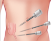
What are the complications of inguina hernias and post-operative complications for their repair?

A

Inguinal Hernia: incarceration, obstruction, strangulation

Post op: (see image)

10
Q

What is the femoral canal made up of?

A

In the anteror thigh and contains lymph vessels, lymph nodes and loose connective tissue

Superior border is the femoral ring that is normal covered by a septum but some omentum or abdominal contents can get through and cause a hernia

11
Q

What are some risk factors for a femoral hernia and why are they a high risk of strangulation?

A

- Risk Factors: female, pregnancy, raised intraabdominal pressure, increasing age

  • More prone as narrow neck and rigid borders of femoral canal with concave lacunar ligament
  • More common in women because of the wider anatomy of the pelvis
12
Q

What are the clinical features of a femoral hernia?

A
  • Small lump in the groin medial to femoral pulse
  • Will present as emergency
  • Vomiting
  • Often irreducible due to tightness of femoral ring

Sometimes femoral hernia can roll above inguinal ligament and appear as inguinal hernia

13
Q

What are some differential diagnoses for a femoral hernia?

A
  • Inguinal hernia
  • Femoral canal lipoma
  • Lymph node
  • Saphena varix (will disappear on lying and have palpable thrill)
  • Athletic pubalgia
14
Q

How are femoral hernias investigated?

A
  • Clinical diagnosis
  • Pre-op assessment as will need surgery
  • Can do US or CT abdomen pelvis
15
Q

How are femoral hernias managed?

A

- Surgery within 2 weeks of presentation due to risk of strangulation

  • Operation involves reducing hernia and reducing size of femoral ring by suture pectineal and inguinal ligaments or putting in mesh plug

- High or Low approach (Inguinal ligament). Low less likely to damage inguinal structures but less space to remove any compromised bowel. High approach used in emergency

16
Q

What are the complications with femoral hernias and complications with their surgical repaire?

A
  • Strangulation
  • Obstruction
  • Bowel resection if strangulation
  • Wound infection
  • Cardiorespiratory complications
17
Q

What is an epigastric hernia and what causes them?

A
  • Occurs in the upper midline through the line albea
  • Usually due to raised chronic intraabdominal pressure (obesity, pregnancy, ascites)
  • Usually affect men and often asymptomatic
  • Midline mass that disappears on lying back
18
Q

How can you distinguish between divarication of the recti and an epigastric hernia?

A

Divarication is a cosmetic condition due to weakening and widening of the linea alba

Divarication is just stretched linea alba, no defect, so will not feel muscle tear

19
Q

What is a paraumbilical hernia and how do they present?

A

Herniation through the linea alba around the umbilical region (not the actual umbilicus).

Due to chronic raised intraabdominal pressure and they have a lump around the umbilical region

Extremely common and often contain pre-peritoneal fat and sometimes bowel but rarely strangulate

20
Q

What is a spigelian hernia?

A
  • Hernia that occurs at the semilunar line around the level of the arcuate line (lateral border of the rectus where the aponeuroses fuse)
  • Small tender mass at the lower lateral edge of the rectus abdominis
  • High risk of strangulation so urgent surgical repair
21
Q

What is an obturator hernia and how will they present?

A
  • Hernia of the pelvic floor through the obturator foramen into the obturator canal
  • Common in elderly women who have lost a lot of weight

- Mass in upper medial thigh and may have features of bowel obstrution

  • May have positive Howship-Romberg sign due to compression of obturator nerve
22
Q

What are Littre and Lumbar hernias?

A

Littre: herniation of a Meckels diverticulum, often into inguinal canal and often becomes strangulated

Lumbar: posterior hernia that occurs spontaneously or iatrogenically, posterior mass with back pain

23
Q

What is a Richter’s hernia?

A

Any hernia site but the anti-mesenteric border becomes strangulated so only part of the lumen of the bowel is in the hernial sac

Tender irreducible mass and obstruction symptoms

Urgent surgical intervention

24
Q

What are the contents of the inguinal canal in males?

A
25
What is the definition of the following terms: - Diarrhoea - Acute and Chronic diarrhoea - Dysentry - Traveller's diarrhoea
Risk factors are poor food preparation, immunocompromised and poor personal hygeine
26
How can you tell what causative organism caused gastroenteritis?
Time between ingestion of food and symptoms Bacterial toxins = hours Virus = days Bacteria = weeks Parasites = months
27
What are the presenting features of gastroenteritis and what are some questions you should ask in the history of a patient presenting with this?
- Cramp-like abdominal pain and diarrhoea (with possible mucus and blood) - May have vomiting, night sweats and weight loss - May have pyrexia and dehydration
28
How is gastroenteritis investigated and managed?
**Ix:** none unless blood/mucus in stool, immunocompromised or persistent, then do a stool culture **Mx:** rehydration, education to prevent in the future, exclusion from work for 48 hours from last episode of D+V, if food posioning report as notifiable disease
29
What are some important causes of dysentry you should consider when a patient presents with gastroenteritis?
- Campylobacter - Shigella - Salmonella - Norovirus
30
What are some viral infective causes of gastroenteritis?
**- Norovirus:** most common viral gastroenteritis in adults, usually abdominal cramps, D+V and lasts 1-3 days **- Rotavirus:** common in young children and resolves in about a week **- Adenovirus:** common in kids
31
What are some bacterial infective causes of gastroenteritis?
- Campylobacter - Shigella - E.Coli - Salmonella ALL GRAM NEGATIVE
32
What are some bacteria that produce toxins that cause gastroenteritis?
Usually acute onset D+V lasting 24 hours
33
What are some parasitic causes of gastroenteritis?
More common in traveller's diarrhoea
34
What is the most common cause of hospital-acquired gastroenteritis and how does this organism cause symptoms?
**C.Difficile (Gram-positive)** usually arising following use of **broad spectrum abx** that disrupts normal microbiota so C.Diff can overgrow Produces **exotoxin A and B** that cause an immune response from the bowel so **inflammatory exudate on colonic mucosa** **Severe bloody diarrhoea** and risk of **toxic megacolon** (dilated bowel with risk of perforation)
35
How is C.Diff investigated and managed?
**Ix:** stool culture and C.Diff Toxin (CDT) testing **Mx:** IV fluid rehydration, oral metronidazole (or vancomycin in severe cases) Have low threshhold for treatment with immunocompromised patients regardless of the cause of the gastroenteritis
36
What are some non-infective causes of gastroenteritis?
**- Radiation colitis** **- IBD** (Crohn's and UC) **- Microscopic colitis** **- Chronic ischaemic colitis** (usually affects watershed area around splenic flexure and is seen on endoscopy as blue swollen mucosa)
37
What is the pathophysiology of angiodysplasia?
Causes GI bleeds and it caused by formation of **arteriovenous malformations** between previously healthy blood vessels, **usually in caecum and ascending colon** Most common cause of small bowel bleeds and second most common cause of rectal bleeding in over 60s Can be **acquired** or **congenital**
38
What are the clinical features of angiodysplasia and what are some differential diagnoses?
**- Painless rectal bleeding** AND **- Anaemia** - If upper GI bleed melena and haematemesis - If lower GI haematochezia **_- Differentials:_** oesophageal varices, GI malignancies, diverticular disease, coagulopathies
39
How is suspected angiodysplasia investigated?
**Lab tests:** blood tests including FBC, U+E's, LFTs, clotting, G+S or crossmatch **Imaging:** ***endoscopy or colonoscopy*** to rule out malignancy. may need ***capsule endoscopy*** if in small bowel. can do mesenteric angiography to plan for intervention
40
How is angiodysplasia managed?
**_Conservative_** ## Footnote - If haemodynamically stable ***- Bed rest and IV fluids*** - Potential ***tranexamic acid*** **_Radiographically_** - If persistent or severe ***- Endoscopy*** that subjects bleeding vessel to argon and electrical current ***- Mesenteric angiograph***y for small bowel that cannot be reached on endoscopy. Embolisation of vessel after catheterisation **_Surgically_** ***- Bowel resection*** (high mortality so only do if necessary)
41
Patients with angiodysplasia are at risk of a major GI bleed, what are some risk factors associated with a poor outcome for a upper GI bleed?
- Advancing age - Liver disease - Patient present in hypovolemic shock - Current inpatients
42
What are some complications of angiodysplasia treatment?
**- Rebleeding** post therapy - Risk of **small bowel perforation** in endoscopy - Risk of **haematoma formation, arterial dissection, thrombosis** and **bowl ischaemia** in mesenteric angiography
43
What are GEP-NETs and how are they classified?
**Gastroenteropancreatic neuroendocrine tumours** that originate from neuroendocrine cells in the tubular GI tract and pancreas and they have the potential to be malignant Most located in small intestine, with the rest in the stomach and rectum
44
What are the risk factors for GEP-NETs?
- Genetic - MEN1 - VHL disease - Tuberous sclerosis complex (TSC)
45
How do GEP-NETs present?
- Non-specific symptoms like vague abdominal pain, N+V, andominal distension - Unintentional weight loss - Can be functioning or non functioning depending on hormonal hypersecretion
46
How are GEP-NETs investigated?
- Chromogranin A and 5-HIAA levels - Routine bloods including FBC, LFTs, pancreatic peptides - Genetic testing - Endoscopy or CT enterocylsis depending on location - If metastatic with unknown primary do whole body somatostatin receptor schintography SSRS
47
How are GEP-NETs managed?
Often palliative as metastatic on presentation **Poorly differentiated:** surgical resection then chemo if localised or if metastatic just palliative chemo **Well differentiated:** localised disease and any liver metastases should be resected
48
What is a carcinoid crisis?
49
What is the pathophysiology of appendicitis?
Usually affects people between aged 10-30 Usually due to **luminal obstruction** of appendix from a **faecolith, lymphoid hyperplasia**, impacted stool or rarely a tumour When obstructed **commensal bacteria in appendix multiply** so acute inflammation. **Reduced venous drainage and localised inflammation** leads to **increased pressure** in the appendix and in turn **ischaemia** If ischaemia untreated can lead to **necrosis** and then **perforation**
50
What are some risk factors for appendicitis and some differential diagnoses?
**Risk factors:** FHx, caucasian (but ethnic minorities more likely to perforate if get it), summer **Differentials:** see image
51
What investigations are done if appendicitis is suspected?
**_Lab Tests:_** urinalysis to exclude UTI, serum b-hCG to exclude pregnancy, FBC to look for raised inflammatory markers and for pre-op assessment **_Imaging:_** often not needed as clinical diagnosis but if uncertainty can do USS first line (especially for children as lower radiation) or CT
52
How is appendicitis risk stratified?
**_Men:_** Appendicitis inflammatory response score **_Women:_** Adult Appendicitis score **_Children:_** Shera score Helps to work out how probable the diagnosis is appendicitis
53
How is appendicitis managed?
**- Laparascopic appendectomy** is gold standard **- If appendiceal mass give antibiotics** then interval appendectomy 6-8 weeks later - After removal **send to histology** to rule out malignancy and then look around rest of abdomen to look for any other pathology and look for Meckel's diverticulum
54
What are some complications with appendicitis?
**- Perforation** and peritoneal contamination **- Surgical site infection** **- Appendix mass** where omentum and small bowel adhere to appendix **- Pelvic abscess** with fever and palpable RIF mass that can be confirmed on CT then percutaneous drainage
55
What are some clinical features of appendicitis and what features can you elicit on examination?
**- Dull poorly localised peri-umbilical pain** that **migrates to RIF** and is **sharp and well localised** - Can have **vomiting, anorexia, nausea, constipation** **- Rebound tenderness** and percussion pain over **McBurney's point** **- Guarding** if perforated but this will also show tachycardia and hypotension
56
How does acute appendicitis vary in children compared to adults?
- Often present in atypical manner e.g diarrhoea, urinary symptoms, left sied pain - Always examine urinary, cardiorespiratory and genitals if male - Children under 6 who have had symptoms for over 48 hours more likely to have perforated
57
What is the aetiology of colorectal cancer?
- Fourth most common cancer and second highest mortality - Mostly **adenocarcinomas** - Progression of normal mucosa to colonic adenoma (polyp) to invasive adenocarcinoma (**adeno-carcinoma sequence)** **- APC** and **HNPCC** predisposes people to colorectal cancer
58
What are some risk factors for colorectal cancer and what are some differential diagnoses?
- 75% are sporadic with no risk factors - Increasing age - FHx - IBD - Low fibre diet - High processed meat intake - Smoking - High alcohol intake **Differentials**: IBD and haemorrhoids
59
What are the clinical features of colorectal cancer
Change in bowel habit, rectal bleeding, weight loss, abdominal pain, iron deficiency anaemia **Right sided:** abdominal pain, occult bleeding/anaemia, mass in RIF, presents late **Left sided**: PR bleeding, change in bowel habit, tenesmus, mass in LIF or on PR
60
According to NICE guidelines, what patients should be sent for urgent investigations for suspected colorectal cancer?
* ≥40yrs with unexplained weight loss and abdominal pain * ≥50yrs with unexplained rectal bleeding * ≥60yrs with iron‑deficiency anaemia or change in bowel habit * Positive occult blood screening test
61
What investigations are done if colorectal cancer is suspected?
**- FBC**, LFTs, clotting (may show microcytic iron deficiency anaemia) **- CEA tumour marker** to monitor disease progression - Gold standard is **colonoscopy with biopsy** - Once diagnosed stage with **CT Chest/Abdo/Pelvis** to look for metastases, can use **MRI/Endo-anal US** to assess any rectal cancers depth of invasion and suitability for trans-anal resection
62
What staging system is used for colorectal cancer?
Duke's Staging or TNM
63
What are the different management options for colorectal cancer?
Only definitive cure is surgery **_Surgical_** **Regional colectomy** to ensure removal of primary tumour with adequate margins and lymphatic drainage followed by **anastomosis or formation of stoma** **_Chemotherapy_** - Usually used ***adjuvantly*** in ***advanced disease*** - FOLFOX regime **_Radiotherapy_** - Used in **rectal cancer neoadjuvantly** (if **threatened circumferential resection** on MRI) to shrink tumour to increase chance of complete resection - Not used in colon cancer due to risk of damage to small bowel **_Palliative_** ***- Endoluminal stenting*** to relieve acute bowel obstruction but risk of perforation, incontinence and migration and cannot be used in rectal as tenesmus ***- Stoma formation*** to relieve same - Resection of metastases
64
What are the different colonic resections used for colorectal cancer?
**_Right hemicolectomy:_** for ***caecal*** or ***ascending colon tumours.*** ileocolic, right colic and right branch of middle colic (SMA branches) are divide and removed **_Left hemicolectomy:_** ***descending colon tumours***. left branch of middle colic vessels, inferior mesenteric vein and left colic vessels divided **_Sigmoidcolectomy:_** ***sigmoid colon tumours***, IMA fully dissected out **_Anterior resection:_** ***high rectal tumours \>5cm from anus.*** favoured as leaves anal sphincter. often perform defunctioning loop ileostomy to protect anastomosis but can be reversed **_Abdominoperineal Resection (AP):_** ***low rectal tumours \<5cm*** from anus. Excision of distal colon, rectum and anal sphincters so needs ***permanent colostomy***
65
What is Harmann's procedure?
Used in emergency bowel surgery (e.g perf or obstruction) **Complete resection of recto-sigmoid colon with formation of end colostomy and closure of rectal stump.** Done when condition means primary anastomosis is not ideal
66
What are the four different manifestations of diverticulum?
**- Diverticulosis –** the presence of diverticula (asymptomatic, incidental on imaging) **- Diverticular disease –** symptoms arising from the diverticula **- Diverticulitis –** inflammation of the diverticula **- Diverticular bleed –** where the diverticulum erodes into a vessel and causes a large volume painless bleed More common in men and developing countries
67
What is the pathophysiology of diverticular disease?
**Aging bowel** becomes weakened and **increase in intraluminal pressure from stool** passing through causes **outpouching of mucosa** through **weaker bits of bowel wall** (the junctions between triangular muscles) ## Footnote - Bacteria can overgrow in outpouchings causing **inflammation** - In chronic cases **fistula** can form (colovesical and colovaginal) **- Simple** or **Complicated** (abscess presence or free perforation)
68
What are some risk factors and differentials for diverticular disease?
**Risk factors:** age, low fibre intake, obesity, smoking, FHx, NSAID use **Differentials:** IBD, bowel cancer, mesenteric ischaemia, gynaecological causes, renal stones
69
What are the clinical features of the following: - Diverticulosis - Diverticular disease - Acute diverticulitis
**Diverticulosis:** Asymptomatic found incidentally on imaging **Diverticular disease:** Intermittent colicky lower abdominal pain that can be relieved by defecation. Altered bowel habit, nausea, flatulence **Acute diverticulitis:** Acute sharp abdominal pain usually localised in LIF and worsened by movement. Localised tenderness and systemic symptoms e.g pyrexia, anorexia. If perforated signs of peritonitis
70
What is important in a drug history when suspecting diverticulitis?
If taking corticosteroids or immunosuppressants can mask symptoms of diverticulitis even if perforated Pain may be in lower right quadrant or suprapubic
71
What is a diverticular abscess and how is it managed?
Complicated diverticulitis Managed with **IV antibiotics** but if doesn't gett better do **radiological drainage** If complicated multi-loculated abscess will need **laparoscopic washout** or **Hartmann's**
72
How is acute diverticulitis investigated?
**Lab Tests**: FBC, CRP, U+E's, consider faecal calprotectin, G+S, venous blood gas, urine dipstick to rule out urological causes **Imaging:** ***CT abdomen pelvis*** then ***flexible sigmoidoscopy*** or ***CT colonography***
73
Why should you not perform a colonoscopy with a suspected presentation of diverticulitis?
Increased risk of perforation
74
How is acute diverticulitis staged?
**Hinchey Classification** based on CT findings helps clinical management
75
How is diverticular disease managed?
- Uncomplicated can be managed as outpatient with **analgesia** and **fluid intake** and **arrange colonoscopy** to exclude any masked malignancies - If diverticular bleed manage conservatively as often self-limiting - If significant bleed appropriate resuscitation with blood products, if fails embolisation or surgical resection
76
How is acute diverticulitis managed?
**Conservative:** Abx, IV fluids, analgesia. Symptoms often improve after 2-3 days, if deterioraion repeat imaging to look for disease progression **Surgical** (if perforation with faecal peritonitis or overwhelming sepsis): Hartmann's procedure with reversal of colostomy at later date
77
What are some complications of diverticular disease?
**- High rate of recurrence:** may opt for elective segmental resection **- Diverticular stricture:** from repeated acute inflammation so bowel scarred and fibrotic. can cause large bowel obstruction, needs sigmoid colectomy **- Fistula formation:** colovesical or colovaginal
78
What antibiotics are used for acute diverticulitis?
5 days Co-amoxiclave or combination of cefalexin with metronidazole if allergic Avoid NSAIDs and codeine due to risk of perforation
79
What is the pathophysiology of Crohn's disease?
**Remitting and relapsing** IBD with **bimodal age distribution** of 15-30 and then again at 60-80 Can affect **any part of GI** from mouth to anus but usually affects **distal ileum** and **proximal colon.** **Transmural inflammation** with **cobble stone appearance** and **skip lesions**
80
What is the aetiology of Crohn's?
Unknown but **family history, smoking, white european** (Ashkenazi Jews) and **appendicetomy** all increase the risk of developing
81
What are some of the differences between UC and Crohn's?
- Crohn's has **perianal disease** but UC doesn't - UC has **bloody diarrhoea** but Crohn's doesn't - UC **continuous** but Crohn's is skip lesions - **Smoking** is protective for UC but risk factor for Crohn's
82
What are some clinical features of Crohn's disease?
- Episodic colicky **abdominal pain** and **diarrhoea** - Diarrhoea is chronic and can contain blood and mucus - Systemic **malaise, anorexia** and **low grade fever** - Malnourishment **- Oral aphthous ulcers** **- Perianal disease** On examination look for abdominal tenderness, mouth or perianal lesions, signs of malabsorption/dehydration, extraintestinal manifestations
83
What are some extra-intestinal features of Crohn's?
**- MSK:** enteropathic arthritis and metabolic bone disease **- Skin:** erythema nodosum and pyoderma gangrenosum **- Eyes:** anterior uveitis, iritis **- HPB:** PSC, cholangiocarcinoma, gallstones **- Renal:** renal stones
84
What are some investigations that can be done if you suspect Crohn's?
**_Supportive_** ## Footnote **- Routine bloods:** look for anaemia, low albumin due to malabsorption, raised inflammatory markers **- X-ray/CT** in acute setting to rule out obstruction or toxic megacolon **- Faecal calprotectin** and consider **stool sample** **_Diagnostic_** - Gold standard **colonoscopy with biopsy** - CT abdomen pelvis - MRI to look for fistulae and perianal disease - Can do proctosigmoidoscopy under anaesthesia to examine and treat perianal fistula
85
How is Crohn's managed?
**Acute attack:** ***Fluid resus, nutritional support, prophylatic heparin*** and ***anti-embolic stockings*** as prothrombotic state when in IBD flare **Inducing remission:** ***Corticosteroids*** and ***immunosuppressants*** like ***azathioprine***. ***Can trial biologics*** like infliximab as rescue therapy **Maintaining remission:** ***Azathioprine*** as monotherapy, can consider biologics if this fails or add in methotrexate. ***Smoking cessation***. ***Colonoscopic surveillance*** due to risk of malignancy. Referred to ***IBD nurse specialist*** and offered ***enteral nutritional support*** **Surgical (when medical management fails, severe complications or growth impairment in younger pt):** see image but as patients are high risk need to do preoperative optimisation. Take ***bowel-sparing aproach to avoid short gut syndrome***
86
What drug should you avoid in an acute attack of Crohn's or UC and why?
- Anti-motility drugs like loperamide as can precipitate toxic megacolon - Also avoid colonoscopy due to risk of perforation - Refer to gastroenterologist
87
What are some of the complications of Crohn's?
**_Gastrointestinal_** ## Footnote - Fistula - Stricture formation leading to bowel obstruction - Recurrent perianal abscesses and fistula - GI malignancy **_Extraintestinal_** - Malabsorption and growth delay - Osteoporosis due to malabsorption and long term steroids - Increased risk of gallstones due to less bile salts reabsorbed in terminal ilium - Increase risk of renal stones due to malapsorption of fats so calcium stays in lumen but oxalate still absorbed freely so oxalate stones
88
When should emergency surgery for Crohn's be carried out?
- Cases not responding to medical management - Bowel perforation - Toxic megacolon Pay close attention to nutritional status!!!!
89
What is the pathophysiology of UC?
**Remitting and relapsing disease** that is usually in **Caucasians** with **bimodal age distribution** of 15-25 and then 66-65. Unknown aetioloy but **FHx** and **environmental triggers** are risk factors. **Smoking** is protective factor
90
What are the clinical features of UC?
- Insidious onset of **bloody diarrhoea** **- PR bleeding** and **mucus discharge** - Increased frequency and urgency of defecation **- Tenesmus** - Abdominal pain - May have **anorexia** and **low grade pyrexia** Clinical exam usually remarkable but if severe abdominal pain consider toxic megacolon and perforation
91
How is the severity of a UC exacerbation graded?
Truelove and Witt Criteria
92
What are some extra-intestinal manifestations of UC?
**MSK:** enteropathic arthritis or nail clubbing **Skin:** erythema nodosum **Eyes:** episcleritis, anterior uveitis, iritis **HPB:** primary sclerosing cholangitis
93
What are some differentials for UC?
- Crohn's (UC is more bloody stools) - Chronic infections (Schistosomiasis, Giardiasis, TB) - Mesenteric Ischaemia - Radiation colitis - Malignancy - IBS - Coeliacs
94
What investigations should you do if you suspect UC?
**Lab Tests**: Routine bloods (FBC, U+Es, CRP, LFTs, clotting) to look for anaemia or deranged LFTs if acute flare, low albumin (malabsorption), raised CRP/ESR **Faecal Calprotectin and Stool Sample:** Raised in IBD but not IBS and should be done on patients with recent onset lower GI symptoms **Imaging:** Gold standard diagnostic is ***colonscopy with biopsy*** but avoid in acute exacerbation (sometimes flexi sig sufficient) If ***acute flare do AXR or CT*** to assess for toxic megacolon and/or perforation
95
If someone is having an acute flare of UC what will be seen on AXR?
- **Mural thickening and thumbprinting** due to severe inflammation **- Lead pipe colon** can be seen on barium studies with toxic megacolon
96
How is UC managed?
**Acute attack:** Fluid resus, nutritional support, prophylatic heparin **Inducing remission:** Corticosteroids and immunosuppressants e.g Sulfasalazine, with biologics trialled as rescue therapy, e.g Infliximab **Maintaining remission:** Immunomodulators like sulfasalazine or biologics if first line fails. Colonoscopic surveillance due to risk of malignancy. IBD nurse specialist and enteral nutritional support **Surgical:** around 30% will need this in their life, on another flashcard
97
How is UC surgically managed?
**- Total proctocolectomy** with require ileostomy is curative - Some people go for subtotal colectomy and preserve the rectum May need acute surgical treatment if refractory to medical management or toxic megacolon or bowel perforation or dysplastic cells found on routine monitoring so reduces risk of colonic carcinoma
98
What are some complications of UC?
**- Toxic megacolon** (needs decompression of bowel ASAP due to high risk of perforation) **- Colorectal carcinoma** **- Osteoporosis** **- Pouchitis** (abdominal pain, bloody diarrhoea, nausea that needs to be treated with metronidazole and ciprofloxacin)
99
How does toxic megacolon present?
In IBD when bowel cannot get rid of air and faeces so dilates - Abdominal distension - Abdominal pain - Fever - Rapid heart rate - Shock - Guarding/rigidity
100
What is the pathophysiology of a pseudoobstruction?
**- Ogilvie syndrome:** dilatation of the colon due to an adynamic bowel in the absence of a mechanical obstruction, often affects caecum and ascending colon - Thought to be due to interruption of the autonomic nervous supply resulting in absence of smooth muscle action - Can lead to toxic megacolon, bowel ischaemia, perforation
101
What are the clinical features of a pseudoobstruction?
- Abdominal pain and distension - Constipation - Late vomiting - On examination abdomen is distended, tympanic but soft and non-tender. If focal tenderness it is a sign of ischaemia so warning sign
102
What are some investigations done when a pseudoobstruction is suspected?
**- Blood tests**: to assess for endocrine or biochemical causes e.g U+Es, Ca, Mg, TFTs **- AXR:** only shows distension, cannot rule out mechanical obstruction **- Abdominal Pelvis with IV contrast:** definitive diagnosis as can rule out mechanical obstruction and look at any complications
103
How is a pseudoobstruction managed?
**_Conservative_** - Make patient NBM - Start IV fluids and fluid balance chart - If vomiting place NG tube to help decompression - If does not resolve in 24-48 hours **insert flatus tube endoscopically** - If above doesn't work trial **neostigmine** - Consider nutritional support **_Surgical (if non-responding or perforation)_** - If non responding but no perforation/iscaemia may need segmental resection +/- anastomosis - Can do caecostomy or ileostomy to decompress bowel long term
104
What is the difference between a paralytic ileus and pseudoobstruction?
Pseudoobstruction is limited to colon and ileus is small and large bowel
105
What is a volvulus and why does it cause issues?
**Twisting a loop of intestine around its mesentry** so there is a closed **loop bowel obstruction** This **compromises blood supply** so leads to necrosis and perforation Mostly occurs at **sigmoid** as has a **long mesentry** that gets bigger with age
106
What are some risk factors for developing a volvulus?
- Increasing age - Neuropsychiatric disorders - Live in nursing home - Chronic constipation or laxative use - Male - Previous abdominal operations
107
What are some clinical features of a volvulus and what are some differentials?
- Features of bowel obstruction **- Colicky pain, abdominal distension, absolute constipation** - Compared to other bowel obstructions there is a **faster onset** and **higher degree of abdominal distension** - Abdomen tympanic to percussion - If signs of peforation or peritonism surgical emergency as this indicates ischaemia or perforation **Differentials:** bowel obstruction, severe constipation, pseudoobstruction, sigmoid diverticular disease
108
What are some investigations to do when a patient presents with a bowel obstructon potentially caused by volvulus?
**- Routine bloods** including electrolytes to rule out pseudo-obstruction - Initial **CT abdomen pelvis with contrast.** Will show very dilated sigmoid colon with **'whirl sign'** due to twisting on mesentry - Can perform **AXR** and will have **'coffee bean sign'** from LIF
109
How is a sigmoid volvulus managed?
**_Conservative_** ***- Rigid sigmoidoscopy with flatus tube insertion*** **_Surgical_** - ***Laparotomy for Hartmann's procedure*** - If recurrent volvulus may have elective sigmoidectomy to prevent further recurrence
110
What are some complications of a sigmoid volvulus?
- Bowel ischaemia and perforation - Risk of recurrence (90% of patients) - Complications from stoma if place High mortality from surgery as often old and frail and has been a delay to getting them to surgery
111
What is the aetiology of a caecal volvulus and how are they managed?
- Second most common volvulus and occurs in bimodal age distribution age 10-29 (**intestinal malformation and excessive exercise**) and age 60-79 (**chronic constipation, dementia, distal obstruction)** **- CT imaging** diagnosis with distended caecum and mesenteric swirl and small bowel obstruction **- Laparotomy and Ileocaecal resection always!!!!**
112
What are some common complications of appendicectomy?
113
How long after an appendectomy can you go back to work and drive?
- Can drive after 24 hours of anaesthetic if not taking strong painkillers but recommended not to drive for 2-4 weeks after laparoscopic and 4-6 weeks after open - Can return to work as soon as feeling well enough but can take 4-6 weeks. Avoid strenous activity and heavy lifting for 6 weeks
114
What are some differentials for RUQ pain?
- Acute cholecysitis - Pyelonephritis - Pneumonia - Hepatitis - Small bowel obstruction
115
What is choledocholithiasis?
Gallstone in the CBD
116
When should gallstones be operated on?
When they are symptomatic or risk of gallbladder cancer
117
What are some differentials for LIF pain?
- Diverticulitis - IBD - Ureteric colic - Testicular tumour - Inguinal hernia - UTI - PID
118
What might you find on abdominal exam with diverticulitis?
- Sharp pain in LIF that is worse on movement - Focal tenderness and/or guarding in LIF - May feel palpable mass in LIF - Decreased appetitie, nausea, pyrexia - If perforated will have signs of peritonitis (lying still) BE CAREFUL IF PATIENT TAKING STEROIDS OR IMMUNOSUPPRESSANTS AS WILL MASK SYMPTOMS OF THIS AND PERFORATION
119
What are the indications for surgery in IBD?
- Perforation - Lack of improvement with medical management - Fulminant colitis - Massive hemorrhage - Haemodynamic instability - Fistulas and Abscesses
120
Where are low and high risk geographical areas for diverticular disease?
**Low:** Africa and Asia **High**: Western countries due to the lack of fibre
121
What are some complications of diverticulitis and what are the indications for surgery?
- Perforation - Diverticular strictures - Fistula formation - PR bleeding - Sepsis Surgery when evidence of perforation, sepsis not responding to antibiotic therapy, or failure to improve despite conservative management
122
What is a major risk when resecting a rectal tumour?
Risk of damaging superior hypogastric plexus so urinary and erectile issues
123
What antibiotics are used to treat diverticulitis?
Co-amoxiclav and Metronidazole
124
What is the advantage of a flexi sigmoidoscopy over colonoscopy?
Doesn't need bowel preparation
125
What investigations should you do when a patient presents with haemorrhoids?
- Proctoscope to help grade haemorrhoid - \>40 with new onset then flexible sigmoidoscopt as can be presentation of colon cancer
126
How long off of work should someone have following an inguinal hernia repair?
Non-manual work after 2-3 weeks for open, or 1-2 weeks laparoscopic