5 - Hepatobiliary Flashcards

1
Q

What is a splenic infarct?

A

Occlusion of the splenic artery or one of its branches resulting in tissue necrosis

Often not complete necrosis due to collateral supply from splenic artery and short gastric (left gastroepiploic)

2
Q

What is the aetiology of a splenic infarct?

A

- Haemotological Disease: Lymphoma, Myelofibrosis, Sickle cell, CML, Polycythemia Vera, Hypercoagulable states

- Thromboembolism: endocarditis, AF, post MI mural thrombus,

- Rare causes: trauma, vasculitis, liver surgery, panreatectomy

3
Q

How do haemtological diseases cause splenic infarction?

A

Congestion of the splenic artery with abnormal cells

4
Q

What are the clinical features of a splenic infarct?

A
  • Often asymptomatic
  • LUQ pain/tenderness that radiates to the left shoulder (Kehr’s sign)
  • Fever
  • N+V
  • Pleuritic chest pain
5
Q

What differential diagnoses can be considered when a patient presents with symptoms of a splenic infarct?

A
  • Peptic ulcer disease
  • Pyelonephritis
  • Ureteric colic
  • Left sided basal pneumonia
6
Q

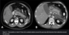
What investigations should be done if you suspect a splenic infarct and what will the findings be?

A

- Gold standard: CT abdomen with IV contrast

Will show segmental wedge shaped infarct towards hilum of the spleen. Whole spleen hypoattentuated if splenic artery occluded not a branch

- Routine bloods: FBC, LFTs, U+E’s, coagulation screen

7
Q

What is the immediate and long term management for a patient with a splenic infarct?

A

Immediate:

  • Analgesia
  • IV fluids
  • Manage underlying condition
  • Consider long term anticoagulation
  • Avoid splenectomy due to OPSI

Long-term:

- Vaccination against encapsulated bacteria NHS

  • Prophylatic low dose abx (Penicillin V)
8
Q

What are the complications of a splenic infarct?

A

- Splenic rupture: do splenectomy

- Splenic abscess: when cause of infarct was non-sterile embolus e.g infective endocarditis. May be seen on CT scan but confirmed with explorative surgery

- Pseudocyst formation

- Autosplenectomy: causes asplenism. Repeated splenic infarcts leads to progressive fibrosis and atrophy of the spleen. Common in sickle cell as when crises there is splenic artery occlusion. Leaves patient susceptible to infection

9
Q

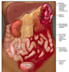
What are some of the causes of splenic rupture?

A

- Blunt trauma: seatbelt injuries, falls onto the left side

- Iatrogenic

- Splenomegaly: EBV, haemtological malignancy. Cause capsule to stretch and thin so susceptible to rupture

10
Q

What are the clinical features of splenic rupture?

A
  • Abdominal pain
  • Hypovolemic shock
  • LUQ tenderness/Peritonism
  • Kehr’s sign due to diaphragm irritation
11
Q

What investigations do you do for a suspected splenic rupture?

A

Haemodynamically unstable with peritonism: immediate laparotomy

Haemodynamically stable: urgent CT chest-abdomen-pelvis with IV contrast

12
Q

How do you grade splenic injuries?

A
  • American Association for the Surgery of trauma.
  • Used to help guide which patients should have conservative maagement and which should have surgical
13
Q

How should we manage patients with splenic rupture?

A

All: assessed and resuscitate with ATLS protocol

Haeodynamically unstable/Grade 5: Urgent laparotomy

Haemodynamically stable/Grade 1-3: Conservative. Move to high dependency area on strict bed rest and regular abdominal exams for deterioration. CT repeated 1/52 post injury. Increasing tenderness or peritonitis re-image/laparotomy/embolisation. Prophylatic vaccinations on discharge

14
Q

What are the main complications of conservative treatment and embolisation treatment for splenic rupture?

A
  • Ongoing bleeding
  • Splenic necrosis
  • Splenic abscess or cyst formation
  • Thrombocytosis (give aspirin if platelet count rises)

- OPSI so give prophylatic Penicillin V and vaccinations to prevent sepsis

15
Q

What is bile made from?

A

Stored in the gallbladder until released to duodenum:

  • Cholesterol
  • Phospholipids
  • Bile pigments from Hb metabolism
16
Q

What are the three main types of gallstones?

A

- Cholesterol: linked to poor diet and obesity

- Pigment : due to excess bille pigment production, seen in haemolytic anaemias

- Mixed: both cholesterol and bile pigments

17
Q

What are the risk factors for gallstones?

A

5 F’s:

  • Fat
  • Female
  • Fertile
  • FHx
  • Forty

Also: pregnancy, COCP (oestrogen causing more cholesterol in bile), haemolytic anaemia, malabsorption (e.g Crohn’s)

18
Q

What are the clinical features of biliary colic?

A

- Sudden, dull, colicky pain in RUQ that may radiate to back or epigastrium

- Precipitated by eating fatty foods due to release of CCK causing gallbladder contraction

  • Can be N+V
  • Settles with pain relief and no inflammatory response
19
Q

What are the clinical features of acute cholecystitis?

A

- Constant pain and tenderness in RUQ/epigastrium

  • Signs of inflammation e.g fever or lethargy

- Murphy Sign +

- Guarding may suspect peritonitis/sepsis

20
Q

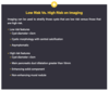
What investigations should you do if someone presents with RUQ pain that you suspect to be gallbladder related?

A
  • Lab tests (see image):
  • FBC, CRP, LFTs, Amylase, Pregnancy

- Imaging:

  • Transabdominal US 1st line
  • MRCP Gold standard if US inconclusive
21
Q

What would you see on ultrasound with gallstone pathology?

A
  • Gallstones or sludge
  • Thickened gallbladder wall if inflamed
  • Bile duct dilatation (stone in the distal bile ducts)
22
Q

How is biliary colic managed?

A

- Analgesia

- Lifestyle advice (weight loss, low fat diet, exercise)

- Elective laparoscopic cholecystectomy within 6 weeks due to high risk of recurrence/complications

23
Q

How is acute cholecystitis managed?

A
  • IV Abx (Co-amoxiclav +/- Metronidazole)
  • Analgesics
  • Antiemetics
  • IV fluids

- Laparoscopic cholecystectomy within a week, better within 72 hours of presentation

- Percutaneous cholecystostomy if not fit for surgery/not responsive to abx. Drains infection

24
Q

What is the difference between MRCP and ERCP?

A

MRCP - Identifies any biliary obstruction

ERCP - Identifies any biliary obstruction and allows for intervention

25
If a patient presents with RUQ post-cholecystectomy, what is the likely pathology and what should you do?
- Retained CBD stone - Use US abdomen but if unremarkable do MRCP
26
What are some complications of gallstones?
- Mirizzi Syndrome - Gallbladder empyema - Chronic cholecystitis - Bouveret’s Syndrome - Gallstone ileus
27
What is Mirizzi Syndrome and how is it managed?
- Stone located in **Hartmann's pouch** or the cystic duct, that **compresses the common hepatic duct** - Causes **obstructive jaundice** - Confirmed with **MRCP** and manage by **laparoscopic cholecystectomy**
28
What is gallbladder empyema and how is it managed?
**- Gallbladder filled with pus** - Patient unwell and **often septic**, same clinical picture as acute cholecystitis - Diagnose with **US** or **CT** - Manage with **laparoscopic** (often switches to open) **cholecystectomy** or p**ercutaneous cholecystotomy**
29
What is chronic cholecystitis, what are the complications of this and how is it managed?
- History of recurrent or untreated cholecystitis - Ongoing RUQ or epigastric pain w/wo N+V **- Complications:** gallblader carcinoma, biliary enteric fistula **- Diagnose with CT** or often histologically post-cholecystectomy. - Treat with **elective cholecystectomy**
30
What is Bouveret’s Syndrome and Gallstone Ileus?
Inflammation of gallbladder can form **cholecystoduodenal fistula** allowing gallstones into small bowel, resulting in **bowel obstruction** **- Bouveret’s Syndrome –** stone impacts proximal duodenum, causing a gastric outlet obstruction **- Gallstone Ileus –** stone impacts terminal ileum (narrowest part of small bowel), causing a small bowel obstruction
31
What is cholangitis and what are some of the causes of this?
**Infection of the biliary tract** due to **biliary outflow obstruction.** Obstruction causes stasis of fluid and increased intraluminal pressure that allows **bacteria to colonise the billiary tree** **_Common Causes:_** Gallstones, ERCP/Iatrogenic, Cholangiocarcinoma **_Rare causes:_** Pancreatitis, Primary Sclerosing Cholangitis, Ischaemic Cholangiopathy, and parasitic infections
32
What are the most common infective organisms in cholangitis?
- Escherichia Coli - Klebsiella species - Enterococcus
33
What are the clinical features of cholangitis?
**- Charcot's Triad** **- Reynold's Pentad** **- Pruitis** **- Pale stool/Dark urine** - Tachycardia - History of gallstones, ERCP, meds like COCP and fibrates
34
What investigations should you do when you suspect cholangitis to find the underlying cause and what will the findings be?
**- Routine bloods:** FBC (raised WBC) and LFTs (raised ALT, gamma GT and bilirubin) **- Blood cultures:** take before broad spectrum abx **- US of biliary tract:** bile duct dilatation \>6mm **- ERCP:** gold standard diagnostic and therapeutic. May do MRCP before
35
How is cholangitis managed?
- Sepsis 6 with I**V broad spectrum abx** (Co-amoxiclav) - Endoscopic biliary decompression with **ERCP** or percutaneous transhepatic cholangiograpy (**PTC**) 2nd line **- Elective cholecystectomy** if the cause was gallstones
36
What are the complications of ERCP?
- **Repeated cholangitis** - Pancreatitis - Bleeding (more common when sphincterotomy performed) - Perforation
37
What is the prognosis for cholangitis and what factors increase mortality?
- 5-10% for those given early abx therapy **- Increased mortality:** delayed diagnosis, liver failure, cirrhosis, CKD, hypotension, female, over 50
38
What is a cholangiocarcinoma and what is the common histology and location of these?
- Cancer in the biliary system but usually extrahepatic **- Common location**: bifurcation of right and left hepatic ducts (Klatskin tumour). Slow growing that invades locally and metastases to lymph nodes then peritoneal cavity, liver, lung **- Histology**: 95% adenocarcinoma, SCC, lymphoma, sarcomas, small cell cancers
39
What are the main risk factors for cholangiocarcinomas?
- Primary sclerosing cholangitis - Ulcerative colitis - Infective (Liver flukes, HIV, hepatitis virus) - Toxins (Chemicals in rubber and aircraft industry) - Congenital (Caroli’s disease, choledochal cyst) - Alcohol excess - Diabetes mellitus
40
What are the clinical features of cholangiocarcinomas?
**- Usually asymptomatic until later disease** - Post hepatic jaundice - Pruitis - Pale stools/dark urine - Cachexia - Sometimes: RUQ pain, malaise, early satetity, weight loss
41
What is Courvoisier’s Law?
In the presence of **painless jaundice** and an **enlarged gallbladder/RUQ mass,** there is likely an **obstructing pancreatic or gallbladder malignancy** not gallstones Sign only present if tumour obstructing distal to cystic duct
42
What are some differentials for cholangiocarcinoma?
Think of other causes of post-hepatic jaundice: - obstructive choledocholithiasis - bile duct strictures - external compression from extra-biliary tumour - benign biliary tumours - pancreatic tumours - primary biliary cirrhosis - primary sclerosing cholangitis.
43
What investigations need to be done when you suspect a cholangiocarcinoma and what do you expect to find?
**_Bloods_** **_-_** Raised ALP, Gamma GT, Bilirubin - Tumour markers **CEA/CA19-9** **_Imaging_** - US **- MRCP GOLD STANDARD** will show strictures in biliary tree - ERCP (can take biopsies) - CT for staging as can look for distant metastases - Angiography for pre-op planning of hepatic arteries
44
The only cure for cholangiocarcinomas is complete surgical resection, what are some of the procedures used?
**Intrahepatic or Klatskin tumour:** partial hepatectomy and reconstruction of the biliary tree **- Distal common duct tumour:** pancreaticoduodenectomy (termed a Whipple’s procedure). **- Radiotherapy** as neo/adjuvant therapy
45
Most patients with cholangiocarcinoma will require palliative treatment. What are some of the options for this?
**Stenting:** with ERCP to relieve obstructing symptoms but may need to be replaced every few months as prone to occlusion **Surgical bypass:** if obstruction not relieved by stenting **Palliative radiotherapy and chemotherapy** to slow tumour growth e.g cisplatin
46
What are some of the complications with cholangiocarcinomas?
- Increased risk of **biliary tract sepsis** due to obstruction **- Secondary biliary cirrhosis** - Prognosis 12-18 months to live after diagnosis - Distal tumours better prognosis than proximal
47
What are simple liver cysts and some of the clinical features of these?
**Fluid-filled epithelial-lined sacs** within the **liver**, most commonly occurring in the right lobe Though to be **congenitally malformed bile duct cells**, failing to connect to the extrahepatic ducts **Symptoms:** mostly asymptomatic and found on imaging, some have ab pain, nausea and early sateity due to mass effect
48
How are simple liver cysts investigated?
**Bloods:** LFTs normal but may have raised gamma GT and tumour markers CEA/CA19-9 **US**: imaging modality of choice. well defined, thin walled, spherical lesions with no septation and strong posterior wall acoustic enhancement
49
How are simple liver cysts managed?
**- \<4cm asymptomatic:** no intervention - \>**4cm asymptomatic:** follow up US scans at 3,6,12 months to check for growth. If size of cyst remains unchanged for 2-3 years no intervention unless becomes asymptomatic **- Symptomatic:** ultrasound-guided aspiration or laparoscopic de-roofing (lower rates of recurrence)
50
What is polycystic liver disease and what is it caused by?
Presence of **≥20 cysts** in **liver parenchyma**, each of which are **≥1cm in size.** Caused by **ADPCKD** or **ADPLD** Structures are **not connected to intrahepatic bile ducts** so **do not drain** and lead to dilatation and cyst formation filled with bile fluid
51
What are the clinical features of polycystic liver disease?
**- Asymptomatic** **- Abdominal pain** and **hepatomegaly** as increases in size - May have urinary symptoms if kidneys involved - Eventual liver cirrhosis and portal hypertension
52
How do we investigate polycystic liver disease?
**Bloods:** normal LFTs **Imaging:** US with \>20 cysts that have the same morphology as simple cysts
53
How do we manage polycystic liver disease?
- Can be **left alone** whilst asymptomatic **- Somatostatin analogues e.g Ocreatide** for short term relief of symptoms as reduces cyst volume **_- Surgery:_** * Laparoscopic de-roofing of cysts * US-Guided Aspiration (high recurrence so not done) * Resection of liver * Transplant
54
What would be an indication for surgery in polycystic liver disease?
(1) intractable symptoms (2) inability to rule out malignancy on imaging alone (3) prevention of malignancy.
55
What are the clinical features of cystic neoplasms in the liver and what is the histology of these?
**Features:** asymptomatic as grow very slowly, abdominal pain, anorexia, bloating, fullness, nausea **Histology:** cystadenomas (non-invasive mucinous cystic neoplasms). Premalignant biliary epithelium that can become cystadenocarcinomas
56
What are the investigations and management for cystic neoplasms?
**- Bloods:** normal LFTs but may have mildly raised CEA, ALP, CA19-9 **- US:** can tell the different between simple and complicated cysts **- CT imaging with contrast:** to diangose and look for metastases **- Management:** liver lobe resection whether adenoma or adenocarcinoma and histological diagnosis
57
What investigative procedure should you avoid if you suspect a cystic neoplasm of the liver?
Aspiration or biopsy should be avoided as can cause peritoneal seeding
58
What are the different features of liver cysts on US that help us to distinguish the type of cyst?
**Malignancy:** septations, wall enhancement, nodularity **Abscess:** debris within cyst, loculation (also malignant sign) **Hyatid Cyst:** calcification, daughter cysts around main lesion
59
What are hyatid cysts?
Liver cysts formed by infection by the **tapeworm** Echinococcus granulosus. Eggs are passed **faeco-orally** (dogs) and **pass into the hepatic portal system** where they form cysts High prevalence in South America, North Africa, Central Asia
60
What are some clinical features of hyatid cysts and what investigations can be done to diagnose this?
**- Asymptomatic** for years as slow growing **- Vague abdominal pain** due to mass effect or rupture - May cause jaundice, cholangitis, vomiting, dyspepsia **_- Bloods:_** LFTs normal, eosinophillia, positive antibody titre **_- US:_** calcified, spherical lesion with multiple septations. Anechoic with snowflake inclusions **_- CT Imaging with contrast_**
61
How are hyatid cysts managed?
- If asymptomatic can monitor **- Surgical cyst deroofing** **- Antiparasitics** e.g albendazole, mebendazole and/or praziquantel - DO NOT ASPIRATE AS RUPTURE CAN CAUSE ANAPHYLAXIS
62
What is the pathophysiology of a liver abscess?
**Polymicrobial bacterial infection** spreading from the **biliary o**r **gastrointestinal tract**, either via contiguous spread or seeding from portal and hepatic veins **Common organsims:** E. Coli, K. pneumoniae, and S. constellatus, Fungal in immunocompromised **_Causes:_** cholecystitis, cholangitis, diverticulitis, appendicitis, or septicaemia
63
What are the clinical features of a liver abscess?
**- Fever, rigors, abdominal pain** - Bloating, nausea, anorexia, weight loss, fatigue and jaundice - On examination will have **RUQ pain** with **hepatomegaly** - If ruptures may have signs of sepsis
64
What investigations should you do if you suspect a liver abscess, and what will they show?
**- Bloods:** raised WBC, raised ALP **- Blood and fluid cultures:** should be sent off **- US:** poor defined lesions with hypo- and hyper-echoic areas, with gas bubbles and septations **- CT with contrast:** similar findings to US with surrounding oedema
65
How are liver abscesses managed?
DEPENDS ON SIZE - Fluid resuscitation **- Antibiotics** **- US/CT guided aspiratio**n w or w/o catheter drainage **- Cyst deroofing** - Surgery if ruptures TREAT ANY UNDERLYING CAUSE TOO
66
What is an amoebic abscess?
Most common extra-intestinal manifestation of **Entomoeba Histiolytica** (from spread via portal system) Spreads faeco-oral and once in colon invades mucosa to get to portal system. Common in developing world e.g South America, India
67
What are the clinical features of an amoebic abscess?
**- Vague symptoms** of abdominal pain, nausea, fever or rigors, weight loss, and bloating - May have prodrome of **abdominal pain** and **diarrhoea** **- History of recent travel (**\<6 months) to an endemic area
68
How are amoebic abscesses investigated and managed?
**_Ix:_** - Bloods: raised WBC, deranged LFTs, E.Histolytica antibodies - Blood cultures - Stool samples: for antibodies - US/CT: poor defined lesions **_Mx:_** - Antibiotics (***Metronidazole*** or ***Tinidazole***) - Surgical drainage for large cysts that do not respond to abx - Luminal agent (***Paromycin***) to clear colon of amoebiasis
69
90% of liver cancer is due to metastases. The other 10% of cases is due to Hepatocellular carcinomas. What are some of the causes of HCC?
6th most common cancer due to chronic inflammatory processes: **- Viral hepatitis** (most common) - Chronic alcoholism - Hereditary haemochromatosis - PBS - Aflatoxin (cereals and nuts)
70
What are some risk factors for developing hepatocellular carcinomas?
- Viral hepatitis (Hep B and C) - Smoking - \>70 - Aflatoxins - Family history of liver disease
71
What are the clinical features of hepatocellular carcinomas and some differentials for this?
**- Main symptom:** liver cirrhosis **- Irregular enlarged craggy tender liver** - Dull ache in RUQ - Fatigue, fever, weight loss, lethargy **- Late:** ascites and jaundice **_Differentials:_** infectious hepatitis, cardiac failure (smooth liver), benign hepatocellular adenoma, other causes of liver cirrhosis
72
What are some labatory tests that can be done when diagnosing HCC?
**- LFTs:** ALP, AST, ALT, Bilirubin all deranged **- Low platelets** and **prolonged clotting** **- Alpha-Fetal Protein (AFP)** may be raised and can be used to monitor progression
73
How is HCC diagnosed with imaging?
**- US:** inital imaging of choice. Mass \>2cm with raised AFP is diagnostic **- MRI liver scanning** or **percutaneous fine-needle aspiration** may be used if US suggestive but not sure. Be careful of seeding **- CT staging scan**
74
What is the staging tool used to stage HCC?
**Barcelona Clinic Liver Cancer staging system** (BCLC) Helps to decide which treatment is suitable
75
What are two risk assessment tools used once a diagnosis of HCC has been made?
**Child-Pugh:** calculates prognosis of patients with liver cirrhosis. Looks at serum bilirubin, albumin, INR, degree of ascites, and evidence of encephalopathy **MELD:** calculates same prognosis but better and also predicts likelihood of patient tolerating a liver transplant. Looks at creatinine, bilirubin, INR, sodium, and the use of dialysis (at least twice per week)
76
Surgery is the only curative treatment for HCC. What is the surgical management for hepatocellular carcinoma?
**- Surgical resection:** if no cirrhosis and good baseline health. High 5 year recurrence rate **- Transplation:** only if fufill the Milan criteria. One lesion \<5cm or three lesions \<3cm, no extrahepatic manifestations, no vascular infiltration
77
What is some non-surgical management used in the treatment of HCC?
_- **Image guided ablation:**_ if early stage BCLC 0 or A. Put US waves into tumour to cause necrosis _**- Alcohol ablation**:_ if small **_- Transarterial Chemoembolisation (TACE):_** BCLC stage B (a large multinodular tumour. High concentrations of ***chemotherapy*** are injected ***directly into hepatic artery*** and an ***embolising agent*** is then added to induce ischaemia. Radiological techniques used so only embolise branches of arteries supplyin tumour not whole liver
78
What is the prognosis with HCC?
- Depends on extent of underlying malignancy - Median survival 6 months
79
What malignancies metastasise to the liver and how do they present?
- **Bowel** (via the portal circulation), **breast, pancreas, stomach,** and **lung** - Hepatomegaly and ascites - Jaundice and upper abdominal pain
80
What investigations and management is done for secondary cancers of the liver?
**_Ix:_** **- Bloods:** derange LFTs with raised ALP **- US** and **CT** to find staging and source of metastases **- Biopsy** not advised due to seeding **_Mx:_** - Often palliative - Surgery if metastases confined to liver and primary tumour under control. Can do TACE or selective internal radiotherapy
81
How can you tell the difference between acute and chronic pancreatitis?
**Limited damage to the secretory function** of the gland, with no gross structural damage developing. Repeated acute can lead to chronic
82
What is the aetiology for acute pancreatitis?
(See image) **Most common:** Excess alcohol consumption and Gall stones Cause not found in some patients
83
What is the pathophysiology of acute pancreatitis?
- Premature and **exaggerated activation of digestive enzymes** - **Pancreatic inflammatory response** (Increasing vascular permeability and fluid shifts, third spacing) - Pancreatic enzymes in blood stream cause fat autodigestion leading to **fat necrosis** and **hameorraghe** into retroperitoneal space - Free fatty acids react with calcium to form chalky deposits and **hypocalcaemia** - End stage is complete **necrosis of the pancreas**
84
What are the clinical features of acute pancreatitis?
- **Sudden onset severe epigastric pain** that can **radiate to back** - Nausea and vomiting - Guarding - Cullen's/Grey-Turners if retroperitoneal haemorraghe - Tetany from hypocalcaemia - Jaundice if gallstones aetiology
85
What are some differential diangoses for acute pancreatitis?
- AAA - Renal calculi - Chronic pancreatitis - Aortic disection - Peptic Ulcer disease
86
What are some labatory investigations to diagnose acute pancreatitis?
**Serum Amylase:** 3x upper limit of normal **LFTs:** to see if any cholecystic involvement. Raised ALT likely cause is gallstones **Serum Lipase:** more accurate than amylase as remains elevated for longer
87
Other than aucte pancreatitis, what may cause a raised serum amylase?
- Bowel perforation - Ectopic pregnancy - DKA
88
What are some of the risk tools used to assess the severity of pancreatitis?
**- Modified Glasgow Criteria** within 48 hours of admission **- Score of 3 or more** have severe pancreatitis and a high-dependency care referral is needed - PANCREAS mneumonic (see image) **Others:** APACHE II score, Ranson Criteria, and Balthazar score (CT scoring system)
89
What imaging is done if you suspect acute pancreatitis and what are the findings?
**_- US Abdomen:_** to find cause **_- AXR:_** not routinely done but may have ***sentinel loop sign***. Do ***CXR*** to look for pleural effusion or ARDS **_- Contrast enhanced CT:_** done if complications or can't find underlying cause if 48 hours after admission there is ***pancreatic oedema and swelling***, or non-enhancing areas suggestive of ***pancreatic necrosis***. If signs of persistent inflammatory response/organ failure do not CT for severity till 6-10 days after admission
90
What are the three steps of management for low severity acute pancreatitis?
- Fluids - Analgesics - Nutrition support (enteral feeding)
91
What is the management for acute pancreatitis?
**- Manage underlying cause** (e.g ERCP and spincterotomy/cholecystectomy for gallstones) **- Supportive treatment** (see image OI OI) **- ONLY Broad spectrum abx** (Imipenem) IF for prophylaxis against infection in cases with **confirmed pancreatic necrosis** - If severe case treat in high dependency unit
92
What are some systemic complications of acute pancreatitis?
- DIC - ARDS - Hypocalcaemia - Hyperglycaemia as Islets of Langerhan's destroyed and insulin production halted
93
What are some local complications of acute pancreatitis and how are they managed?
**_Pancreatic Necrosis_** - Suspect if patient has signs of persistent systemic inflammation for more than 7-10 days after onset - Necrosis prone to infection so suspect if ***patient deteriorates*** and ***raised inflammatory markers*** - Confirm by ***CT imaging*** then ***fine needle aspiration biopsy*** - Treat with ***pancreatic necrosectomy*** 3-5 weeks after onset so walled off necrosis **_Pancreatic Pseudocyst_** - ***Collection of fluid*** containing pancreatic enzymes, necrotic material and blood. ***Lack epithelial lining*** but surrounded by vascular and fibrous that's why psuedo - Often in lesser sac blocking gastro-epiploic foramen - Often found incidentally on image or mass effect e.g biliary or gastric outlet obstruction - Can ***haemorraghe, rupture*** or ***become infected*** - May ***resolve spontaneously*** but if still there after 6 weeks ***surgical debridement*** or ***endoscopic drainage***
94
What is chronic pancreatitis and the aetiology of this?
Chronic fibro-inflammatory disease causing **progressive and irreversible damage** to the pancreatic parenchyma 4x more likely in men and more likely aged \>40 **Main causes:** chronic alcohol abuse or idiopathic **Rare causes:** metabolic (hyperlipidaemia, hypercalcaemia), infection (HIV, mumps, coxsackie), hereditary (CF), autoimmune (e.g. AIP or SLE), anatomical (malignancy or stricture formation), or congenital anomalies
95
What are the clinical features of chronic pancreatitis?
**- Chronic Epigastric Pain** (radiating to back, eased by leaning forward) - Exocrine/endocrine insufficiency (see image - Type 3c DM) - Often pseudocysts present and may have mass effect e.g biliary obstrucution
96
What lab investigations should be done for patients with abdominal pain you suspect to be chronic pancreatitis?
**- Urine dip** **- Routine bloods:** FBC/CRP **- Serum amylase/lipase:** not raise **- LFTs:** for obstructive jaundice **- BM:** to see if endocrine dysfunction **- Faecael elastase:** low with exocrine dysfunction. Enzyme released from pancreas into bowel that is not degraded
97
What imaging is done if you suspect chronic pancreatitis?
**CT imaging:** may show pancreatic calcification or atrophy and any pseudocysts present. Can show malignancies that can cause chronic pancreatitis **US or MRCP:** image biliary tree and pancreas but if normal doesn't exclude chronic pancreatitis **Secretin Stimulation test** or **Endoscopic US (EUS)** if diagnostic uncertainty
98
How is chronic pancreatitis managed?
**- Treat reversible underlying cause** e.g alcohol cessation or statins for hyperlipidaemia **- Analgesics** (neuropathic not opiates like acute) - Exocrine dysfunction give **enzyme replacement** (like Creon) and fat soluble vitamins - Endocrine dysfunction may need **insulin regimes** and annual check of HbA1c **- ERCP** for diagnostic and stone removal/sphincterotomy - Endosonography-guided celiac plexus blockade or thoracoscopic splanchnicectomy for analgesia **- Steroids** to reduce symptoms of autoimmune pancreas. High dose prednisolone then low dose maintenance
99
What is the prognosis with chronic pancreatitis?
- Endocrine and exocrine insufficiencies - Pancreatic malignancy risk once had for over 20 years
100
What is the pathophysiology of pancreatic cancer?
- 90% are **ductal carcinoma of the pancreas.** - 10% Exocrine (Pancreatic Cystic Carcinoma) or Endocrine (Derived from Islet Cells of Pancreas) **- Direct invasion of local structures** typically involves the spleen, transverse colon, and adrenal glands. **- Lymphatic metastasis** typically involves regional lymph nodes, liver, lungs, and peritoneum and common at diagnosis
101
What are some of the risk factors for pancreatic cancer?
- Smoking - Chronic pancreatitis - FHx - Late onset DM (\>50)
102
What is the clinical presentation of pancreatic cancer?
Features depend on site of tumour, at tail insidious onset + not symptomatic until late * *- Obstructive Jaundice** * *- Abdo Pain** (Secondary to invasion of coeliac plexus) * *- Weight loss/**Cachetic - Acute Pancreatitis - Thrombophlebitis Migrans - Abdominal mass/enlarged gallbladder
103
What is Courvoisier’s Law?
104
What investigations are done when we suspect pancreatic cancer?
**_Lab_** ***- FBC*** (anaemia and thrombocytopenia) ***- LFTs*** (raised bilirubin, ALP and gamma GT showing obstructive jaundice) ***- CA19-9*** not to diagnose but to measure response **_Imaging_** - ***Initial USS*** to look for ***bile duct dilatation*** or ***pancreatic mass*** ***- CT to diagnose*** followed by ***chest-abdomen-pelvis CT scan*** or ***PET*** to stage ***- Endoscopic US*** for ***fine needle aspiration biopsy*** or ERCP for biopsy
105
What is the curative treatment for pancreatic cancer?
**_Surgical resection:_** - Whipples Procedure (pancreaticduodenectomy) or - Distal Pancreatectomy (if tumour of tail) CONTRAINDICATED IF METASTASES **_Adjuvant chemotherapy:_** - Following surgery give 5-fluorouracil
106
What is a Whipple's procedure and what are some complications of this procedure?
Removal of the ***head of the pancreas,*** the ***antrum*** of the stomach, the **1st and 2nd parts of the duodenum**, ***common bile duct,*** and ***gallbladder.*** All share blood supply of ***gastroduodenal artery*** ***Tail of the pancreas*** and the ***hepatic duct*** are ***anastomosed to the jejunum*** to drain bile and ***stomach*** anastomosed too **_Complications:_** pancreatic fistula, delayed gastric emptying, and pancreatic insufficiency
107
In metastatic disease for pancreatic cancer, what is the chemotherapy regime?
**FOLFIRINOX** (folinic acid, 5-fluorouracil, irinotecan, and oxaliplatin) If not well enough to tolerate this give ***gemcitabine therapy***
108
Pancreatic cancer has a very poor prognosis as it is a highly metastatic cancer. What are the palliative care options with pancreatic cancer?
**Percutaneous or ERCP _Biliary stent_:** to relieve obstructive jaundice and pruitis **Palliative chemotherapy:** gemcitabine based. used if good performance status **Enzyme replacements (inc Lipases e.g Creon):** for exocrine insufficiency to stop malnourishment and steatorrhea
109
What are some endocrine tumours of the pancreas?
Functional (symptoms due to secretion) or Non-functional (symptoms due to metastatic spread) - Gastrinoma (Zollinger-Ellison) - Insulinoma - Somatostatinoma - Glucagonoma - VIPoma
110
What is multiple endocrine neoplasia 1 syndrome (MEN1)?
Hereditary condition associated with tumours of the endocrine. Used to be called Wermer syndrome - Hyperparathyroidism due to parathyroid tumours - Endocrine pancreatic tumours - Pituitary tumours
111
How are endocrine tumours of the pancreas investigated and managed?
**_Ix:_** - Combination of CT, MRI and/or Endoscopic Ultrasound. - Intra-arterial calcium with digital subtraction angiography **_Mx:_** - If small non-functioning just observed - If large functioning then primary tumour and metastases resected - Somatostatin analgoues can help control effects of hormone oversecretion
112
What are some low risk and high risk pancreatic cysts?
Cysts identified by histology, risk of malignancy and secretions In general **mucinous cysts** are **high risk** and **serous cysts** are **low risk**
113
What are some clinical features of pancreatic cysts?
**- Mostly asymptomatic** - **Abdominal/back pain** (compression), **post obstructive jaundice, vomiting** - If turn malignant may have general features of malignancy such as weight loss, loss of appetite, change in bowel habits
114
What are pancreatic pseudocysts?
Often following pancreatitis The inflammatory reaction produces a necrotic space in the pancreas that fills with pancreatic fluid, however this lacks an epithelial lining Often asymptomatic
115
How are pancreatic cysts investigated and how do we stratify them as low risk/high risk?
Often picked up on imaging. If found do **pancreatic protocol CT scan** or **magnetic resonance cholangiopancreatography** to further assess. May then do **Endoscopic US scan with Fine Needle Aspiration (EUS-FNA)** to further assess and work out low risk/high risk
116
How are pancreatic cysts managed and what is the prognosis?
**_Low risk:_** left alone with ***surveillence*** every 5 years due to malignancy risk **_High risk:_** ***resection*** is first line with follow up MRI every 2 years If non-malignant excellent prognosis but if malignant 60% 5 year survivial
117
What can cause air in the billiary tree?
- Post ERCP due to sphincterotomy - Following passage of gallstone - Scarring from acute pancreatitis
118
Why shouldn't you do a CT for gallstones?
Will not see them if they are largely cholesterol based as not calcified Will only see fluid and inflammation around the gallbladder
119
What is gallbladder hydrops?
Gallstone stuck in Hartman's pouch which in turn chronically blocks the cystic duct. Gallbladder gets enlarged and creates mucus pouch Abdominal pain with palpable gallbladder without any signs of infection
120
How long after acute pancreatitis caused by gallstones should there be a cholecystectomy?
2 Weeks
121
What are some causes of chronic pancreatitis?
122
What is the prognostic scoring system used for acute pancreatitis?
Modified Glasgow Score. If \>3 suggest HDU/ITU input as severe P A N C R E A S