12 - T&O Spine, Hip and Thigh Flashcards

1
Q

What are the different types of fractured neck of femur and what are the causes?

A

NOF is a fracture anywhere from subcapital region of femoral head to 5cm below the lesser trochanter

High energy: RTA in the young

Low energy: Fall from standing height in elderly

Think pathological fractures if no trauma

Intra or Extracapsular

2
Q

Why is the mortality with NOF fractures so high in the first year?

A

Those with poor mobility before fracture, high age and co-morbidities at higher risk

Patient tends to die from complications such as pnuemonia etc

3
Q

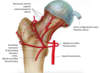
What is the blood supply to the femoral head?

A

Retrograde from MCFA from deep femoral artery from external iliac

Some blood from ligamentum arteriosum but only enough blood to supply head in children

Displaced intracapsular fractures can disrupt blood supply causing avascular necrosis

4
Q

Extracapsular fractures can be classified as intertrochanteric and subtrochanteric. What are the different classifications of intracapsular fractures?

A

Garden Classification

I and II are non-displaced

III and IV are displaced

5
Q

How will a #NOF present?

A
  • Pain in groin, thigh or referred to knee
  • Inability to weight bear
  • Shortened and externally rotated
  • Pain on pin rolling and axial loading
  • Unable to do straight leg raise
6
Q

What are some investigations that need to be done if you suspect a #NOF?

A
  • AP and Lateral Hip and Pelvis X-Ray

- Full neurovascular exam of the limp

  • FBC, U+Es, CK if long lie for rhabdo

- Group and Save

  • Urine dip, CXR and ECG in elderly
7
Q

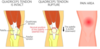
How is a #NOF managed?

A

- A to E

- Opioid or regional anaesthesia (fascia-iliaca block)

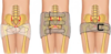
- Surgical (see image) with urgent physio and mobilisation after

If displaced intracapsular needs arthroplasty (full not hemi if active but will dislocate more) due to risk of AVN. If non-displaced can try screws to see if will repair without AVN as metal work has a life span

8
Q

What are some post op complications with #NOFs?

A

Immediate: pain, bleeding, leg length discrepancies, neurovascular damage

Long term: joint dislocation, aseptic loosening, peri-prostethic fracture, deep/prosthetic joint infection, mortality, AVN, malunion-nonunion

Make sure to get early physio, ortho-geriatricians and OTs involved

9
Q

Why should you not use NSAIDs in fracture healing?

A

Need inflammatory process for bone healing so will prolong healing time

10
Q

What is the Nottingham Hip Score?

A

Tool used by surgeons to work out the 30 day mortality risk of a patient following a #NOF

High lactate is also a marker of mortality in #NOF

11
Q

What are some of the symptoms and risk factors of hip osteoarthritis?

A

Symptoms

  • Dull pain in groin or buttock that is exacerbated by movement and relieved by rest
  • Stiffness and crepitus worse after resting
  • May have antalgic gait but if severe may have fixed flexion defority and Trendelenbery gait
  • Passive movement painful and reduced range of motion
12
Q

What are some differential diagnoses for hip OA?

A
  • Trochanteric bursitis
  • Gluteus medius tendinopathy
  • Sciatica
  • Femoral neck fracture
13
Q

What investigations are done to diagnose and classify hip OA?

A
  • X-ray Pelvis to show at least 2 typical OA features
  • MRI gold standard if not sure from X-ray
  • Can use WOMAC tool to monitor disease progression. Looks at pain, stiffness and function
14
Q

How is hip OA managed?

A

Conservative:

  • NSAIDs for pain
  • Weight loss, exercise, smoking cessation
  • Physiotherapy

Surgical (if above doesn’t work)

  • Hip hemi/arthroplasty
15
Q

What are some complications with a hip arthroplasty?

A
  • Thromboembolic disease
  • Bleeding
  • Dislocation
  • Infection
  • Loosening of prosthesis
  • Leg length discrepancy
  • Need for revision hip arhtroplasty after 15-20 years
16
Q

What is the pathophysiology of a femoral neck shaft fracture?

A
  • Often due to high energy trauma
  • Associated with neurovascular injury
  • Large blood loss (up to 1.5L) as highly vascularised due to haemopoetic role. Supplied by penetrating branches of profunda femoris
  • Often transverse fracture in proximal femur
17
Q

How does a femoral shaft fracture present and how is it classified?

A

- Pain in thigh and/or hip/knee

- Inability to weight bear

  • Assess skin as may be open or threatened (tethered, white, non-blanching)

- Proximal fragment in flexion and external rotation (iliopsoas and gluteus medius/minimus)

  • Full neurovascular exam needed
  • May have signs of hypovolemia
18
Q

What investigations are done when a femoral shaft fracture is suspected?

A

- Routine urgent bloods including coagulation and G+S

- Plain Film Radiograph AP and Lateral of femur, hip, knee

- CT scan if polyfracture or concurrent #NOF suspected

19
Q

How is a fractured femoral shaft managed before surgery?

A

Initial

  • ATLS protocol with A to E and appropriate fluid resus

- Adequate pain relief (opioid or regional block)

  • If open fracture abx prophylaxis and photography

- Immediate reduction and immobilisation using in-line traction so haematoma forms in right place

  • If too many co-morbidities or undisplaced femoral shaft fracture then long-leg cast and no surgery
20
Q

How is a femoral shaft fracture managed surgically after traction splinting?

A

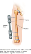
Within 24-48 hours needs antegrade intramedullary nail (retrograde if hip replacement)

If unstable polytrauma or open fracture then external fixation until intramedullary nail can be done

Early mobilisation after nailing decreases complications. If bilateral fractures more pulmonary complications

21
Q

When should you not use traction splinting for a fracture?

A
  • Hip or pelvic fracture
  • Supracondylar fracture
  • Fractures of ankle or foot
  • Partial amputation
22
Q

What are some complications of a femoral shaft fracture?

A
  • Neurovascular injury (pudendal or femoral nerve)
  • Mal-union, delayed union non-union (higher risk if smoker or post op NSAID use)
  • Infection (especially open)
  • Fat embolism
  • Hip flexor or knee extensor weakness
23
Q

What is the pathophysiology and risk factors for a quadriceps tendon rupture?

A

Rupture usually occurs at site of insertion on superior patella. Mechanism is following sudden excessive loading of quadriceps e.g landing from a jump

Risk factors: increasing age (>40), CKD, Diabetes, RA, medication like corticosteroids and fluoroquinolones

Differentials: patella tendon rupture, patella fracture, femoral shaft fracture

24
Q

How will a quadriceps tendon rupture present?

A

Symptoms

  • May hear a pop or feel a tearing sensation then…
  • Pain in anterior knee or thigh
  • Difficult to weight bear

Examination

  • Localised swelling
  • Tender palpable defect at top of patella
  • If complete tear inability to straight leg raise or extend knee
25
How is a suspected quadriceps tendon rupture investigated and diagnosed?
Diagnosis can be made clinically but definitive diagnosis with **US imaging** **Plain film radiograph** will show caudally displaced patella (patella baja) and can rule out patella fractures **MRI** if not sure from US
26
How is a quadriceps tendon rupture managed?
**_Partial tear:_** nonoperatively if extensor mechanism in tact. **Immobilisation of knee in brace** then **intensive rehab** **_Complete tear:_** If at insertion then **longitudinal drill holles** or **suture anchors**, if intra-tendinous tear then **end-to-end sutures**. Post op **immobilise in brace** then rehab at 6 weeks
27
What is a distal femur fracture and how are they classified?
**Fracture extending from distal metaphyseal-diaphyseal junction to articular surface of femoral condyles** Either high energy trauma in young, low energy in elderly due to malignancy/osteoporisis or peri-kneeprosthetic fracture
28
How does a distal femur fracture present and what are some differentials you should consider?
**- Severe pain in distal thigh** following fall or traumatic injury **- Inability to weight bear** - **Swelling and ecchymosis of distal thigh** - May be haemarthrosis **knee effusion** if fracture extends into joint - Full neurovascular exam **DD:** tibial plateau fracture, haemarthrosis, tibial shaft fracture
29
How is a suspected distal femur fracure investigated?
- ATLS protocol so **urgent bloods with G+S** **- Serum Ca/Myeloma screen** if suspect pathological fracture **- AP and Lateral plain radiograph** of knee and femur **- CT** if intraarticular involvement
30
How is a distal femur fracture managed?
- If **minimal displacement or very co-morbid** then non-operative **long period of immobilisation** and non weight bearing in **brace** **-** If significant mal-alignment in A+E then **initial realignement and immobilisation with skin traction** before surgery **_- Surgical:_** ***retrograde intramedullary nail*** (if proximal extra-articular or simple intra-articular) or ***open reduction internal fixation (ORIF)*** ***External fixation*** if severe comminuted or open fracture
31
What are the complications of a distal femoral fracture?
- Malunion - Non union (often in metaphyseal area) - Secondary OA if intrarticular extension of fracture - Knee pain/Stiffness
32
What is the pelvic ring and the true pelvis?
**Pelvic ring:** two inominate bones (ilium, ischium, pubis), the sacrum and their supporting ligaments **True pelvis:** rectum, bladder, uterus, iliac vessels, lumbosacral nerve roots Pelvic fracture can cause life threatening haemorraghe, neurological deficit, urological trauma, bowel injury
33
How do pelvic ring fractures present and what should you check on examination?
Usually high energy from blunt trauma like RTA or ralls from height so concurrent injuries **Pelvic deformity with significant pain and swelling around pelvis. External rotation and shortened limbs** Do **full neurovascular assessment** of lower limvs including checking **anal tone** as sacral nerve roots and iliac vessels can be injury Need to check for **urethral injury, open fractures** (in rectum and vagina), **abdominal injury, ecchymosis or haematomas** around perineum/scrotum/labia Also check chest, head spine, acetabulum and long bones
34
How do low energy pelvic fractures present and how are they managed?
Often affecrs ASIS due to sartorius, AIIS due to rectus femoris and ischial tuberosity due to hamstrings
35
How are pelvic fractures investigated and classified?
- ATLS guidelines - **3 plain film radigraphs** to see whole ring (AP, inlet view, outlet view) OR **- CT** **Young and Burgess** or **Tile Classification**
36
How are pelvic fractures managed?
**- ATLS** to identify life threatening injuries **- Pelvic binder** if any pelvic trauma to reduce bleeding and hypovolaemic shock due to blood loss intraabdominally or retroperitoneal venous plexus - Immediate surgery **if haemodynamically unstable** for **interventional radiology or trauma laparotomy +/- retroperitoneal packing** **-** Surgery for combination of **anterior and posterior stabilsation** depending on Young and Burgess score
37
What are some indications for stabilsation surgical management of a pelvic fracture?
- Life threatening haemorraghe - Unstable fractures (APC1 and LC1 only stable ones) - Open fractures - Fracture with urological injury
38
What are some complications of pelvic fratures?
- Urological injury (more in men) - Thromboembolic disease (PE, DVT) - Long standing pelvic pain - Infection
39
How does an acetabular fracture occur and how does it present?
Same as other hip fractures high/low energy **Hip or groin pain** with **inability to weight bear.** Need to do secondary survey for associate injuries e.g hip dislocation, NOF Check **neurovascular status, any open fracture, any Morel-Lavallee lesions** on overlying skin
40
What investigations are done if you suspect an acetabular fracture and how are they classified?
**- Plain film radiographs with antero-posterior view, Judet view (obturator oblique and iliac oblique)** **- Gold standard is CT** as in trauma setting - Classified by **Judet and Letournel classification** into either **elementary** or **associated**
41
42
How are acetabular fractures managed?
**- ATLS guidelines** for life-threatening injuries as high energy trauma **- No pelvic binder** needed as no risk of haemorraghe like with pelvic ring fracture but any **hip dislocation should be reduced urgently to prevent further damage to acetabulum** **- Un/Minimally displaced** can be conservatively managed with **non-weight bearing for 6-8 weeks** **- Displaced** needs **fracture fixation** as precursor to **total hip replacement.** Anterior approach for anteriorly displaced fractures and vice versa
43
What are some complications of acetabular fractures?
- Secondary OA - Venothromboembolism - Rarely damage to sciatic and obturator nerves
44
What is the difference between radiculopathy and radicular pain?
**Radiculopathy:** conduction block in axons of spinal nerve or its roots so weakness and/or anaesthesia. State of neurological loss that may/may not be associated with radicular pain **Radicular Pain:** pain from damage or irritation of the spinal nerve tissue, especially dorsal root ganglion
45
What is the aetiology of radiculopathy?
**Usually due to nerve compression by:** - Intervertebral disc prolapse (lumbar spine) - Degenerative disease of the spine (spinal canal stenosis) - Fracture - Malignancy - Infection (Herpes Zoster, Pott's disease, extradural abscess)
46
How may radiculopathy present and what are some red flags that could indicate emergency/sinister pathology?
**Paraesthesia, numbness and weakness** in a dermatomal/myotomal distribution. Can also have radicular pain which is burning, deep, strap-like or narrow pain Check perianal dermatomes, anocutaneous reflex and rectal pressure sensation with pinprick as diminished in CES
47
What are some differentials for radicular pain?
- Referred pain (e.g MI to left shoulder, HBD to right shoulder) - Myofascial pain (hip and shoulder muscles) - Thoracic outlet syndrome - IT band syndrome - Meralgica Paraesthetica - Piriformis syndrome - Greater trochanteric bursitis
48
What is the management of radiculopathy?
- Depends on underlying cause, only surgery if CES - If unremitting pain despite non-surgical management or if new or progressive myelopathy then surgery **- Analgesia** and **Neuropathic pain modulators** (Amitriptyline, Gapapentin, Pregabalin) **- Benzodiazepenes** (Diazepam) for muscle spasms **- Physiotherapy**
49
What is myelopathy?
- Injury to the spinal cord by compression - Radiculopathy is nerve root, myelopathy is spine
50
What chart is used for a spinal cord injury?
ASIA Chart
51
What is the pathophysiology of degenerative disc disease?
Related to aging where there is progressive dehydration of the nucleus pulposis and tears in the annulus fibrosis
52
How may degenerative disc disease present?
**Early:** exam may be remarkable or local spinal tenderness, contracted paraspinal muscles, hypomobility, painful extension of back or neck **Late (when instability):** pain more severe and may include radicular leg pain or paraesthesia and eventually scoliosis. Pain can be reproduced on raising extended leg (***Lasegue Sign)*** Complete neurological exam needs to be done to rule out spinal cord compression and CES
53
Degenerative disc disease diagnosis is largely clinical. When should imaging for degenerative disc disease take place?
- Red flags present - Radiculopathy with pain for more than 6 weeks - Evidence of spinal cord compression - Imaging would alter management **MRI spine is gold standard**
54
How is degenerative disc disease managed?
**- Adequate simple pain relief** **- Encouraging mobility and physiotherapy** - If pain continues \>3months then refer to pain clinic NO EVIDENCE TO SUPPORT SURGICAL INTERVENTION LIKE SPINAL FUSION
55
How are cervical fractures classified?
**AO classification** **_Upper Cervical (C1 or C2)_** - Region involved (1-3) - Injury type (A-C) **_Subaxial_** - Injury type (A, B, C, F)
56
How does a cervical fracture present and what are the three main types of cervical fracture?
- May have neck pain - Neurological involvement - If vertebral artery involved may cause POCS stroke **Differentials:** whiplash, cervical dislocation, cervical spondylosis
57
How are patients with cervical spine injury (possible fracture) investigated?
Depending on Canadian C Spine Rules: **- CT in adults** **- MRI in children** MRI can look at soft tissue injuries, ligaments and intervertebral discs
58
How are cervical fractures managed?
- If suspected 3 point C-Spine immobilisation to prevent damage to spinal cord **_Non operative (Stable)_** **- Rigid collar** until inital assessment **- Halo vest** for definitive treatment - If operation is high risk use **traction devices** **_Operative (Unstable)_** - Fuse injured segment to uninjured segments above and below for stabilisation
59
Where is the most common fracture in the spine and how are they classified?
Thoracolumbar juntion (T11-L2) as this is a zone of mechanical transition
60
What is a Burst and Chance fracture?
**Burst** ## Footnote - Compressive force through anterior and middle column of vertebrae so retropulsion of bone into spinal canal - Potential spinal cord injury - Can involve one end plate (incomplete burst) or both (complete) **_Chance_** - Excessive flexion of spine and involves all three spinal columns - Compression and distraction - Unstable injuries and need surgical intervention to stabiise - Occur after RTAs when person only wearing lap belt
61
How will a thoracolumbar fracture present?
Back pain after trauma but may not be present if other distracting pain Often occurs in low energy trauma in osteoporotic patients Need to do full neurological exam as may involve spinal cord
62
What investigations should you do if you suspect a thoracolumbar fracture?
- Plain film radiograph first line (AP and lateral) - CT if x-ray abnormal or neurological signs
63
How are thoraco-lumbar fractures managed?
**- Immobilsation** whilst assessing - If **stable** can do **extension bracing** or **lumbar corsets** with **analgesia** and **physio** - If **unstable** look at **TLICS** score to decide whether surgery. Will have **decompression and instrumented spinal fusion** with pedicle screws and rods
64
What is the pathophysiology of cauda equine syndrome?b
**- Disc herniation:** L4/L5, L5/S1 **- Trauma:** vertebral fracture and subluxation **- Neoplasm:** primary or metastatic (BLTKP) **- Chronic spinal inflammation:** ankylosing spondylitis **- Iatrogenic:** haematoma secondary to spinal anaesthesia
65
How does CES present and what examinations should you do if you suspect this?
**LMN signs:** - Bilateral reduced lower limb sensation - Bladder or bowel dysfunction - Lower limb motor weakness - Back pain - Impotence - So check for bladder retention and perianal numbness **PR and post-void bladder scan**
66
How is CES classified in stages?
**CES with retention (CESR)** Back pain with uni/bilateral sciatica, lower limb motor weakness, perianal numbness, loss of anal tone and loss of urinary control **Incomplete CES (CESI)** As above but instead of urinary retention just altered urinary sensation (loss of desire to void, diminished sensation, poor stream, need to strain) **Suspected CES (CESS)** Cases of severe back and leg pain with variable neurological symptoms and suggestion of sphincter disturbance
67
How is a suspected case of CES investigated?
Gold standard: whole spine MRI
68
How is CES managed?
**Early neurosurgical review with urgent surgical decompression** (within 24h) May be started on **high dose steroids** (dexamethasone) to reduce any localised swelling If trauma is the cause **immobilsation**
69
What are some red flags with back pain?
70
What are the most common cancers affecting the spine?
- Metastases BLTKP - Myeloma
71
What is the difference between vascular and neurogenic claudication?
72
What are the differences in UMN and LMN lesions?
73
How can you spot a NOF on x-ray?
**Shenton’s line disruption:** loss of contour between normally continuous line from medial edge of femoral neck and inferior edge of the superior pubic ramus **Lesser trochanter may be more prominent** due to external rotation of the femur
74
How does sciatica present and what tests can you do on examintation?
- Sudden pain from lower back to foot often due to disc prolapse at L4/L5 or L5/S1 - Do straight leg raise and this will cause leg pain NOT back pain (Lasegue's sign) - Weakness and numbness in L5 or S1 (EHL, Soleus/Gastrocnemius)
75
Where is the hip capsule?
Capsule sits on intertrochanteric line on the front then sits 2/3 of the way along the neck of femur on the back Intertrochanteric fracture is extracapsular
76
How is sciatica treated?
- Usually self-resolving within 4-6 weeks - NSAIDs, amitriptylline - Physio - Encourage to stay active - If above doesn't work can do epidural steroid/local anaesthesia injections (nerve root block) or decompression surgery (mini-disectomy or laminectomy)
77
How does mechanical back pain present on examination and how do you manage it?
- Remarkable exam where pain is relieved by rest but worse with exercise. Pain can radiate down legs but not below knee - Most recover in 1 week, encourage to stay active and no bed rest. Give NSAIDs - Do not X-ray until 12 weeks of symptoms unless red flags
78
What are some red flags for hip pain?
79
What is the cause of a fixed flexion deformity (positive Thomas test)
- OA - RA - Congenital abnormality - After hip replacement - Trauma - Neurological disorders
80
What is femoroacetabular impingement?
- Bony spurs occur around the femoral head or the acetabulum causing abnormal contact with hip on movement. - Risk of OA - Stiffness, limping and groin pain which can be sharp or dull ache
81
What is Perthe's disease?
Rare childhood disease where the blood supply to the head of the femur is temporarily disrupted leading to AVN - Child often develops a limp or runs funny - Pain in the hip or groin or referred to leg - Pain worse on activity and relieved by rest.
82
What are the differences between SUFE and Perthe's disease?
83
What is the ATLS protocol?
- Primary Survery (A to E) - Secondary Survery (full history, examination and investigations) Secondary survey does not begin until primary is complete and patients vitals are stabilising
84
What is classed as a massive haemorraghe and what is the protocol to treat this?
- Loss of more than one blood volume within 24 hours (around 5L) - 50% of total blood volume lost in less than 3 hours - Bleeding in excess of 150 mL/minute.
85
What can cause false talar shift on an x-ray?
If foot is plantarflexed during x-ray
86
What should you do if the area around a fracture is too swollen for surgery?
External fixation to allow swelling to go down before definitive management
87
Why do you need to be careful with ankle fractures in diabetic patients?
- Vascular compromise so slower healing - Neuropathy so likely to weight bear through pain Keep immobilised twice as long as normal patients and keep NWB as long as possible
88
How would you interpret this x-ray and how would the limb appear if you looked at it?
- Left displaced intracapsular neck of femur fracture - Disruption to Shenton's line - Limb would be externally rotated and shortened - Need to do hip hemiarthroplasty as would be risk of AVN with fracture fixation due to retrograde blood supply
89
What are some investigations you should do for an 80 year old man who has presented with a NOF? (excluding pelvic X-ray)
- G+S - ECG - CXR - U+Es - Glucose - FBC DO THIS FOR THE ELDERLY