13 - T&O Knee and Leg Flashcards
What are some risk factors for knee OA?
Knee is the most commonly affected joint by OA!
- Female
- Obese
- Previous injury
- Ligament laxity

How does knee OA present and what are some differentias for this?
- Pain in the knee that can radiate to hip and thigh
- Exacerbated by exercise and relieved by rest
- Joint stiffness
- Reduce range of movement
- Crepitus
Differentials: meniscal or ligament injury, crystal arthropathies, patellofemoral arthritis

What are some investigations you should do when you suspect knee OA?
- Plain film radiograph AP and lateral
- Skyline view for patella involvement
- If suspect other diagnosis e.g ligaement injury then MRI

How is knee OA classified?
Kellgren and Lawrence system

How is knee OA managed?
Conservative
weight loss, smoking cessation, regular exercise, NSAIDs, physiotherapy to slow disease progression
Surgical
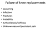
- total knee replacement (lasts 10 years)
- partial unicondylar knee replacement if disease localised to medial or lateral condyle. has faster recovery but may need full replacement at one stage

What is patellofemoral arthritis and how is it managed?
OA affecting articular cartilage along the trochlear groove and the underside of the patella. May occur with patella dysplasia or previous patella fracture
Symptoms: anterior knee pain worse when pressure on patella (e.g climbing stairs), joint stiffness, swelling
Dx: skyline plan film radiograph
Mx: conservative then patellofemoral replacement. (if OA in other parts of knee will need TKR)

How does an ACL tear present?
- History of twisting the knee whilst weight bearing (non contact change of direction on flexed knee)
- Unable to weight bear
- Rapid joint swelling (haemarthrosis due to ligament being vascular)
- Pain
- Instability if delayed presentation

What special tests can diagnose an ACL tear?
- Lachmans test: put knee in 30 degrees flexion then one hand stabilises the femur and the other pulls the tibia foward, check both knees for comparison
- Anterior drawer test: flex knee to 90 degrees, place thumbs on joint line and index fingers on hamstring tendons posteriorly, force then applies

What investigations should you do if you suspect an ACL tear and what are some differentials?
- Plain film radiograph AP and lateral: exclude bony injuries, joint effusion, lipohaemarthrosis. (Segond Fracture usually means ACL tear)
- MRI of knee: gold standard, can pick up any associated meniscal tears (usually medial)

What is a Segond fracture?
Bony avulsion of the lateral proximal tibia that is most likely caused by an ACL tear

How is ACL managed?
- Immediate RICE
Conservative (less active patients)
- Patient can often weight bear so cricket pad knee splint for comfort and send home
- Rehabilitation to strengthen quadriceps that stabilise the knee
Surgical (more active)
- Arthroscopic reconstruction with tendon or artifical graft. Often done after some time and prehabilitation. Doesn’t reduce risk of OA
- Sometimes acute repair can be done if MRI favourable, do GA and arthroscopy and resuture ends of torn ligament

What is the complication of ACL tears and ACL reconstruction surgeries?
Post-traumatic OA
What is the function of the MCL and how can injuries to the MCL be classified?
Most commonly injured ligament of knee. Acts as valgus stabiliser of knee so when forces are applied to lateral knee it tears

How does an MCL tear present and what are some differentials?
- History of trauma to lateral knee or valgus stress with external rotation (skiing)
- May hear pop then immediate medial joint line pain
- Swelling a few hours later
- Tender along joint line
- Can still weight bear
Differentials: fractures, menismcal injury, multi-ligament tears

What special tests can aid your diagnosis of a MCL injury?
Valgus stress test
Will have increased laxity and reproduction on painwhen testing MCL
Do flat and then in 20/30 degress flexion
How is a suspected MCL tear investigated?
- Plain film radiograph AP and Lateral to exclude dracture
- Gold standard MRI

How are MCL tears managed and what are some complications that can arise with an MCL tear?
Grade I: RICE with NSAIDs. Strength training and return to full exercise within 6 weeks
Grade II: Analgesia with knee brace. Weight bearing/strength training and return to exercise within 10 weeks
Grade III: Analgesia with knee brace and crutches. If distal avulsion surgery. Return to exercise within 12 weeks
Complications: instability in joint, damage to saphenous nerve

What is the role of the medial meniscus and the pathophysiology of injury to this structure?
Shock absorber of the knee joint and increases the articulating area. It is connected to the MCL
- Trauma related injury (young person twisted knee whilst weight bearing)
- Degenerative disease

How does a meniscal tear present and what will you find on examination ?
Symptoms

- Tearing sensation
- Intense sudden onset pain
- Slow swelling over 6-12 hours
- If bucket handle may be locked in flexion
Examination
- Joint line tenderness
- Joint effusion
- Limited knee flexion
- Mcmurray test (may be too painful)
How are meniscal tears investigated and managed?
Ix
- Plain film radiograph to exclude fractures
- Gold standard: MRI
Mx
- RICE if <1cm
- Arthroscopic surgery if large and symptomatic

What are some complications of meniscal tears and arthroscopy to treat them?
Meniscal Tear: OA
Arthroscopy: DVT, damage to saphenous nerve/vein, damage to peroneal nerve, damage to popliteal vessels
How do patella fractures present?
Often in 20-50 year old males due to either direct trauma or rapid eccentric contraction of quadriceps
- Anterior knee pain following trauma (e.g dashboard injury)
- Pain worse on movement
- Cannot straight leg raise
- Swollen and bruised
- Palpable patella defect

What else can cause a palpable defect in the patella apart from a fracture?
Bipartite patella

What are the investigations and management for patella fractures

Ix
- Plain film radiographs three views (AP, Lateral, Skyline)
- CT if comminuted
Mx
Conservative: if non/minimally displaced then put in brace or cylinder cast with early weight bearing in extension
Surgical: if displacement or damage to extensor mechanism then open reduction and internal fixation (ORIF) with tension band wiring. If simple vertical/transverse fracture can screw fix not wiring