17 - T&O Wrist and Hand Flashcards
What is the pathophysiology of distal radius fractures?
Fracture of the distal metaphysis of the radius with/without articular involvement
Usually due to FOOSH as distal radius takes 80% of axial load and impaction from supinated/pronated lunnate and scaphoid
- Usually fragility fractures due to osteoporosis or aged 5-15 years
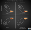
What are the different classifications of distal radial fractures?
Colles’
- Extra articular fracture with dorsal angulation and dorsal displacement, within 2cm of articular surface
- Due to FOOSH and often fragility fracture
- Often cause ulnar styloid avulsion fracture
Smith’s
- Extra articular fracture with volar angulation with/without volar displacement
- Often due to falling backward and planting hand behind the body
Barton’s
- Intrarticular fracture of the distal radius with associated dislocation of the radio-carpal joint
- Can be volar (more common) or dorsal

What are the risk factors for a distal radius fracture?
Factors related to osteoporosis

How does a distal radius fracture present and what are some differentials?
- Immediate pain, deformity and swelling around fracture site
- Need to assess neurovascular compromise (nerve function, cap refill, pulses) and joint above and below for any injuries
- Differentials: forearm fracture (Galeazzi and Monteggia), carpal bone fractures, tendonitis, wrist dislocation

How are distal radius fractures investigated?
Plain radiographs with the following measurements means distal radius fracture:
- Radial height <11mm
- Radial inclination <22 degrees
- Radial (volar) tilt >11 degrees
Can do CT or MRI for operative planning

How are distal radial fractures managed?
- Resuscitate and stabilise
- Closed reduction in A+E by traction and manipulation under anaesthesia (haematoma block or Bier’s block)
- Below-elbow back slab and repeat radiographs in a week to check for displacement
- If no displacement just physiotherapy
- If displacement surgical management (see image)

What are the complications of distal radius fractures?
- Malunion (shortened radius causes reduced wrist motion, wrist pain and reduced forearm rotation. Treat with osteotomy)
- Median nerve compression/Carpal tunnel syndrome
- OA
- EPL rupture

What is the pathophysiology of why a scaphoid fracture results in avascular necrosis?
Usually in men aged 20-30 due to high energy injury
- Scaphoid has proximal pole, waist and distal pole
- Branch of radial artery enters in distal pole and travels in retrograde fashion to proximal pole
- More proximal scaphoid fracture more risk of AVN

How does a scaphoid fracture present and what are some differentials?
- After high energy trauma sudden onset wrist pain
- Tenderness in floor of anatomical snuffbox
- Pain on palpating scaphoid tubercle
- Pain on telescoping the thumb
Differentials: distal radial fracture, alternative carpal fracture, fracture of base of 1st MC, ulnar collateral ligament injury, wrist sprain, De Quervains Tenosynovitis
How are scaphoid fractures investigated, diagnosed and managed?

Ix
- Scaphoid series of plain radiographs (AP, Lateral Oblique)
- If negative initial imaging but high clinical suspicion immobilise wrist in thumb splint for 10-14 days then repeat radiographs
- If still negative imaging but clinical findings still there do MRI
Mx
- If undisplaced strict immobilisation in plaster with thumb spica splint.
- If undisplaced in proximal pole high risk of AVN so surgery particularly if patient’s dominant hand
- All displaced fixed surgically with percutaneous variable pitched-screw

What are some complications with scaphoid fractures?
- AVN (risk increasing with more proximal fractures)
- Non-union (due to poor blood supply)
Can fix above with internal fixation and bone grafts but still do not fix the cause

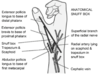
What is the normal volar tilt and inclination of the distal radial articular surface?
Volar tilt: 10 to 25 degrees
Inclination: 23 degrees (13 to 30)

How do you measure volar tilt and radial inclincation of the distal radius?
Volar tilt: on the lateral projection of the wrist as an angle of the distal radial surface with respect to a line perpendicular to the shaft
Radial inclination: is measured by drawing a line perpendicular to the long axis of the radius and a tangential line from the radial styloid to the ulnar corner of the lunate fossa

What are some risk factors for carpal tunnel syndrome?
Compression of the medial nerve at the carpal tunnel so pain, numbness and paraesthesia of lateral 3.5 digits
More common in women aged 45-60
Risk Factors: female, increasing age, pregnancy, previous injury to wrist, diabetes, RA, hypothyroidism, repetitve hand movements

How does carpal tunnel syndrome present and what might you find on examination?
- Pain, numbness and paraesthesia in median nerve sensory distribution with palmar sparing as palmar cutaneous branch comes off before carpal tunnel
- Symptoms worse during night and relieved by shaking or hanging wrist over edge of bed
- On examination in early stages reproduction of sensory symptoms by Tinel’s and Phalen’s test
- In late stages weakness of thumb abduction wasting of thenar muscles due to denervation

What are some differential diagnoses for carpal tunnel syndrome?
- Cervical radiculopathy C6 (however will have neck pain and entire arm affected)
- Pronator teres syndrome (median nerve compressed by PT, symptoms will extend into forearm and affect palm)
- Flexor Carpi Radialis Tenosynovitis (tenderness at base of thumb)

How is carpal tunnel syndrome investigated and managed?
Ix
- Usually clinical diagnosis but can do nerve conduction studies to confirm median nerve damage
Mx
Conservative: wear wrist splint at night, physiotherapy, corticosteroid injections
Surgical: carpal tunnel release surgery by cutting through flexor retinaculum to reduce pressure on median nerve, can be done under local

What are some carpal tunnel syndrome complications and some complications of release surgery?
- Untreated CTS: permanent neurological impairment that will not improve with surgery
Surgery: persistent CTS symptoms if ligament not completely released, infection, scar, nerve damage, trigger thumb
What is the pathophysiology of Dupuytren’s contracture?
- Contraction of longitudinal palmar fascia
- Starts as painless nodules and fibrous cords at MCP and interphalangeal joints eventually leading to contraction

What are some risk factors for developing Dupuytren’s contracture?
- Smoking
- Alcoholic liver cirrhosis
- Diabetes mellitus
- Heavy manual work/vibration
- Idiopathic

How does Dupuytren’s contracture present and what are some differentials?
- Thickened band or firm nodule adherant to skin may be palpable
- Skin blanching may occur on extension of digits
- Ring and little finger often involved
- Hueston’s palm table top test
Differentials: stenosing tenosynovitis, ulnar nerve palsy, trigger finger (nodules associated with finger motion)

How is Dupuytren’s contracture managed once a clinical diagnosis is made?
Conservative (early stages):
- Hand therapy, injectable collagenase clostridum histolyticum
Surgical (if functional impairment, MCP joint contracture >30 degrees, PIP contracture or rapidly progressive disease):
- Removal of diseased fascia called a fasciectomy

What is De Quervain’s Tenosynovitis and what are some risk factors for this?
Inflammation of the tendons in the first extensor compartment (EPB and APL) of the wrist resulting in wrist pain and swelling
Risk factors: female, age 30-50, pregnancy, occupations or hobbies involving repetitive movements of the hands

How does De Quervian’s tenosynovitis present on examination?
- Pain near the base of the thumb
- Grasping or pinching movements painful and difficult
- Swelling and palpable thickened over tendon group
- Positive Finkelstein test


