!! 5 - Gross Topography Flashcards
What view of the brain is this?
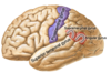
Lateral
What view of the brain is this?

Sagittal
What view of the brain is this?

Coronal
Telencephalon
Cerebrum
(cerebral cortex, cerebral hemispheres, hippocampus, amygdala, basal ganglia)

A) Lateral Ventricle
B) Fornix
C) Thalamus
D) 3rd ventricle
E) Massa intermedia
F) Pineal gland
G) Posterior Commissure
H) Hypothalamic sulcus
I) Hypothalamus
J) Anterior Commissure
K) Cerebral Aqueduct
L) 4th Ventricle
M) Foramen of Luschka
N) Choroid Plexus
O) Superior Colliculi
P) Inferior Colliculi
Q) Mamillary body
R) Interventricular foramen of Monro
Occipital Lobe Function, Location
- Function: Visual info processing
- Location:
- Posterior/caudal to imaginary line from parieto-occipital sulcus to pre-occipital notch
Calcarine Sulcus
- Divides the occpital lobe into two
Primary Visual Cortex (V1)
- Occipital lobe
- Superior and inferior to calcarine fissure
Association Visual Cortex (V2)
- NOT actually part of occipital lobe
- Two major pathways:
- WHERE pathway: parietal lobe
- WHAT pathway: temporal lobe
Parietal Lobe Functions
- Sensory integration
- Spatial orientation
- Language
- “Where” visual processing
Postcentral Gyrus
- Parietal Lobe
- AKA Primary Somatosensory cortex
- Uses somatotopic organization for sensory (skin) touch
Supramarginal Gyrus
- Parietal Lobe
- Basically the gyrus around the edge of the lateral fissure
- Language processing

Angular Gyrus
- Parietal Lobe
- Language processing

Temporal Lobe Functions
- Hearing
- Language
- Visual “What” processing
- Memory
- Recognition
- Reaction system
Temporal Lobe Key Features
- Transverse temporal gyrus (Heschl’s)
- Superior temporal gyrus
- Wernicke’s Area
- Limbic Lobe
- Parahippocampal gyrus
- Hippocampus
- Amygdala
Transverse Temporal Gyrus
- AKA Heschl’s Gyrus
- Located in roof of superior temporal gyrus (must be pulled away from the insula to see)
- Primary auditory cortex

Wernicke’s Area Location, Function
- Temporal Lobe
- In superior temporal gyrus
- ONLY ON SPEECH DOMINANT (LEFT) SIDE
- Anteriolateral to the supramarginal and angular gyri
- Function: deconde verbal info into sound (comprehension)

Medial Temporal Components
Some of Limbic Lobe
- Parahippocalmpal Gyrus
- Hippocampus (memory)
- Amygdala (emotional responses)

Frontal Lobe Functions, Key Features
- Intelligence, personality, motivation, executive control, motor command
- Anterior to central sulcus, superior to lateral fissure
- Contains:
- Precentral gyrus/primary motor cortex
- Inferior frontal gyrus (Broca’s area) on speech dominant side

Precentral Gyrus
- Frontal lobe
- Primary motor cortex
Broca’s Area
- Inferior frontal gyrus
- ONLY ON SPEECH DOMINANT SIDE
- Motor speech (speech production)
Limbic Lobe Function, Components
Function: emotions, basic drives, memory, smell
Includes frontal, parietal, and temporal lobes
Components:
- Cingulate gyrus
- Emotion formation
- Processing, learning, memory
- Uncus
- Smell
- Parahippocampal gyrus
- Memory coding and retrieval
- Hippocampus
- Memory formation and recall
- Amygdala
- Emotion, emotional association learning
- Insula
- Taste, visceral sensation, emotional pain
- Interoception

Cingulate Gyrus Location, Function
- Limbic Lobe component
- Superior to corpus callosum (frontal)
- Functions:
- Emotion formation and processing
- Learning
- Memory
Uncus Location, Function
- Limbic lobe component
- Located at medial, anterior edge of the temporal lobe
- Function: smell


