1.9: The Eye and Raised ICP Flashcards

1
Q

What is raised ICP? Types?

A

This is raised pressure in the cranial cavity Chronic or acute

2
Q

Causes of raised ICP?

A

Increased pressure in fluid around brain (CSF) or increased pressure within brain itself

  • Brain Tumour
  • Head injury
  • Hydrocephalus
  • Meningitis
  • Stroke
3
Q

Is raised ICP a medical emergency? If yes, why?

A

Yes Because it can cause damage to the brain, spinal cord and the eyes

4
Q

Describe the components of ICP?

A

ICP is the sum of:

  • Blood
  • Brain
  • CSF
5
Q

Describe how the ICP is kept constant?

A

If one component increases, the others decrease to compensate and keep the ICP constant (and vice versa)

6
Q

Describe the monro-kellie hypothesis?

A

If one component of ICP increases, the others decrease the compensate and keep the ICP constant (and vice versa)

7
Q

Describe the cranial cavity in regards to ICP?

A

The cranial cavity is an enclosed space and is unable to expand as the pressure increases

8
Q

Describe the relationship between ICP and volume?

A

As volume increases, compensatory mechanisms are able to prevent the pressure from rising After compensatory mechanism fail, a small change in volume will cause a large change in pressure (exponential)

9
Q

Increased pressure can lead to damage Give examples?

A

Damage to tissue

Shifts to tissue

Damage to brain, spinal cord and eye

Herniation

Constriction of blood vessels

10
Q

Describe a statistic that links raised ICP and visual symptoms?

A

65% of patients with ICP will complain of some visual symptoms

11
Q

Examples of visual symptoms caused by raised ICP?

A

Transient Blurred Vision Doubled Vision Loss of Vision Papilloedema (Swelling of optic discs due to raised ICP) Pupillary changes Can affect one eye or both

12
Q

Optic nerves are not actually nerves but they are…?

A

CNS Tracts

13
Q

What covers optic nerves?

A

3 Meninges: - Dura - Arachnoid - Pia

14
Q

What is the space between the Arachnoid and Pia called?

A

Sub-arachnoid space

15
Q

What are meninges?

A

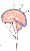
These are the three membranes than envelope the brain and the spinal cord - they are the protective coverings

16
Q

Describe the Dura Mater? Sensory supply?

A

This is the tough, outer meningeal layer Sensory supply is CN V Encloses dural venous sinuses

17
Q

Describe the arachnoid mater?

A

This is the spidery mother Has lots of little projections called ‘arachnoid granulations’

18
Q

Describe the subarachnoid space?

A

This is space below the arachnoid mater There is circulating CSF and blood vessels

19
Q

Describe the pia?

A

This is the faithful mother Adheres to the brain Also adheres to vessels and nerves that are entering/leaving the brain

20
Q

Label this diagram?

A

A = Pia B = Arachnoid Layer C = Dura D = Dura

21
Q

How do you access the subarachnoid space?

A

Lumbar puncture between L3/L4 or L4/L5

22
Q

Where does the subarachnoid space end?

A

At the level of S2 (Part of the Sacrum)

23
Q

Label this diagram?

A

A = Subarachnoid Space B = Dura C = Arachnoid D = Pia

24
Q

The subarachnoid space communicates with another series of CSF filled spaces in the brain. What are these called?

A

The Ventricles

25
Q

Name the ventricles of the brain?

A

3rd 4th Left Lateral Right Lateral

26
Q

Label this diagram?

A

A = Third Ventricle B = Left Lateral Ventricle C = Right Lateral Ventricle D = Subarachnoid Space E = Central Canal of the Spinal Cord F = 4th Ventricle G = The Cerebral Aqueduct H = Pituitary Gland I = Optic Chiasm J = Sinus

27
Q

Describe the circulation of CSF?

A
  1. Produced by the choroid plexus of both lateral ventricles 2. Passes into the third ventricle 3. Goes via the cerebral aqueduct into the fourth ventricle 4. Passes into subarachnoid space 5. Some goes into central canal of spinal cord 6. Most is reabsorbed from the subarachnoid space by the arachnoid granulations 7. Reabsorbed into the dural venous sinuses
28
Q

Describe how raised ICP is transmitted?

A

This is transmitted along the subarachnoid space of the optic nerve sheath

29
Q

Describe how raised ICP leads to swollen discs?

A

Raised ICP compresses the optic nerve It also compresses the central retinal artery and vein This leads to bulging, swollen discs

30
Q

Describe damage to CN III?

A

This is susceptible to damage (Eg: Compression) Leads to the eye being ‘down and out’ with a dilated pupil

31
Q

Oculomotor nerve damage causes the eye to be in what position?

A

Down Out Dilated pupil

32
Q

Describe herniation/raised ICP?

A

Raised ICP can compress/stretch oculomotor nerve if medial temporal lobe herniates through tentorial notch

33
Q

Describe damage to the trochlear nerve (CN IV)?

A

This has a long intracranial course and is susceptible to injury: - Stretching - Compression Results in superior oblique paralysis Eye cannot move inferior

34
Q

Describe damage to CN VI? (Abducens)

A

Long intracranial course and is susceptible to stretching Results in lateral rectus paralysis Eye cannot move laterally