1:2. Orbit and the Eye Anatomy Flashcards

1
Q

Name the bones (not the labels)

A

Beige = Frontal

Blue = Zygomatic

Purple = Maxilla

Red = Sphenoid

Yellow = Ethmoid

Dark Blue = Lacrimal

Green = Nasal

2
Q

Name the labels (not the bones)

A –> F

A

A = Superior Orbital Fissue

B = Optic Canal

C = Orbital Plate of the Frontal Bone

D = Supraorbital Notch/Foramen

E = Orbital Plate of Ethmoid

F = Infraorbital Foramen

3
Q

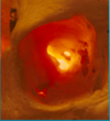
Name all the same labels on this skull?
A

Superior Orbital Fissure

Optic Canal

Orbital Plate of the Frontal Bone

Orbital Plate of the Ethmoid Bone

Supraorbital Notch

Infraorbital Foramen

4
Q

Difference between a foramen and a notch?

A

Foramen is a complete circle

Notch is uncomplete - like a C shape

5
Q

The bony orbit is described as a four sided pyramid

The apex is the orbital canal

What does the base make up?

What is it composed of?

A

The base makes up the orbital rim

Composed of superior, inferior, medial and lateral orbital margins

6
Q

How is the eye protected from a direct blow?

A

Orbital Margins - superior orbital margin is further forward than inferior so eye is protected

Orbital rim - nothing bigger than the orbital rim will come into contact with the actual eye

7
Q

Describe orbital blowout fractures?

A

The medial wall and orbit floor are very thin

In trauma, the orbital margins and rim don’t fracture as they are strong

The medial wall and floor are very thin though and can break

Can cause an orbital blowout fracture

8
Q

Complications of orbital blowout fracture?

A

Orbital contents can become trapped

Infraorbital neurovascular bundle can be damaged

9
Q

If the infraorbital neurovascular bundle was damaged in a blowout fracture, what would happen?

A

Loss of sensation to face

10
Q

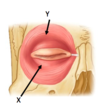
The external eyelid is split into two parts:

Name them

Identify them
A

Orbital Part

Palpebral Part

X = Palpebral

Y = Orbital

11
Q

Name the muscle that surronds the eye in the external layer?

A

Obicularis Oculi

12
Q

What is the orbital septum?

A

This is a sheet of fascia below the eyelid

Found in the middle layer

This separates the superficial part of the eye from the deep part to prevent the spread of infection

13
Q

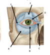
Label these parts of the eyelid

Which layer is this?
A

A = Orbital Septum

B = Inferior Tarsus

C = Medial Palpebral Ligament

D = Superior Tarsus

E = Tendon of Levator Palpebrae Superioris

F = Lateral Palpebral Ligmanent

Internal Layer

14
Q

What is the Tarsal gland?

Where is it found?

A

These secrete an oily/waxy substance that prevents the eyelids from sticking together and prevents tears from overflowing and streaming constantly

Found in the tarsal plates of the tarsi

15
Q

Describe the Tarsi?

A

Found above and below the eye

Plates of fibrous connective tissue that give the eyelid its half moon shape

Give protection to eye and attachment for muscles

16
Q

Where does the Levator Palpebrae superioris attach?

A

The superior Tarsi

17
Q

Name the surface anatomy of the eye

A

A = Location of the lacrimal duct

B = Pupil

C = Blood Vessel (Conjuctival vessel)

D = Lacrimal Lake

E = Puncta

F = Lower eyelid, lined with Conjuctiva

G= Conjunctival Fornix

H = Sclera, covered by conjuctiva

I = Limbus/Corneoscleral Junction

18
Q

What innervates the lacrimal gland?

A

CN VII (Facial Nerve)

Parasympathetic

19
Q

Describe how the lacrimal apparatus works?

A

CN VII Parasympathetic innervation

Lacrimal gland produces lacrimal fluid

Washes across eye

Collects in lacrimal lake

Drains out of Puncta

Drains into lacrimal sac

Drains along nasolacrimal duct

Eventually reaches inferior meatus

20
Q

Where does the lacrimal fluid end?

A

Inferior Meatus

21
Q

Coloured bit in the centre of the eye?

Black bit in the very centre of the eye?

White bit in the eye?

Clear, transparent bit that covers the coloured bit?

A

Iris

Pupil

Sclera

Cornea

22
Q

What control is the pupil under?

A

Autonomic

23
Q

Describe the layers of the eye?

A

Fibrous, Outer Part

Uvea = Middle Layer

Retina (Inner Layer)

24
Q

Describe the outer layer of the eye

A

Tough and fibrous

Composed of two parts

Sclera - the white part where extra-ocular muscles attach. Makes up about 5/6ths of the eye

Cornea = Clear, colourless, front of the eye. Makes up most of the refractive power - the ability to focus light

25
Q

Describe the middle layer of the eye

A

The Uvea

This is the vascular layer

Has three parts:

  • Iris
  • Ciliary Body
  • Choroid
26
Q

Describe the function of the three parts of the middle layer of the eye (the uvea)

A

Iris = Changes size of pupil

Ciliary Body = Attaches to iris. Changes shape of lens and secretes aqueous humour

Choroid = Provides nutrition and gas exchange

27
Q

Describe the inner layer of the eye?

A

This is photosensitive

Contains:

  • Retina
  • Macula
  • Optic Disc
28
Q

Describe the segments of the eye?

  • Names?
  • Where is the divide?
  • Humour?
A

Dividied into anterior and posterior

LENS is the borders

Anterior = In front of Lens

Posterior = Behind lens

Anterior = Aqueous Humour

Posterior = Vitreous Humour

29
Q

Describe the anterior segment of the eye?

A

Divided into two chambers - anterior and posterior

Anterior: Between cornea and iris

Posterior: Between iris and suspensory ligaments

Both contain aqueous humour

30
Q

Describe the posterior segment of the eye?

A

This is everything found behind the lens (2/3rds of the eye)

Contains vitrous body which contains vitreous humour

Vitreous humour is gel like

Vitrous humour holds the lens and the choroid against the sclera

31
Q

Describe floaters?

A

Commonly seen in the vitreous body

Caused by floating lumps of vitreous humour

32
Q

Describe the circulation of aqueous humour?

A
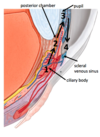
  1. Produced by ciliary body
  2. Circulates within posterior chamber to nourish lens
  3. Passes through pupil into anterior chamber
  4. Nourishes cornea
  5. Reabsorbed into the scleral venous sinus (Canal of Schlemm)
33
Q

What is the posterior part of the retina called?

A

Fundus

34
Q

Optic Disc:

  • Site of what?

Macula

  • Site of what?

Fovea

  • Site of what?
A

Optic Nerve Formation (CN II)

Only point of entry/exit for blood vessels and nerves of CN II

Blind spot due to no photoreceptors

Macula

Site of greatest number of cones

Fovea

Depression in the macula

Site of most acute vision

35
Q

If one branch of the retina is disrupted, what happens?

If the central artery/end vein is disrupted, what happens?

A

Only the area supplied by that branch affected will lose vision

If central artery is affected, all vision will by lost as entire retina is affected

36
Q

Describe the layers of the retina?

A
  1. Photoreceptors (MOST POSTERIOR)
  2. Ganglion Cells (Anterior to photoreceptors)
  3. Axons of the ganglion cells (Most anterior)
37
Q

Describe the passage of light entering the eye (not including brain)

A

Comes through cornea

  • Axons of ganglion
  • Ganglion
  • Photoreceptors
  • Optic Nerve
38
Q

Describe the visual pathway?

A

Light enters eyes

  • Temporal or nasal retina
  • Optic Nerve
  • Optic Chiasma
  • Optic Tract
  • Optic Radiation
  • Visual Cortex of the Occipital Lobe
39
Q

Where is light from the right visual field processed?

Where is light from the left visual field processed?

A

Right –> Left

Left –> Right

40
Q

Describe the visual pathway of light from the left visual field:

  • Left Eye
  • Right Eye
A

Left Eye:

Left Nasal Retina

Left Optic Nerve

Optic Chiasma

Right Optic Tract

Right Optic Radiation

Right side of the visual cortex

Right Eye:

Right Temporal Retina

Right Optic Nerve

Optic Chiasma

Right Optic Tract

Right Optic Radiation

Right Visual Cortex

41
Q

Describe the arterial blood supply to the orbit?

A

The Opthalmic artery is the first branch of the internal carotid

Gives off branches which supply the eye and oculomotor muscles

42
Q

Describe the special relationship between light and superior/inferior

A

All light coming from superior hits the inferior part of the eye

All light coming from inferior hits the superior part of the eye

43
Q

Describe where the internal carotid becomes the opthalmic?

A

The internal carotid passes through the carotid canal

Passes through the cavernous sinus

THEN the opthalmic artery comes off

44
Q

Describe the branches of the opthalmic artery?

A

Nasal Cavity Branches (Supply Kiesselbach’s area)

Forehead/Scalp Branches

Ciliary Branches (Supply the choroid and uvea - middle vascular layer)

Central Artery of the Retina

45
Q

Describe the route of the central artery of the retina?

A

Branches from the opthalmic artery

Enters the optic nerve

Supplies retina

46
Q

Describe what happens if the central artery of the retina is occluded?

A

The retina begins to die off as this is its only blood supply

47
Q

What vein(s) drain the retina?

A

Central Vein only

48
Q

Describe the venous drainage of the eye?

A

Retina = Central Vein

Everything else is mainly Superior Opthalmic Vein

  • Forehead/Scalp vein
  • Facial Vein
  • Inferior Opthalmic vein

ALL DRAIN INTO THE SUPERIOR OPTHALMIC VEIN

Drains into the cavernous sinus

49
Q

Describe the danger triangle of the face?

A

From corners of mouth to nose

Squeezing spots in this area can pierce veins and lead to infections in bloodstream

Can lead to infections surronding the brain

50
Q

Name the 7 extra-ocular muscles?

A

`Superior Rectus

Inferior Rectus

Medial Rectus

Lateral Rectus

Superior Oblique
Inferior Oblique

Levator Palpebrae Superioris

51
Q

Describe where the 7 extra-ocular muscles insert?

A

First 6 (Rectus and Obliques) attach onto the sclera

The levator palpebrae superioris attachs onto the superior tarsus

52
Q

Name the parts of the eye (A–> I)

YELLOW IS MEANT TO BE F

A

A = Inferior Rectus

B = Lateral Rectus

C = Medial Rectus

D = Superior Rectus

E = Levator Palpebrae Superioris

F = Superior Oblique

G = Inferior Oblique

H = Superior Tarsus

I = Trochlea

53
Q

What is the way to remember cranial nerve innervation of extra-ocular muscles?

A

LR6 SO4 AO3

Lateral Rectus = 6 (Abducens)

Superior Oblique = 4 (Trochlear)

All others = 3 (Oculomotor)

54
Q

Superior oblique is supplied by what?

Anatomically, how does this make sense?

A

Trochlear Nerve

Trochlea is the name of the small pulley that Superior Oblique passes through

55
Q

What are the 6 movements on the eye?

A

Adduction

Abduction

Elevation

Depression

Intorsion

Extorsion

56
Q

Do all the muscles have just one movement?

Eg: Superior Rectus

A

No, they have a primary movement and also secondary

Superior Rectus causes elevation of the eye

Also causes adduction and medial rotation (think the eye muscles attach medially)

57
Q

How do you clinically test the lateral rectus?

A

Abduction of the eye

58
Q

How do you clinically test the medial rectus?

A

Adduction of the eye

59
Q

How do you clinically test the superior rectus?

A

Abduct the eye first

Elevate the eye

60
Q

How do you clinically test the inferior rectus?

A

Abduct the eye

Depress the eye

61
Q

How do you clinically test the superior oblique?

A

Adduct the eye

Depress the eye

62
Q

How do you clinically test the inferior oblique?

A

Adduct the eye

Elevate the eye

63
Q

Describe synergestic movement?

Give two exampels

A

When two or more muscles work together to perform a movement

Eg: Superior Rectus and Inferior Oblique work together to elevate the eye

Eg: Inferior rectus and superior oblique work together to depress the eye

64
Q

Describe antagonistic movement?

Give one example

A

This is when two or more muscles are competiting to try and move the eye in different directions:

Eg: Superior rectus tries to medially rotate the eye, inferior oblique tries to laterally rotate the eye

65
Q

Describe pure elevation and pure depression

A

Elevation:

Superior rectus and inferior oblique both try to move the eye upward (syngerstically) They both try to rotate in different directions (anatagonistically). This cancels each other out and results in pure elevation

Depression:

Inferior rectus and superior oblique both try to move the eye downwards (syngerstically). Both try to adduct/abduct the eye differently (antagonists). Cancels each other out and results in pure depression

66
Q

https://www.bmc.med.utoronto.ca/anatomia/intro.swf

Go onto this website and view their anatomy dissection

A

x

67
Q

https://cim.ucdmc.ucdavis.edu/eyes/version1/eyesim.htm

Use this interactive eye simulator and quiz

A

x