19 Pathology of Glomerular Disease Flashcards
Glomerulus
- Site of…
- Capillary beds
- Surrounded by…
- Visceral epithelial cells
- PT epithelial cells
- Site of ultrafiltration of plasma resulting (following tubular modification) in the formation of urine
- First of two capillary beds in the kidney that connects the AffAs & EffAs
- Second capillary bed is the peritubular capillary plexus in the cortex & the vasa recta in the medulla
- Surrounded by Bowman’s capsule
- Covered by parietal epithelial cells
- Visceral epithelial cells (podocytes) cover the glomerular capillary surfaces
- Formed as the parietal epithelial cells
- PT epithelial cells reflect over the vascular and tubular poles
- Podocytes
- Important part of the filtration barrier of the glomerulus
- Cover the entire glomerular BM

Basement membrane overlying glomerular capillaries
- 3 layers (from outer to inner)
- Endothelial cell
- Overlying mesangial areas, the BM has 2 layers
- Deep to the BM is the…
- Mesangial matrix contains mesangial cells that have 3 functions
- 3 layers (from outer to inner)
- Lamina rara externa
- Lamina densa
- Lamina rara interna
- Endothelial cell
- Internal to the BM
- Unique flat cell with numerous holes or fenestrations that retard cells but allow plasma to freely enter the BM
- Overlying mesangial areas, the BM has 2 layers
- Lamina rara externa
- Lamina densa)
- Deep to the BM is the…
- Mesangial matrix
- Contiguous with the lamina rara interna
- Mesangial matrix contains mesangial cells that have 3 functions
- Tether BM to mesangium –> formation of glomerular segments with multiple peripheral capillaries & a central mesangial unit
- Phagocytosis of cell debris & material that gets deposited in the mesangium & lamina rara interna
- Production of cytokines that stimulate glomerular cellular proliferation in response to injury
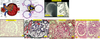
Assessing glomeruli in a renal biopsy
- Histologic sections
- Glomerular mesangial areas
- Glomerular capillaries
- Glomerular capillary walls
- Urinary space
- H&E stain
- Methenamine silver stains
- PAS stains
- Renal biopsy paraffin histologic sections
- Histologic sections
- 2D representation of a 3D structure
- Glomerular mesangial areas
- Relatively inapparent
- Contain < 3 mesangial cells / mesangial area
- Exception: hilum has a greater # of mesangial cells
- Glomerular capillaries
- Patent
- Glomerular capillary walls
- Glomerular BM + endotehlial cell + podocyte
- Thin & expanded
- Urinary space
- Empty
- H&E stain
- Limited by its inability to distinguish cytoplasm of endothelial cells, podocytes, & mesangial cells from GBM & mesangial matrix
- Methenamine silver stains
- Stain type IV collagen in glomerular & tubular BMs, bowman’s capsule, & extraglomerular blood vessels
- PAS stains
- Stain polysaccharides in glomerular & tubular BMs, bowman’s capsule, & extraglomerular blood vessels
- Renal biopsy paraffin histologic sections
- Cut at 2-3 microns in thickness
- Thicker sections –> cell overlapping –> appearnace of hypercellularity

Clinical classifications
- Primary
- Secondary
- Onset
- Single occurence
- Chronic
- Primary
- Limited to the kidney
- Systemic
- Secondary glomerulonephropathies
- Onset
- Acute
- Insidious (chronic)
- Sublincial (detected as a lab abnormality only)
- Single occurence
- Resolves w/ no clinical or pathologic sequelae
or - Organizes w/ a persistent & stable deficit
- Resolves w/ no clinical or pathologic sequelae
- Chronic
- Periods of alternating activity & inactivty (relapses & remissions)
- Frequently –> progressive loss of glomeruli & progressive renal dysfunction
Glomerular syndromes
- Acute nephritic syndrome
- Rapidly progressive glomerulonephritis
- Nephrotic syndrome
- Chronic renal failure
- Asymptomatic hematuria or proteinuria
- Acute nephritic syndrome
- Hematuria, azotemia/ARF, variable proteinuria, oliguria, edema, & hypertension
- Reversible lesion that doesn’t –> glomerular scarring
- Renal biopsy: proliferative glomerular disorder w/o necrotizing lesions / crescents
- Rapidly progressive glomerulonephritis
- Often presents like an acute nephritic syndrome w/ proteinuria & ARF
- Progressive w/o therapy
- –> glomerular scarring w/ loss of functional glomeruli
- In some pts, the disease is more slowly progressive
- Renal biopsy: glomerular necrotizing lesions / crescents
- Nephrotic syndrome
- >3.5 gm proteinuria / day, hypoalbuminemia, hyperlipidemia, hyperlipiduria, & edema
- Renal biopsy: non-proliferative glomerular disorder w/ consistent podocyte injury manifested by foot process fusion, in addition to other disease specific pathology
- Chronic renal failure
- Renal impairment (azotemia) progressing gradually to renal failure over a period of years
- May be associated with…
- All forms of progressive glomerular disease
- Glomerular diseases which occur as a single episode –> significant loss of glomeruli w/ subsequent progressive hyperfiltration injury & glomerular loss occurring in the remaining glomeruli
- Asymptomatic hematuria or proteinuria
- Non-progressive subclinical hematuria or proteinuria
- Detected on urine evaluation during a routine physical exam
- Glomerular hematuria: sub-nephrotic proteinuria
Glomerulopathy:
Approach to morphologic classification
- Establish…
- Define…
- Define…
- Assess…
- Establish glomerulus as primary target of injury
- Define distribution of glomerular injury
- Subcapsular vs. juxtamedullary vs. random
- Diffuse vs. focal
- Global vs. segmental
- Peripheral (capillayr loop) vs. central (mesangial) vs. extraglomerular
- Define light microscopic pattern of glomerular injury
- Assess for involvement of other renal compartments

Morphologic (pathologic) classification:
Light microscopy
- Normal subcapsular glomerulus including Bowman’s space/capsule measures…
- Juxtamedullary glomeruli may measure…
- Compare the structure of the glomerulus to that of a tree
- Trunk & branches –>
- Leaves –>
- More peripheral mesangium (in a 2-3 micron section)…
- More centrally towards to the hilum…
- Capillary loops
- Capillary walls
- Light microscopic (H&E) classification of glomerular disorders
- Specific etiologic diagnosis
- Renal biopsy
- For those disorders which characteristically diffusely involve glomeruli…
- For early stage focal glomerular disorders…
- For non-random focal processes…
- Normal subcapsular glomerulus including Bowman’s space/capsule measures…
- 250 microns in max diameter
- Juxtamedullary glomeruli may measure…
- Up to 300 microns
- Compare the structure of the glomerulus to that of a tree
- Trunk & branches –> mesangium
- Leaves –> capillary loops
- More peripheral mesangium (in a 2-3 micron section)…
- Inapparent matrix
- < 3 mesangial cells
- More centrally towards to the hilum…
- Both mesangial matrix & cellularity are increased
- Capillary loops
- Peripheral structures
- Widely patent
- Capillary walls
- Combination of endothelial & epithelial cells & BM
- Thin and uniform
- Light microscopic (H&E) classification of glomerular disorders
- Descriptive: based on the qualitative & quantitative morphologic alterations from the normal glomerulus
- Iintended to define the distribution & pattern of glomerular injury
- Specific etiologic diagnosis
- Based on the integration of the clinical & serologic data + light (H&E & special histochemical stains), IF, & EM
- Renal biopsy
- Represents a sampling of a pathologic process occurring in the kidney
- May or may not be representative of this process
- For those disorders which characteristically diffusely involve glomeruli…
- Sampling which includes a single glomerulus may be sufficient to define the disease process
- For early stage focal glomerular disorders…
- Adequate sampling is critical for establishing the diagnosis
- > 10 non-sclerotic glomeruli are arbitrarily required for an adequate biopsy only if the focality of the glomerular process is random
- For non-random focal processes…
- E.g. glomerular disorders which preferentially affect subcapsular or juxtamedullary glomeruli
- Full thickness cortical (with medullary) sampling is optimal to evaluate glomeruli at all levels within the cortex
- Best accomplished by examination of > 2 1.5 cm needle biopsy cores
Morphologic (pathologic) classification:
Light microscopy:
Distribution of injury
- Diffuse vs. focal
- Global vs. segmental
- Mesangial vs. peripheral vs. extraglomerular
- Diffuse vs. focal
- Diffuse: > 50% of glomeruli affected
- Focal: < 50% of glomeruli are affected
- Global vs. segmental
- Global: entire glomerulus
- Segmental: subtotal of glomerular involvement by a lesoin affecting > 1 anatomic segments
- Mesangial vs. peripheral vs. extraglomerular
- Mesangial: mesangium
- Peripheral: GBM + podocytes
- Extraglomerular: bowman’s space

Morphologic (pathologic) classification:
Light microscopy:
Patterns of injury:
Increased ECM
- Sclerosis
- Types
- On silver stain, 2 patterns of obsolescence are seen
- Sclerosis (ex.)
- Fibrillar collagen (scar)
- Mucopolysaccharides
- Non-collagen proteins
- Immune complex deposits
- Types
- Mesangial
- Peripheral (capillary loop) BM
- Bowman’s space/capsule
- Obsolescence (entire glomerulus is eosinophilic and hypocellular on H&E stain)
- On silver stain, 2 patterns of obsolescence are seen
- Tuft is collapsed and fibrosis is present only in Bowman’s space
- Ischemic pattern
- Fibrosis replaces part of or entire glomerulus and fills Bowman’s space
- Organization of a necrotizing inflammatory GN or crescent
- Tuft is collapsed and fibrosis is present only in Bowman’s space

Morphologic (pathologic) classification:
Light microscopy:
Patterns of injury:
Increased cellularity
- Intraglomerular
- Mesangioproliferative
- Endocapillary proliferative = mesangiocapillary proliferative
- Membranoproliferative
- Exudative
- Extraglomerular
- Pure epithelial
- Crescent
- Intraglomerular
- Mesangioproliferative
- Iincreased cellularity & matrix confined to mesangial areas
- > 3 mesangial cells / mesangial area
- Endocapillary proliferative = mesangiocapillary proliferative
- Increased mesangial & capillary loop cellularity
- Increased mesangial matrix
- Capillary wall thickening
- Capillary luminal occlusion secondary to endothelial swelling &/or BM thickening
- Membranoproliferative
- Special forms of mesangiocapillary proliferative GN
- Distinctive features on silver stains
- Double GBM contours
- Mesangial cell ingrowth into contiguous subendothelial GBM (mesangial interposition)
- Exudative
- Mesangiocapillary proliferative GN
- Increased numbers of glomerular intracapillary neutrophils
- Mesangioproliferative
- Extraglomerular (glomerulus peripheral to the BM)
- Pure epithelial
- Visceral &/or parietal)
- Correlates with podocyte injury
- Crescent
- Results from glomerular necrotizing lesion (glomerular capillary vasculitis)
- Ttransmural glomerular capillary wall breaks
- Bleeding into Bowman’s space
- Earliest crescents have blood and fibrin in Bowman’s space (fibrinous crescent)
- Cytokines produced by incoming monocytes cause proliferation of parietal epithelial cells (cellular crescent)
- Recruitment of fibroblasts –> fibrosis in the affected area of the glomerular tuft & adjacent Bowman’s space (fibrocellular –> fibrous crescent)
- Pure epithelial

Morphologic (pathologic) classification:
Light microscopy:
Patterns of injury:
Other
- Hyalinosis
- Plasma protein insudation in mesangium, capillary BM, arterioles &/or Bowman’s capsule
- Due to endothelial or epithelial injury w/ serum protein leakage & entrapment
- In diabetes, hyalinosis lesions are designated as
- Fibrin caps (glomerular tuft)
- Capsular drops (Bowman’s capsule).
- Necrosis or necrotizing lesion
- Necrotizing glomerular capillaritis associated w/ inflammatory cells, karyorrhectic nuclear debris & fibrin
- Organization –> segmental or global sclerosis / scar
- Glomerular foam cells
- Frequently seen in glomerular – associated proteinuric disorders
- Correspond to resorption of protein & lipoprotein in macrophages within the glomerular tuft
- Mesangiolysis
- Disruption of mesangium
- Because several capillary loops are tethered to one mesangial area, mesangiolysis may –> single large capillary loop (microaneurysm)
Morphologic (pathologic) classification:
Light microscopic description
- LM classification/description (ex.s)
- Etiological specificity
- Specific diagnosis requiresintegration of…
- LM classification/description = distribution pattern(s) + morphologic pattern(s) of injury (ex.s)
- Focal segmental glomerulosclerosis with hyalinosis.
- Focal segmental endocapillary proliferative and necrotizing glomerulonephritis with focal cellular crescents.
- Diffuse global endocapillary proliferative and exudative glomerulonephritis.
- Diffuse mesangioproliferative glomerulonephritis.
- Etc.
- Etiological specificity
- A particular LM description may be shared by a variety of etiologically distinctive glomerular disorders
- Conversely, a specific disorder (e.g. lupus) may display >1 glomerular morphologic pattern of injury
- Depends upon factors such as the time course in the illness, physical properties of deposited materials (e.g. immune complex deposits), host-specific immune response to injury, etc.
- Specific diagnosis requires integration of…
- Clinical & serologic data
- LM
- IF
- EM
Morphologic (pathologic) classification:
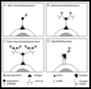
Immunofluorescence microscopy
- Used to assess…
- Uses…
- Panel of stains usually includes…
- Procedure
- A positive signal with a particular antibody indicates…
- Used to assess…
- Evidence of antibody-mediated immunologic injury
- Uses…
- A panel of commercially prepared fluorescein labeled antibodies which recognize specific antigens
- Panel of stains usually includes…
- Ig heavy chain classes (IgG, IgM, IgA) & light chains (kappa, lambda)
- Complement components of the shared (C3), classical (C1q, C4) and alternative (properdin) pathways
- Fibrinogen
- Albumin
- Procedure
- One frozen section slide is prepared for each specific antibody used
- The fluorescein labeled antibody is incubated with the frozen section tissue
- A positive signal with a particular antibody indicates…
- Presence & location of that antigen within the tissue
Morphologic (pathologic) classification:
Immunofluorescence microscopy:
Distribution
- Glomerular
- Extragomerular
- Glomerular (including glomerular tuft and Bowman’s space/capsule)
- Diffuse vs. focal
- Global vs. segmental
- Mesangial vs. peripheral (GBM + podocytes)
- Bowman’s space
- Extraglomerular components
- Tubules (cytoplasm and tubular basement membrane)
- Interstitium
- Blood vessels

Morphologic (pathologic) classification:
Immunofluorescence microscopy:
Patterns
- Granular
- Linear
- Homogeneous, smudgy, or irregular
- Intracellular droplet staining
- Absence
- Granular
- Discrete immune complex deposits
- Linear
- Uniform distribution of targeted antigen
- Weak BM linear staining for albumin and IgG is always present
- Corresponds to small amounts of these proteins that get into the BM (background staining)
- Specific pathologic staining only if albumin stain is less intense than another more intense stain
- E.g. IgG in anti-GBM / Goodpasture disease
- Homogeneous, smudgy, or irregular
- Protein trapping / insudation within an area of sclerosis (usually IgM & C3).
- Deposition of a non-immune complex protein (e.g. amyloid)
- Intracellular droplet staining
- Protein resorption droplets within glomerular epithelial cells or tubular cells
- Absence of staining
Morphologic (pathologic) classification:
Immunofluorescence microscopy:
Staining profile
- Used to assess…
- Uses…
- Panel of stains usually includes…
- Procedure
- A positive signal with a particular antibody indicates…
- Focal segmental glomerulosclerosis
- Homogeneous
- Segmental IgM and C3
- IgA nephropathy
- Granular mesangial IgA, C3
- Membranous nephropathy
- Granular peripheral staining for IgG and C3
- Amyloid
- Smudgy mesangial + GBM staining for deposited amyloid protein
- Lupus
- Granular mesangial and/or peripheral loop IgG, IgM, IgA, C3, C1q, C4
- “Full house”

Morphologic (pathologic) classification:
Electron microscopy
- Transmission EM
- Features assessed
- Cellular alterations
- ECM changes
- Electron dense deposits
- Transmission EM
- Permits resolution of ultrastructural anatomy & morphologic alterations, which are below the resolution of LM or IF
- Provides the highest sensitivity for diffuse glomerular disorders
- However, b/c only 1-2 glomeruli are usually examined, focal processes may be missed.
- Features assessed
- Cellular alterations
- Podocyte foot process effacement (fusion)
- Intracellular inclusions (e.g. viruses)
- ECM changes
- BM (increase, remodeling (e.g. duplication, splitting, breaks, etc.), thinning, interruption)
- Mesangium.
- Electron dense deposits
- Immune complexes, protein insudates, protein deposits w/ organized substructure (e.g. amyloid)
- Location
- Mesangial, subendothelial, subepithelial, intramembranous, Bowman’s space/capsule
- Substructural detail
- Some immune complexes, e.g. those occurring in lupus or cryoglobulinemia, have distinctive substructures
- Reaction of the glomerulus to the protein deposits
- E.g. mesangial interposition with new subendothelial basement membrane formation in type 1 membranoproliferative GN
- Subepithelial basement membrane encasement of deposits in membranous GN
- Cellular alterations
Pathogenesis of glomerular disorders
- Determinants of the clinical expression of glomerular disease
- Glomerulopathies
- Glomerular proteinuria
- Glomerular hematuria
- Glomerulopathies presenting with acutely impaired renal function
- Unrecognized slowly progressive chronic renal disorders
- Determinants of the clinical expression of glomerular disease
- Relative quantity (proportion) of injured glomeruli
- Type of injury
- Glomerulopathies
- Either focal or diffuse without impairment of overall glomerular filtration
- May produce proteinuria or hematuria
- Typically don’t present with loss of renal function
- Glomerular proteinuria
- Results from injury to the podocyte or GBM
- Glomerular hematuria
- Results from GBM breaks with bleeding into Bowman’s space
- Glomerulopathies presenting with acutely impaired renal function
- Typically diffuse and proliferative
- Associated with glomerular capillary narrowing & reduced glomerular perfusion / filtration
- Unrecognized slowly progressive chronic renal disorders
- May ultimately present with symptomatic renal failure simulating acute renal failure
- Typically show diffuse chronic changes in all four renal compartments, with glomeruli showing diffuse (non-proliferative) segmental and global glomerulosclerosis
Pathogenesis of glomerular disorders:
Proteinuria
- Proteinuria
- Hematuria
- Renal function
- Normal
- Azotemia
- Proteinuria
- Injury to GBM &/or podocytes w/o capillary wall break
- Hematuria
- Break in glomerular capillary wall
- Renal function
- Normal
- Focal glomerulopathy +/- proliferation
- Diffuse glomerulopathy w/o proliferation
- Azotemia
- Diffuse glomerulopathy w/ proliferation +/- necrosis/crescents (acute)
- Diffuse glomerulosclerosis (chronic)
- Normal

Pathogenesis of glomerular disorders:
Proteinuria
- Glomeruli filter…
- Blood cells, large molecules (proteins) and intermediate-sized negatively charged molecules (proteins)
- Small molecules (water, electrolytes, glucose, small proteins, etc.) & positively charged intermediate sized molecules (proteins)
- Damage to the podocyte filtration slit diaphragms (most important size barrier) or reduction in the negative charge in the GBM &/or podocyte & endothelial cell surfaces
- If the tubules are unable to reabsorb the filtered proteins…
- Albumin
- Selective proteinuria
- If the glomerular permeability defect is greater…
- Low MW proteins irrespective of charge
- Tubular proteinuria
- Overflow proteinuria
- The three types of proteinuria can be distinguished by…
- Glomeruli filter…
- A large volume of blood (blood cells and plasma) each day
- Blood cells, large molecules (proteins) and intermediate-sized negatively charged molecules (proteins)
- Prevented from being filtered b/c of size & charge barriers in the glomerular capillary wall
- Small molecules (water, electrolytes, glucose, small proteins, etc.) & positively charged intermediate sized molecules (proteins)
- Forced through the glomerular capillary walls by hydrostatic & oncotic pressure in the circulating blood within glomerular capillaries
- Damage to the podocyte filtration slit diaphragms (most important size barrier) or reduction in the negative charge in the GBM &/or podocyte & endothelial cell surfaces
- Allows progressively larger & less negatively charged molecules to be filtered
- If the tubules are unable to reabsorb the filtered proteins…
- Proteinuria develops
- Albumin
- Intermediate sized protein in high concentration in the blood
-
Selective proteinuria
- Less severe glomerular capillary wall injury leads to selective proteinuria (albuminuria)
- If the glomerular permeability defect is greater…
- Larger proteins in high concentration in the blood (i.e. Igs) are also filtered
- –> non-selective glomerular proteinuria (albumin + immunoglobulin)
- Low MW proteins irrespective of charge
- Normally filtered by the glomerulus
- Don’t appear in the urine because they’re in small conc in the blood & filtrate, & are easily reabsorbed by proximal tubules
-
Tubular proteinuria
- When there is tubular injury (w/o glomerular injury), filtered low MW proteins aren’t reabsorbed & appear alone in the urine
-
Overflow proteinuria
- Occurs when small proteins not normally present in the blood (e.g. monoclonal free light chain immunoglobulin or Bence-Jones protein occurring in patients with multiple myeloma) are overproduced
- Proteins are freely filtered by a normal glomerulus
- If the concentration of this protein in the filtrate exceeds the tubules’ capacity for reabsorption, the overflow protein appears in the urine (overflow proteinuria)
- The three types of proteinuria can be distinguished by…
- Urine protein electrophoresis

Pathogenesis of glomerular disorders:
Immune mediated glomerular injury
- Clinical glomerular disease is the result of…
- For immunologic diseases, the inciting process involves either…
- Immunoglobulins produce glomerular injury through either…
- Immune complex deposits form when…
- In a naïve host…
- Approximately 1-2 weeks after the introduction of the antigen…
- As the antigen is cleared…
- In immune complex associated glomerular disorders, this process may either…
- Clinical glomerular disease is the result of…
- An initiating pathologic process that may directly injure the glomerulus, or alternatively mediate injury indirectly via the host response
- For immunologic diseases, the inciting process involves either…
- Humoral (type II or III hypersensitivity) immune responses
- Cell mediated (type IV hypersensitivity) immune responses
- Immunoglobulins produce glomerular injury through either…
- Direct antibody dependent cytotoxicity targeted against glomerular cells (type II hypersensitivity)
- Formation of immune complexes forming or secondarily depositing in the mesangium and/or GBM (type III hypersensitivity reaction) (more common)
- Immune complex deposits form when…
- An antigen stimulates the immune system to produce an antigen-specific antibody, initially forming soluble single antigen - antibody complexes that are easily cleared by the reticuloendothelial system (RES; liver, spleen, lymph nodes, bone marrow, etc.)
- In a naïve host…
- There is a latency of several days before antigen-specific antibodies are produced, with the antigen initially present in higher concentration (antigen excess)
- Approximately 1-2 weeks after the introduction of the antigen…
- The antibody conc increases & becomes proportionate to the antigen concentration
- Allows cross-linking of single antigen - antibody complexes
- Allows the formation of insoluble larger immune complex deposits that deposit in vascular walls
- When this occurs, the host develops an immune response and clinical features of an immune complex disorder (small vessel vasculitis and / or glomerulonephritis)
- As the antigen is cleared…
- The relative antibody conc increases (antibody excess)
- The immune complex disorder resolves
- In immune complex associated glomerular disorders, this process may either…
- Occur s a single episode
- May be recurrent

Pathogenesis of glomerular disorders:
Mechs of glomerular immune deposition
- Most immunologic glomerular injury has been shown to occur…
- Two distinctive forms of antibody-associated injury
- Most immunologic glomerular injury has been shown to occur…
- On a humoral basis
- Two distinctive forms of antibody-associated injury
- Injury mediated by circulating antibodies binding in-situ to intrinsic or planted glomerular antigens
- Injury resulting from deposition of preformed soluble circulating antibody-antigen (Ab-Ag) immune complex deposits

Pathogenesis of glomerular disorders:
Glomerular immune complex localization
- Immune complex deposits can form in either…
- The location of intraglomerular immune complex deposits is determined by multiple physical factors of the immune complex deposit, including…
- The intraglomerular location of immune complex deposits determines…
- Immune complex deposits can form in either…
- Glomerular locations
- GBM (subepithelial, subendothelial)
- Mesangial
- Extraglomerular locations
- Tubular BM
- Interstitium
- Extraglomerular blood vessels
- Glomerular locations
- The location of intraglomerular immune complex deposits is determined by multiple physical factors of the immune complex deposit, including…
- Stability of the immune complex deposit in the circulation
- Determined in part by antigen – antibody affinity
- Preformed circulating immune complex deposit vs. dissociated antigen & antibody
- Size and shape of the immune complex deposit
- Charge of the immune complex deposit
- Stability of the immune complex deposit in the circulation
- The intraglomerular location of immune complex deposits determines…
- The clinical presentation & pathologic appearance of the affected glomeruli

Pathogenesis of glomerular disorders:
In-situ immune deposition
- In-situ immune deposition indicates that…
- Two well-characterized models of GN arising on the basis of in-situ immune deposition
- Anti-glomerular basement membrane (anti-GBM) disease
- Heymann’s nephritis
- Subepithelial deposits are formed in-situ, either…
- In-situ immune deposition indicates that…
- The antigen & antibody arrive independently & initially form an immune complex at the site of deposition
- As opposed to a preformed circulating antigen – antibody immune complex that deposits as an intact structure
- Two well-characterized models of GN arising on the basis of in-situ immune deposition
- Anti-glomerular basement membrane (anti-GBM) disease
- Circulating antibodies which recognize an epitope of type IV collagen molecule within the lamina densa bind diffusely & uniformly to this portion of the GBM
- Produce a linear fluorescence pattern w/o discrete electron dense deposits by EM
- Inflammatory host response –> necrotizing / crescentic glomerulonephritis
- Circulating antibodies which recognize an epitope of type IV collagen molecule within the lamina densa bind diffusely & uniformly to this portion of the GBM
- Heymann’s nephritis
- An experimental model of membranous glomerulopathy characterized by a non-proliferative GN w/ diffuse GBM thickening due to numerous subepithelial deposits
- Animals are immunized w/ a preparation of PT brush border
- This preparation contains a 330 kD glycoprotein (GP330) which is also present on the basal surface of the podocyte foot processes
- The resulting circulating GP330 antibody may traverse the GBM & bind to the podocyte foot process cell membrane, either…
- Inducing a complement rxn at the cell surface –> podocyte foot process injury/lysis
- Surface immune complex deposits that form may be shed into the lamina rara externa, altering GBM protein permeability by reducing negative charge in the GBM & inciting the formation of new GBM around the deposits
- Lastly, antibodies may react with previously planted nonglomerular (circulating) antigens
- Typically cationic molecules trapped in the anionic GBM (e.g. endostreptosin or hepatitis B surface antigen)
- Anti-glomerular basement membrane (anti-GBM) disease
- Subepithelial deposits are formed in-situ, either…
- As described above
- Less commonly by dissociation of preformed subendothelial deposits with reassociation in the subepithelial space

Pathogenesis of glomerular disorders:
Mesangial & subendothelial deposits
- Circulating immune complex deposits are physiologically generated…
- Some of these immune complexes…
- While most…
- Most free & adsorbed immune complexes are removed by the…
- Mesangial/subendothelial immune complex deposits usually form by…
- The mesangium…
- When the capacity of the mesangium is overwhelmed…
- Immune complex deposit localization is determined by…
- Preformed immune complex deposits must be…
- Positively-charged, small-intermediate size immune complexes are preferentially deposited in the…
- Later, these immune complex deposits may…
- Deposits localizing to the mesangium are typically…
- Negatively charged immune complex deposits do not deposit in the…
- Circulating immune complex deposits are physiologically generated…
- In small numbers, e.g. during transient bacteremias, etc
- Some of these immune complexes…
- Remain solubilized within the plasma
- While most…
- Become adsorbed to the surfaces of RBCs via the CR1 complement receptor
- Most free & adsorbed immune complexes are removed by the…
- Reticuloendothelial system (RES)
- Mesangial/subendothelial immune complex deposits usually form by…
- Deposition of preformed circulating immune complex deposits which have escaped the RES
- The mesangium…
- Serves as the RES of the kidney
- Normally removes small numbers of circulating physiologically generated immune complex deposits
- When the capacity of the mesangium is overwhelmed…
- Immune complex deposit expand the mesangium and may diffuse into the continuous paramesangial / subendothelial space
- Immune complex deposit localization is determined by…
- The size & charge of the immune complex deposit
- Preformed immune complex deposits must be…
- Initially small enough to traverse the endothelial cell fenestrations
- Positively-charged, small-intermediate size immune complexes are preferentially deposited in the…
- GBM lamina rara interna (subendothelial space) which is rich in negatively charged heparan sulfate proteoglycans
- Later, these immune complex deposits may…
- Enlarge by coalescence
- Deposits localizing to the mesangium are typically…
- Less positively charged (more often IgM or IgA containing)
- Negatively charged immune complex deposits do not deposit in the…
- Mesangium or GBM
Pathogenesis of glomerular disorders:
Mesangial & subendothelial deposits
- Complement activation occurs most intensely with…
- Complement activation may directly injure…via…
- Neutrophil degranuation at endothelial surfaces results in…
- A primary T-cell response may result in…
- Both humoral & cell mediated responses ultimately lead to the generation of…
- Damage to the podocyte filtration slit diaphragms, detachment of the epithelial cells from the GBM, and/or alteration of the size and charge barriers in the GBM results in…
- Transmural breaks in the capillary wall results in…
- Mesangial cells and inflammatory cells may secrete additional cytokines resulting in…
- Complement activation occurs most intensely with…
- IgG-containing immune complex deposits
- Complement activation may directly injure glomerular cells & the GBM via…
- Neutrophil chemotaxis (C5a) & activation
- Immune complexes have antibody Fc components & C3b which bind to receptors on neutrophils & stimulate neutrophil degranulation in glomerular capillaries
- The lytic portion of the complement cascade (C5-9)
- Neutrophil chemotaxis (C5a) & activation
- Neutrophil degranuation at endothelial surfaces results in…
- Vascular wall injury (necrotizing capillaritis)
- A primary T-cell response may result in…
- Cytokine generation w/ stimulation of macrophages & mesangial cells
- Both humoral & cell mediated responses ultimately lead to the generation of…
- Oxidants, cytokines, proteases, growth factors, complement activation, etc., mediating glomerular cell and/or GBM injury
- Damage to the podocyte filtration slit diaphragms, detachment of the epithelial cells from the GBM, and/or alteration of the size and charge barriers in the GBM results in…
- Proteinuria
- The quantity and selectivity of the proteinuria are related to the magnitude of the permeability barrier alteration
- Transmural breaks in the capillary wall results in…
- Leakage of a small amt of blood (blood cells and plasma) manifested as hematuria w/ RBC casts
- B/c the blood loss through these lesions is small, patients don’t become anemic and don’t have significant proteinuria
- Mesangial cells and inflammatory cells may secrete additional cytokines resulting in…
- Glomerular cellular proliferation, & later fibroblast proliferation

Pathogenesis of glomerular disorders:
Glomerular proliferation, immune complex removal, & inflammatory response
- Cellular proliferation correlates w/…
- The site of proliferation is dependent on…
- While the magnitude of the cellular response is in part determined by…
- Cellular proliferation is mediated through…
- The quantity of immune complex deposits is determined by…
- Immune complex deposits form a latticework structure when there is…
- Immune complexes are spontaneously reabsorbed when…
- Empirically, older immune complex tend to contain proportionately greater amounts of…
- Within the mesangium, immune complexes are further removed via…
- Within the subendothelial space, immune complex deposits may be…
- Subepithelial deposits are removed principally via…& are isolated by…
- Cellular proliferation correlates w/…
- The presence of immune complex deposits in the mesangial-subendothelial space, but not in the subepithelial space
- The site of proliferation is dependent on…
- The distribution of the immune complex deposits
- While the magnitude of the cellular response is in part determined by…
- The quantity of immune complex deposits
- Cellular proliferation is mediated through…
- The production of various cytokines
- The quantity of immune complex deposits is determined by…
- The relative rates of their formation vs. removal
- Immune complex deposits form a latticework structure when there is…
- An optimal ratio of Ab:Ag (zone of equivalence), permitting Ab-Ag crosslinking
- Immune complexes are spontaneously reabsorbed when…
- Ab or Ag production ceases or when the relative concentrations of Ab and Ag change
- –> either Ag or Ab excess & dissolution of the immune complex
- Empirically, older immune complex tend to contain proportionately greater amounts of…
- Complement relative to Ig
- Suggests that immune complexes may be removed in part by complement-mediated solubilization (steric disruption of immune complex lattice)
- Within the mesangium, immune complexes are further removed via…
- Mesangial cell phagocytosis & transmigration of immune complexes back into the glomerular capillaries
- Within the subendothelial space, immune complex deposits may be…
- Isolated & eliminated by the ingrowth of mesangium into the continuous lamina rara interna w/ the production of a new subendothelial layer of GBM
- –> capillary wall mesangial interposition
- Seen as tram tracking by silver stain
- Occurs in type I membranoproliferative GNs
- Subepithelial deposits are removed principally via…& are isolated by…
- Removed principally via solubilization
- Isolated by the glomerulus by forming new GBM around each individual deposit
- Spikes –> domes –> train tracks

Pathogenesis of glomerular disorders:
Glomerular proliferation, immune complex removal, & inflammatory response
- The extent to which complement is activated in part determines the…
- Some immune complexes, e.g. IgG-strep Ag occurring in post-streptococcal GN…
- Whereas other deposits, e.g. IgA-Ag deposits occurring in IgA nephropathy…
- As a consequence, glomerular lesions in IgA nephropathy characteristically show…
- Significant complement activation may be reflected by…
- Pts with hypocomplementemia almost always have…
- Subendothelial complement-activating immune complex deposits produce some (but not all)…
- Like complement-activating subepithelial deposits, these immune complex deposits are capable of…
- However, b/c subendothelial ICs are physically accessible to circulating intracapillary neutrophils which contain Fc and C3 receptors on their surfaces, these immune complex deposits can…
- The extent to which complement is activated in part determines the…
- Magnitude of the inflammatory response
- Some immune complexes, e.g. IgG-strep Ag occurring in post-streptococcal GN…
- Readily activate complement with avid chemotaxis
- Whereas other deposits, e.g. IgA-Ag deposits occurring in IgA nephropathy…
- Typically don’t intensely activate complement
- As a consequence, glomerular lesions in IgA nephropathy characteristically show…
- Increased mesangial cellularity w/o an inflammatory response
- Significant complement activation may be reflected by…
- Serum hypocomplementemia
- In particular if the hepatic synthetic capacity is overwhelmed by the rate of complement consumption
- Pts with hypocomplementemia almost always have…
- Proliferative glomerulonephritis
- However, not all proliferative GNs are hypocomplementemic
- Subendothelial complement-activating immune complex deposits produce some (but not all)…
- Necrotizing lesions
- Like complement-activating subepithelial deposits, these immune complex deposits are capable of…
- Inflammatory cell chemotaxis
- However, b/c subendothelial ICs are physically accessible to circulating intracapillary neutrophils which contain Fc and C3 receptors on their surfaces, these immune complex deposits can…
- Directly activate the neutrophils within the circulation
- –> degranulation & the production of a necrotizing capillaritis (glomerulonephritis)

Immune complexes:
Removal
- Spoontaneous solubilization
- Actie elimination
- Mesangium
- Subendothelial
- Subepithelial
- Spoontaneous solubilization
- Ab or A excess
- Complement mediated solubilization
- Actie elimination
- Mesangium
- mesangial cell phagocytosis
- Transmigration of ICs into vasculature
- Subendothelial
- Capillary loop mesangialization / mesangial interposition
- Subepithelial
- GBM surrounds individual ICs w/ incorporation of ICs into GBM
- Mesangium
