12/1 Anatomy of Lower Limb! Flashcards
what is the mnumonic for the lumbar plexus?
Even Eddie Favors Bow Tie Line V-Links Says It Is Great Lunch For Outdoors.

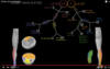
How do we draw the Lumbar Plexus?
Draw an ‘E’ and then another ‘E” and then a ‘F’ and then a bow tie and then a line and connects it with “v-link” and then labal with S.I.I.G.L.F.O for the nerves:
Subcostal; Iioinguinal, iliohypogastric, genital femoral, lateral femoral cutaneous, femoral, and obtorator.
Don’t forget to label the nerve levels: T12, L1, L2, L3, L4.

What skin does the Lateral femoral cutaneous innervate?
the Anterior lateral thigh

what skin does the femoral nerve innervate?
It will innervate the anterior middle thigh, and the medial lower leg thorugh the Anterior femoral cutaneous and the Safinous nerves.

What does sensory of the skin innervation to the front of the lower limb?
the sciatic nerve that becomes the common fibular cutaneous nerve, that becomes the superfical fibular nerve and the deep fibular nerve
and finally at the lateral spot of the foot is the sural nerve.

What innervates the muscles of the thigh?
the Anterior compartment of the thigh is innervated by the femoral nerve, the medial thigh is innervated by the obterator nerve. and the posterior thigh is innervated by the tibial nerve.

what is the primary action of the different compartments of the thigh?
the anterior: Extension
Posterior: Flexion
Medial Adduct.

How do we draw the sciatic nerve, what are the motor and sensory branches and what do they innervate?
Idendtify this muscle its actions, inervations and insertions…

Sartorius, ASIS, the medial to the tibial tuberosity; flex, abduct, and external rotate the hip.
ID this muscle where it is what does and what innervate

Vastaus lateralis, arise from the linea aspera on femer and course down and engulph the patela and insert on tibial tuberosity, Innervate by the femoral nerve.
ID this muscle and where it is, what does, and what innervates it?

the rectus femoris muscle in the anterior comp. of femer => femoral nerve, and extend the knee joint, insert from anterior inferior iliac spine. (also a weak hip flexor since only one of quads to cross the hip joint. engaulph the patella and then tibial tuberosity.
ID muscle, location, innervation, and actions!

Vastus medialis, anterior comp. of thigh, extend the knee and innervated by the femoral nerve, linea aspa to the patella to the tibial tuberostity.
ID muscle, action, location, innervation.

The vastus intermediaus muscle. Extend the knee, anterior thigh comp. femoral nerve. front of the femur, engulph the tibia and insert tibial tuberosity.
what is the major myatome for the extension of the knee and therefore the emphisis of the femoral nerve for the extension of knee
L4
the emphisis of the ant. thigh comp. is on the L4 level!!!!!
Common issertion of the anterio thigh muscles?
the tibial tuberostiy!!!
The five muscles of the medial thigh

ID this muscle, innervation, Action, location,

Adductor Longus muscle, Adduct the thigh, medial comp. of thigh, obtorator nerve. from the pubic bone to the linea aspera of femor
ID
Location
Innervation
Actoin

Adductor brevis muscle
Medial thigh comp. pubic bone to the linea aspera
obtorator nerve
adduct the thigh
ID
Location
Innervation
Action

Adductor magnus muscle
med. comp. of thigh from ischium/pubic to linea aspera and the adductor tubericl
obtorator nerve where it attaches to the linea aspera and tibial nerve where it attaches to the adductor tuberosity.
adduction of the thigh
What is the adductor hiatus?
Why important?
the opening in the adductor magnus where it stretches to atach to the adductor tuberosity.
this is where the femoral artery and vien course!

ID
Location
function
Innervation

gracilis muscle (thin like grass)
medial comp. from the pubis to the medial to the tibial tuberosity.
Addcut the thigh
obtorator nerve.
ID
location
Functino
innervation

Obturator externus
med. thigh comp ext. obtorator membrae, to the greater trochator
extrernal rotator
obtorator nerve.
the common attachment of the medial thigh muscles?
Common function?
common innervation?
the linea aspera
Adduct the hip
Obturator nerve (magnus has some tibial nerve inner.)
What is the major myatome level of the obtorator nerve for adduction of the thigh
L3 level!!!!!

What are the muscles of the posterior thigh?
the muscles of the posterior thigh:

ID
location
innervation
Action

Semitendinosus
Ischial tuberosity to medial the tibial tuberosity
The Tibial nerve
To flex the knee joint and extend the hip
What is this structure?
what are the muscles involved
what is the significance?

Medial part of the knee, medial to the tibial tuberosity, this is the Pes anserinus (foot of the duck)
The Sartorisus m; Gracilis m. Semitend m.
each of the muscles that come to this point are innervated by a different nerve
“SGT FOT”

ID
Innervation
Function
Location

Semimembranosus
Tibial nerve
flex the knee joint and extend the hip
posterior comp. of thigh: from the ischial tuberosity to proximal tibia
ID
Innervation
Function
Location

biceps femoris muscle
tibial nerve and short head is the common fibial nerve.
2 heads: posterior comp. of thigh: from ischial tub. to head of fibula and short is from the linea aspera to the head of the fibula
Flex the knee joing and the long head will also extend the hip
common innervation of the post. comp. of the thigh (hamstring muscles)
the Tibial nerve that comes from the L5-S3 levels at the sacral plexus

What are the ant. comp. lower leg muscles?

ID
Location
Innervation
action

Tibialis anterior muscle
front tibia to the tibia taylor joint and then to the bottom of the foot to the tarsals
Deep perionial nerve
Lifts the medial foot up and innverts the taylor joint.
ID
Location
Innervation
Action

Extensor hallucis longus
Ant. comp of leg: from more lat. on fibula to the tibial taylor joint and down to the big toe (hallusic or great toe
Deep perionial nerve
extends the great toe
ID
Innervation
action
location

extensor digitorum longus
Deep perionial nerve
to extend the digits and dorsal flexion
tibial/fibula to the tibial taylor to the digits 2-5 or the lessor toes.
ID
Location
Action
Innervation

Extensor hallucis brevis
on the dorsum of the foot, to the great toe
to extend the big toe
Deep peronial (fibular) nerve.
ID
action
innervation
location

Extensor digitorum brevis
extend the lessor toes
Deep peronial (fibial) nerve.
dorsal of the foot, but more lateral the dorsum.
ID these muscles!


Common innervation of the anterior comp. leg muscles!
Common action
Deep peronial nerve
dorsal fexion/digit extension
ID the muscles of the lateral compartment of the leg.

ID
Location
Action
Innervation

Peroneus longus
Lateral leg comp. from the head neck of the fibula to the lateral maleolus then to the lateral plantor surface of foot and to the 1st metatorasal
plantar flexor and Everstion (esp. eversion)
superficial peroneal nerve.

ID
Location
Action
Innervation

Peroneus brevis muscle
from the midshaft fibula to behind metatarsals
plantar fexor and Eversion
Superficial peronial nerve
ID muscles
common innervation?

superficial peroneal nerve.

ID these muscles
common action
common innervation

Plantar flex the foot,
Innervated by the Tibail nerve!

ID
Location
Action
Innervation

gastrocnemisu
two heads on lateral condiles of femer to the calcaneus (heel)
plantar flexion and week knee flexors
tibial nerve
ID
Location
Action
Innervation

Soleus muscle
tibia and fibula to the achiles tendon on the calcaneous
plantar flexion
tibial nerve.
the group of superficial muscles on the posterior of the leg is the gastronemius and the soleus or someimes calle dthe
triceps suris
ID
location
innervation
action

Plantaris muscle
from the lateral condile of femur. (the freshman nerve)
tibial nerve.
plantar flexor (weak knee fexor)
(don’t care if you know this one)
ID
Action
Location
Innervation

Popliteus muscle
unlock the knee when the knee is extended and tibia rotates in relation to the femur
Right behind the knee in a little square
tibial nerve.
Id
location
action
innervation

Tibialis posterior muscle
the tibial/fibula and interosius membrane to the medial condial, and to the plantar surface of the foot
plantar flexion and Inversion!

ID
location
innervation
action

flexor digitorum longus
From the tibial to the lesser toes
tibial nerve
plantar fexion and flexion of the lesser toes (curl to the ground)

ID
Location
Innervation
action

flexor hallucis longus muscle
from the mid fibula to the medial maleolus to the bottom of the hallucis
tibial nerve
plantar flexion and curl the big toe to the ground (flexion)

The memory aid for the deep posterior leg muscles
“Tom, Dick, and Harry”

