10/21- Liver failure and portal HTN Flashcards
What is seen here?

Normal liver (left) vs. cirrhosis (right)
Non-invasive alternatives to liver biopsies?
- Fibroscan
- Magnetic Resonance Elastography (MRE)
Describe Fibroscan
- Pros/cons
Fibroscan
- Works best on thin patients
- Tells you how stiff/soft the liver is (stiffness can indicated cirrhosis)
What do these fibroscan results show?

- Not as steep slope on left (stiffer?)
- Steep slope on right (softer?)
How do liver diseases compare on fibroscan results (grades of stiffness)?
Different diseases cause different fibrotic patterns and different densities. Also happen at different times

Describe Magnetic Resonance Elastography (MRE)
Again, indicates stiffness (blue -> red scale)
- Newer technique
- Good for saying yes/no to cirrhosis

What are the 3 types of liver decompensation?
- Synthetic failure
- Portal HTN
- Hepatocellular Carcinoma (HCC)
What are markers of synthetic failure in terms of liver disease (what symptoms do you see)?
Synthetic Failure
- Jaundice
- Prolonged INR (low clotting factors)
- Hypoalbuminemia
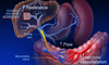
What are signs/symptoms of portal hypertension?
Portal vein entering the base of the liver; dilates; 3x increase in pressure (4->12 mmHg)
- Hypersplenism
- Fluid retention
- Ascites and peripheral edema
- Varices
- With/without bleeding
- Encephalopathy
What are the cardinal signs of liver failure?
If you have any of these, you have liver failure.
Mortality goes up significantly once any of these happen
- Jaundice
- Ascites
- May include SBP
- May include peripheral edema
- Variceal bleed
- Encephalopathy
- Clinical or subclinical
Other signs of liver failure?
- Spider angiomata
- Fed from the middle (blanching)
- Surprisingly common in people with cirrhosis (especially alcoholic)
- Palmar erythema
- Dupuytren contracture
Describe bilirubin synthesis
- RBCs -> hemoglobin -> globin + heme
- Heme is degraded into bilirubin (unconjugated); carried with albumin to liver
- Bilirubin transported into hepatocyte and binds Ligandin
- Glucuronidation -> conjugated bilirubin
- Bilirubin conjugation actually involves clevage site, heme oxygenase conversion into Biliverdin, and then conjugated bilirubin
- Bile excreted into bile duct In the intestine:
- Bilirubin glucuronide (conjugated) converted back into bilirubin (unconjugated) by gut bacteria
- Bilirubin converted into urobilinogen/urobilin
- Urobilin should be reabsorbed and sent to kidney (yellow color of urine)
- Urobilinogen converted to stercobilin and excreted by gut (brown color of stool)
**There is no bilirubin in urine or stool!
What are causes of high unconjugated (indirect) bilirubin?
Excess bilirubin production
- Hemolysis
Failure of conjugation (typ hereditary)
- Gilbert syndrome
- Neonatal jaundice
- Crigler-Najjar
(Other lecture also mentioned decreased uptake such as with Rifampin)
What are causes of high conjugated (direct) bilirubin?
- Biliary obstruction
- Liver damage
- Failed excretion
- Dubin-Johnson
- Rotor
(Other lecture separated this into main liver disease or obstructive causes; important here to also add failed excretion)
How is clinical testing for direct vs. indirect bilirubin done?
Uses a color assay (diazo)
- Conjugated bilirubin is soluble and direct-reacting
- Unconjugated bilirubin is not soluble and only reacts after alcohol is added
After initial and then alcohol stages, the total amount has reacted
- Indirect is calculated by subtracting direct from total
- A small amount of unconjugated reacts without alcohol (5-10% of total), so direct levels might be a tad high
- Normal unconjugated levels are 0, but will get a low amount using this assay
What is cholestasis?
Failure of bile excretion
What are effects of cholestasis?
- Bile contents in the circulation
- Bilirubin -> jaundice
- Bile salts -> pruritis
- Hepatocyte effects
- Obstruction -> alkaline phosphatase
- Damage -> ALT and AST
- Malabsorption of fats and fat-soluble vitamins
- Absence of stercobilin in stool (clay-colored) and urobilin in urine (colored instead by bilirubin, darker?)
Look at these pictures of dilated bile ducts due to obstruction

What is shown here?

ERCP
- Large duct obstruction
- Can see stone and dilated duct leading into liver
What is seen here? Main features?

Normal liver histology
- Portal tract (triad)
- Terminal hepatic venule
- Zones 1-2-3 moving from tract -> venule

What are diseases of small duct obstruction
- Primary biliary cirrhosis (aka non-suppurative
- Primary sclerosing cholangitis
What happens in primary biliary cirrhosis?
- PBC – cells and lymphocytes destroy bile duct
- Granulomas
What happens in primary sclerosing cholangitis?
- Primary sclerosing cholangitis
- Bead signs (dilated and strictured areas
- Thick onion-skin appearance of fibrous tissue obstruction the duct
What is seen here?

Primary biliary cirrhosis
- PBC – cells and lymphocytes destroy bile duct
- Granulomas
What is seen here?

- Primary sclerosing cholangitis
- Bead signs (dilated and strictured areas
- Thick onion-skin appearance of fibrous tissue obstruction the duct
What is seen here?

- Primary sclerosing cholangitis
- Bead signs (dilated and strictured areas
- Thick onion-skin appearance of fibrous tissue obstruction the duct
Look at these key anatomic features of the liver

- Hepatic vein: entering superiorly
- Sinusoid
- Portal vein: entering base of the liver
- Coronary vein: comes int quite high off of the liver
- Splenic vein
What happens vascularly in cirrhosis (think of key anatomic features just mentioned)
Portal HTN- result of increased portal venous inflow and sinusoidal resistance
- Splanchnic vasodilation and increased flow into mesenteric veins (leading to:)
- Increased flow into portal vein
- Distorted sinusoidal architecture -> increased resistance
What is the pathophysiology of ascites and HRS (hepato-renal syndrome)?
- Cirrhosis causes portal HTN and NO overproduction
- NO overproduction causes splanchnic and arterial vasodilation
- This splanchnic arterial vasodilation results in both (1) decreased effective BV and (2) splanchnic lymph production
- Decreased effective blood volume stimulates ADH production, Sympathetic nervous system activation, and the RAAS system
- All of these cause (1) sodium/water retention and (2) decreased renal blood flow
- Ascites results from this sodium/water retention (from decreased effective blood volume) and splanchnic lymph production (both of these are a result of splanchnic and arterial vasodilation)
- HRS results from decreased renal blood flow

What causes hyponatremia?
- Cirrhosis is the leading cause of ____
- Indication of outcomes
- Associations
- Treatment
NOT a shortage of salt
Cirrhosis is the leading causes of dilutional (hypervolemic) hyponatremia
- Water retention exceeds Na retention
- Complications and outcomes are worse in pts with hyponatremia
- May be associated with diuretics
- Treated with WATER restriction
What sign/symptom is a big turning point?
- Prognosis?
Ascites
- 50% mortality at 2 yrs
What causes ascites?
- Complications
- Treatment
- Requires 10 mm transhepatic pressure gradient
- May be infected- spontaneous bacterial peritonitis (SBP)
Treatment: paracentesis
- Direct aspiration of fluid
- Bleeding is not related to coagulopathy
- Fluid shift after paracentesis
What is the Serum-Ascites Albumin Gradient (SAAG)?
- Hydrostatic pressure pushes fluid out
- Osmotic pressure pulls fluid in
- The difference is SAAG (>1.1 g/dL)
- Serum and ascites on the same day
- > 97% accuracy in predicting portal hypertension
How does paracentesis of someone with ascites change the body volume compartments?
- In ascites, there is much flow from blood compartment into body water and ascites
- With paracentesis, you remove the fluid from ascites compartment, but also blood volume (poor perfusion of kidney)
- Want to add albumin to move fluid from body water and maintain blood volume
How does diuretic use of someone with ascites change the body volume compartments?
- Decrease blood volume in an attempt to decrease amount of fluid causing ascites?
- Diuretics are dangerous, because they remove fluid from your most vulnerable compartment (blood), causing decreased perfusion to kidneys

What do diuretics do?
- How to use
- Promote excretion of salt in urine
- Combination is more effective
- Spironolactone 100 – 400 mg/day
- Furosemide 40 – 160 mg/day
- Peripheral edema provides a buffer
- Patients without edema are at risk of renal failure
- May cause hyponatremia
What is refractory ascites?
- Prevalence
- Prognosis
Failure to respond to optimal dosing of diuretics and sodium restriction
- Assuming compliance with 2g Na
- Not taking NSAIDs Happens in under 10% of pts 75% mortality at 1 yr
What are options for treating refractory ascites?
- Serial paracentesis
- TIPS
- Liver transplantation
- Perinovenous shunt (LeVeen, Denver)
How is serial paracentesis used to treat refractory ascites?
What do different amounts of ascites indicate/require?
Pts who require paracentesis >6L every 10 days are not compliant with sodium restriction
- Ascites [sodium] is the same a serum
- 10d oral intake amount = 6 L ascites fluid amount
Albumin infusion
- Prevents renal injury
- Given for paracentesis > 5L TIPS makes ascites easier to control
How does TIPS work?
Transjugular Intrahepatic Porto-systemic Shunt
- Expandable shunt from hepatic vein to portal vein
- Immediately solves the pressure problem
- Downside is that this shunted blood is no longer filtered… -> encephalopathy and confusion

What is Hepatorenal syndrome (HRS)?
- Caused by advanced chronic or acute liver failure with portal hypertension
- Serum creatinine >1.5 mg/dL or clearance under 40 mL /min
- No shock, bacterial infection, nephrotoxic drugs, or massive gastrointestinal or renal fluid losses
- No sustained improvement in renal function following diuretic withdrawal and expansion of plasma volume with 1.5 L of isotonic saline
- Less than 500 mg/dL proteinuria and no ultrasonographic evidence of obstructive uropathy or parenchymal kidney disease
What are the types of hepatorenal syndrome?
Type 1: rapidly progressing
- Doubling of the initial serum creatinine to a level > 2.5 mg/dL OR
- 50% reduction in initial creatinine clearance to a level lower than 20 mL/min in under 2 wks
Type 2: slow course
What is hepatorenal management?
- Triple therapy
- Albumin
- Octreotide (SS analog)
- Midodrine
- TIPS
- Liver transplantation
What is seen here?

Esophageal bleed on endoscopy
Portal hypertension can lead to varices where?
How does it alter blood flow?
High pressure in portal vein reverses direction of blood through coronary veins to stomach and esophagus
- Esophageal varices
- Stomach
What are the different stages of varices/pressures?
- Expansion: HPVG > 10 mmHg (hepatic portal vein gradient?)
- Bleeding: HPVG > 12 mmHg (mean 20 mmHg)
What characteristics of varices is related to risk of bleed?
- Location (gastric > esophageal)
- Risk of bleed is related to the SIZE of varices (directly proportional to wall tension; LaPlace)
- Appearance: red sign (mucosa starting to split apart)
- Variceal pressure > 12 mmHg (not direct ratio, just 12 threshold)

The risk of bleeding with varices is due to ___ NOT ____
The risk of bleeding with varices is due to location (and underlying pressure) NOT coagulopathy!
What are the relative bleeding risks for different variceal locations?
1. Esophageal (most common)
2 .Gastroesophageal (higher risk)
- Lesser curve (GOV type 1)
- Greater curve (GOV type 2)- bleed more than lesser
3. Isolated gastric varices (less common but higher bleeding risk than GOV for type 1. Basically all IGV type 1 bleed)
- Fundus (IGV type 1)- bleed more than other
- Other (IGV type 2)

What is seen here?

Red sign (Red Wales) of varices

What is primary prophylaxis for varices?
- Hepatic venous pressure gradient (HVPG) is affected by inflow and outflow
- No bleeding if HVPG under 12 mm Hg
Treatment:
- Non-selective beta-blockers: reduce splanchnic blood flow
- Vasoconstriction (unopposed alpha)
- Decreased cardiac output
- TIPS
- Variceal ligation (+/- B blocker)
Not:
- Nitrates – higher mortality
- Angiotensin II blockers not helpful
T/F: Most cirrhotics have true clotting defect
False?
What can be done to evaluate in vivo coagulation?
Thrombin generation assay is pretty close
- Measures both procoagulant and anticoagulant activities
- “ETP—endogenous thrombin potential”
What conclusions were found in regards to coagulopathy in cirrhotics?
- Routine coagulation tests (PT, PTT) are inadequate to assess bleeding risk in cirrhotic patients
- Thrombin generation assays incorporating anticoagulant factors provide a more relevant indicator of bleeding risk
- Coagulation is “rebalanced” in cirrhosis
- Abnormal coagulation in cirrhosis is less important than portal hypertension in increasing bleeding risk!
T/F: There is a concern about thrombosis in cirrhosis
True!
May be shifting from worried about bleeding (coagulation defect) to thrombosis
- One study found an increased risk of VTE in patients with cirrhotic and non-cirrhotic liver disease
- An imbalance in coagulation was found favoring procoagulant activity
- Looks like Protein C deficiency
What are different encephalopathy symptoms seen in cirrhotics (more/less common)?
More common:
- Confusion or coma
- Asterixis
- Loss of fine motor skills
- Hyperreflexia
Less common:
- Cognitive deficits detected by special testing
- Babinski sign
- Slow, monotonous speech
- Extrapyramidal-type movement disorders
- Clonus
- Decerebrate posturing
- Decorticate posturing
- Hyperventilation
- Seizures*
What are the symptoms of encephalopathy: stage 0?
- No changes in personality or behavior
- No asterixis
What are the symptoms of encephalopathy: stage 1?
- Trivial lack of awareness
- Shortened attention span
- Impaired addition or subtraction.
- Hypersomnia, insomnia, or inversion of sleep pattern
- Euphoria or depression
- Asterixis may be present
What are the symptoms of encephalopathy: stage 2?
- Lethargy or apathy
- Disorientation
- Inappropriate behavior
- Slurred speech
- Obvious asterixis
What are the symptoms of encephalopathy: stage 3?
- Gross disorientation
- Bizarre behavior
- Semistupor to stupor
- Asterixis generally absent
What are the symptoms of encephalopathy: stage 4?
Coma
What are goals of therapy in treating encephalopathy?
Identify and eliminate precipitating factors
- Infection
- GI Bleeding
- Constipation
- Hypokalemic metabolic alkalosis
- Drugs: benzodiazepines, barbiturates, opiates
Inhibit noxious neurotransmitters
- Reduce production and absorption of nitrogenous substances from the gut
- Increase excretion of nitrogenous substances
What drugs can be used to treat encephalopathy?
- Lactulose
Antibiotics
- Rifaximin
- Neomycin
- Metronidazole
Characteristics of Lactulose?
- Drug class
- Indications
Poorly absorbed disaccharide; changes colon pH and bacterial growth environment
- Prevention and treatment of HE
- Diarrhea (esp), dehydration, hypernatremia
Characteristics of Rifaximin?
- Drug class
- Indications
Non-aminoglycoside, semi-synthetic, nonsystemic antibiotic
- Reduction in risk of recurrence of overt HE in patients 18+ years old
Characteristics of Neomycin?
- Drug class
- Indications
Aminoglycoside antibiotic
- Adjuvant in hepatic coma or intolerance other antibiotics
- Ototoxicity and nephrotoxicity
Characteristics of Metronidazole?
- Drug class
- Indications
Synthetic antiprotozoal and antibacterial agent
- Not FDA approved for HE May cause peripheral neuropathy and DDI with alcohol
Fun fact: on an individual basis, ammonia levels aren’t that indicative of risk of HE event (although there is some broad correlation)
Merp
How to manage PSE?
- Serum ammonia is not usually helpful
- Maintain protein intake as tolerated
- We no longer recommend protein restriction
- Lactulose
- Non-absorbable antibiotics
- Neomycin
- Metronidazole
- Rifaximin
- Experimental therapy – GPB
How to assess liver failure (for possible surgery?)?
Child-Turcotte-Pugh (CTP)

What are the Child-Turcotte-Pugh (CTP) classes based on point score?

What scoring system is better than CTP for evaluating liver failure?
MELD- Model of End Stage Liver Disease
What does MELD measure? Predict?
- Formula based on bilirubin, INR, creatinine; no subjective component
- Predicts mortality at 90 days
- Adopted for liver allocation in 2002
- Sickest get transplanted first
- Avoids certain problems with CTP
- Accurate in about 80% of patients – requires certain exceptions
What is the MELD formula?
6.3 + ([0.957 x log creat] + [0.378 x log bili] + [1.12 x log INR] + 0.643) x 10
When should MELD be renewed? Exceptions?
- 25+ – every 7 days
- 19-24 – every 30 days
- 11-18 – every 90 days
- < 10 – every year
Exceptions:
- Tumors
- Biliary sepsis
- Lung diseases
- Metabolic diseases
- Bleeding
What is the prognostic significance of MELD scores?

Prognostic evaluations of MELD score are affected by what?
Serum sodium!
- With lower sodium, MELD score doesn’t have quite such a bad prognosis (modulated)
Summary of Cirrhosis Complications

Summary of Ascites and HRS

Summary of Variceal Bleeding

Summary of PSE

Summary of Risk Assessment

